



Chitoheptaose
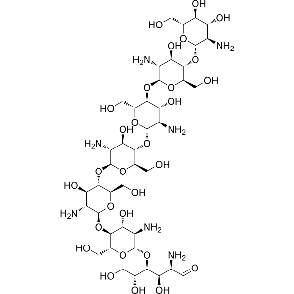
Chitoheptaose is an orally active chitooligosaccharide (degree of polymerization = 7). Chitoheptaose can be extracted from the exoskeletons of crustaceans, such as the shells of crabs, shrimps and lobsters. Chitoheptaose reduces pro-inflammatory cytokines while increasing the levels of anti-inflammatory cytokines (decreasing the levels of pro-inflammatory cytokines IL-1β, IL-17A and IFN-γ, and increasing the level of the anti-inflammatory cytokine IL-10). Chitoheptaose possesses antioxidant, anti-inflammatory and anti-Apoptotic activities. Chitoheptaose improves cardiac parameters, alleviates myocarditis injury, and exerts cardioprotective effects in a rat model of myocarditis. Chitoheptaose can be used in studies related to myocarditis.
For research use only. We do not sell to patients.
- CAS No.: 68232-35-9
- Formula: C42H79N7O29
- Molecular Weight:1146.11
-
Storage:
-20°C, stored under nitrogen
* In solvent : -80°C, 6 months; -20°C, 1 month (stored under nitrogen)

To place orders, for customer services and technical support, please contact: MedChemExpress USA
Tel: 609-228-6898 E-mail: [email protected] [email protected]


-
 Biological Activity
-
Chemical Information
- Protocols
- Purity & Documentation
- References
-
 Help & FAQs




Biological Activity
Description
IC50 & Target
[1]|
IL-1β |
IL-17A |
IL-10 |
In Vitro
Chitoheptaose (100 μM; 24 h prior to 6 h LPS stimulation at 200 ng/mL) exerts anti-inflammatory activity in LPS-stimulated RAW264.7 cells, with activity greater than chitopentaose and chitohexaose, ranking third among tested chitosan oligosaccharides with degree of polymerization >4[1].
MedChemExpress (MCE) has not independently confirmed the accuracy of these methods. They are for reference only. Further protocols information, click here.
-
Cell Line:RAW264.7 macrophage cells
-
Concentration:100 μM
-
Incubation Time:24 h (prior to 6 h LPS stimulation at 200 ng/mL)
-
Result:Reduced levels of proinflammatory cytokines IL-1β, IL-17A, and IFN-γ.
Increased levels of anti-inflammatory cytokine IL-10.
Showed significantly greater anti-inflammatory activity than chitopentaose and chitohexaose, but less than chitooctaose (p < 0.05).
In Vivo
MedChemExpress (MCE) has not independently confirmed the accuracy of these methods. They are for reference only.
-
Animal Model:SPF rats (8-week-old, 200-220 g, experimental autoimmune myocarditis model induced by subcutaneous injection of porcine cardiac myosin emulsified with complete Freund’s adjuvant on day 1 and day 8)[1]
-
Dosage:100 μM
-
Administration:p.o.; daily; 30 days
-
Result:Reduced left ventricular internal diameter in end diastole (LVIDd) and left ventricular internal diameter in end systole (LVIDs) significantly compared with other chitosan oligosaccharides (p < 0.05).
Increased ejection fraction (EF) and fractional shortening (FS) significantly compared with other chitosan oligosaccharides (p < 0.05).
Reduced myocardial histopathological scores and inflammatory cell infiltration more effectively than other chitosan oligosaccharides (p < 0.05).
Lowered serum levels of proinflammatory cytokines IL-1β, IL-17A, and IFN-γ, and increased anti-inflammatory cytokine IL-10 significantly compared with other chitosan oligosaccharides (p < 0.05).
Reduced cardiac tissue levels of reactive oxygen species (ROS) and reactive nitrogen species (RNS) significantly compared with other chitosan oligosaccharides (p < 0.05).
Reduced relative mRNA and protein levels of proapoptotic factors caspase 3 and BAX, and increased relative mRNA and protein levels of antiapoptotic factor BCL-2 significantly compared with other chitosan oligosaccharides (p < 0.05).
Reduced IHC scores for caspase 3 and BAX, and increased IHC scores for BCL-2 significantly compared with other chitosan oligosaccharides (p < 0.05).
Achieved a serum level of 45 μM after treatment.


Chemical Information
-
CAS No. 68232-35-9
-
Appearance Solid
-
Molecular Weight 1146.11
-
Formula C42H79N7O29
-
Color White to off-white
-
SMILES
OC[C@@H](O[C@H]([C@@H]([C@H]1O)N)O[C@@H]2[C@H](O[C@H]([C@@H]([C@H]2O)N)O[C@H]([C@H](O)CO)[C@H](O)[C@@H](N)C=O)CO)[C@H]1O[C@@H]3O[C@@H]([C@H]([C@@H]([C@H]3N)O)O[C@@H]4O[C@@H]([C@H]([C@@H]([C@H]4N)O)O[C@@H]5O[C@@H]([C@H]([C@@H]([C@H]5N)O)O[C@@H]6O[C@@H]([C@H]([C@@H]([C@H]6N)O)O)CO)CO)CO)CO
-
Shipping
Room temperature in continental US; may vary elsewhere.
-
Storage
-20°C, stored under nitrogen
* In solvent : -80°C, 6 months; -20°C, 1 month (stored under nitrogen)

Protocols
-
Apoptosis
Apoptosis, also called programmed cell death, is generally characterized by distinct morphological characteristics.
-
LPS-Induced Endotoxemia/Systemic Inflammation
Lipopolysaccharide (LPS)-induced endotoxemia is a widely used in vivo model of acute systemic inflammation in which LPS, a Gram-negative bacterial endotoxin, activates innate immune signaling primarily through TLR4, leading to rapid and transient induction of pro-inflammatory cytokines such as TNF-α, IL-6, and IL-1β in circulation and tissues. This cytokine surge is commonly used as a measurable readout of systemic inflammatory activation and immune dysregulation, and is typically assessed within hours after intraperitoneal LPS administration in mouse models of endotoxemia. The model captures key features of systemic inflammatory response syndrome, including cytokine release, immune cell activation, and downstream tissue responses, and has been used to evaluate anti-inflammatory interventions such as cytokine modulation, lipid mediators, and immune cell-targeting therapies.
-
How to Select a Suitable Non-Mouse Animal Model
Selecting a suitable non-mouse animal model is a structured decision based on the research question, required anatomy or physiology, disease mechanism, endpoint feasibility, translational relevance, and ethical justification. Non-mouse models are preferred when mice cannot reproduce key human-relevant features, such as organ size, surgical anatomy, cardiovascular physiology, neuroanatomy, immune features, pharmacology, toxicology, or long-term clinical procedures. Candidate species may include rats, rabbits, guinea pigs, ferrets, zebrafish, pigs, sheep, goats, dogs, cats, horses, and non-human primates, but each species must be justified by its specific scientific advantage rather than convenience or tradition. Unresolved questions include how to quantify translational superiority across species, how to balance increased biological relevance against higher ethical burden, and when human-derived systems or new approach methodologies should replace animal use.
-
Apoptosis Solutions
Apoptosis is a regulated, generally non-lytic cell-death pathway that removes unwanted, damaged, infected, or abnormal cells through coordinated morphological changes, caspase activation, DNA fragmentation, and membrane remodeling. The intrinsic apoptosis pathway is controlled mainly by mitochondrial outer membrane permeabilization, BCL-2 family proteins, cytochrome c release, apoptosome formation, caspase-9 activation, and downstream executioner caspase-3/7 activation. The extrinsic apoptosis pathway is initiated by death receptors such as Fas, TNFR, and TRAIL receptors, which recruit adaptor proteins and activate caspase-8 before engaging executioner caspases or mitochondrial amplification through BID cleavage. Apoptosis is linked to many phenotypes, including cancer cell killing, tissue homeostasis, immune regulation, neurodegeneration, infection response, and treatment-induced cytotoxicity; unresolved questions include how apoptosis interacts with necroptosis, pyroptosis, ferroptos
-
Research Protocol for Inflammation-related Diseases
The NLRP3 inflammasome is a cytosolic innate immune signaling platform that integrates priming signals and danger-signal activation to promote caspase-1 activation, maturation of IL-1β and IL-18, and gasdermin D-mediated pyroptotic cell death. The core experimental logic is to determine whether inflammatory disease phenotypes are driven by increased NLRP3 expression, ASC-containing inflammasome assembly, caspase-1 cleavage, GSDMD cleavage, and extracellular release of IL-1β/IL-18 rather than by nonspecific cell injury alone. The pathway is strongly linked to inflammation-related disease phenotypes because monosodium urate crystals activate NALP3/NLRP3 inflammasome signaling in gout-like crystal inflammation, cholesterol crystals activate NLRP3 inflammasomes in atherogenesis models, and DSS-induced intestinal inflammation has been reported to involve NLRP3 inflammasome activity. However, experimental colitis studies also show context-dependent protective effects of NLRP3 inflammasome co
-
Pyroptosis Solutions
Pyroptosis is a lytic inflammatory cell-death pathway executed by gasdermin pores, most classically through inflammasome-mediated activation of caspase-1, cleavage of gasdermin D, membrane pore formation, LDH release, and secretion of IL-1β and IL-18. The canonical pathway is commonly modeled by priming cells with an inflammatory signal such as LPS to induce pro-IL-1β and inflammasome components, followed by an activation signal such as ATP or nigericin to activate NLRP3, ASC speck formation, caspase-1 cleavage, GSDMD cleavage, cytokine release, and pyroptotic membrane rupture. The non-canonical pathway is triggered when cytosolic LPS activates mouse caspase-11 or human caspase-4/5, leading to GSDMD cleavage and pyroptosis, and this can secondarily activate NLRP3-dependent IL-1β release. Pyroptosis is linked to inflammatory injury, infection, cancer, liver disease, ocular disease, placental inflammation, and other disease phenotypes, but unresolved questions include which gasdermin fam
-
How to Choose the Right Model Animal
Choosing the right model animal is a validity-driven decision in which the species, strain, sex, age, genetic background, disease-induction method, outcome measures, and welfare burden must match the scientific question rather than laboratory tradition or convenience. A model should be selected by judging face validity, construct validity, and predictive validity: whether it resembles the human phenotype, whether it reproduces relevant mechanisms, and whether results are likely to predict human biology or treatment response. Animal studies often fail to translate because of species differences, weak disease resemblance, poor experimental design, inadequate reporting, publication bias, and underuse of randomization, blinding, and sample-size justification. Unresolved questions include how to rank competing models objectively, how much human-disease complexity must be reproduced for a given objective, and when non-animal systems such as organoids, ex vivo tissue, or computational models


Purity & Documentation
-
Data Sheet (279 KB)
-
SDS (252 KB)
- English - EN (252 KB)
- Français - FR (252 KB)
- Deutsch - DE (252 KB)
- Norwegian - NO (252 KB)
- Español - ES (252 KB)
- Swedish - SV (252 KB)
- Italian - IT (252 KB)
- Korean - KR (252 KB)
- Portuguese - PT (252 KB)
-
Handling Instructions (2659 KB)
References

Calculators
Concentration (start) × Volume (start) = Concentration (final) × Volume (final)


Chitoheptaose Related Classifications
HY-W145695 Related Classifications
 Powered by Bioz
Keywords
- Chitoheptaose
- 68232-35-9
- Biochemical Assay Reagents
- Apoptosis
- Interleukin Related
- IFNAR
- antiapoptotic factor
- reactive oxygen species
- rat autoimmune myocarditis model
- chitosan oligosaccharide
- proapoptotic factor
- rat myocarditis model
- anti-inflammatory cytokine
- proinflammatory cytokine
- RAW264.7 cells
- reactive nitrogen species
- Inhibitor
- inhibitor
- inhibit