



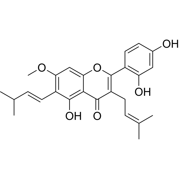
Artocarpin
Artocarpin is an orally active apoptosis inducer. Artocarpin targets NF-κB, Erk1/2, p38 MAPK, AktS473, p53, Akt 1 kinase and Akt 2 kinase. Artocarpin induces reactive oxygen species (ROS) production, mediates p53-dependent and p53-independent apoptotic signaling pathways, induces G1-phase cell cycle arrest, and triggers autophagic cell death. Artocarpin exerts cytotoxic and bactericidal effects on cancer cells, reduces bacterial load, and exhibits anti-inflammatory, analgesic and anti-angiogenic activities.
商品は「研究用試薬」です。人や動物の医療用・臨床診断用・食品用の製品ではありません。
研究用途以外に使用した場合、当社は一切の責任を負いかねます。
- CAS 番号: 7608-44-8
- 分子式: C26H28O6
- 分子量:436.50
-
保管条件:
Please store the product under the recommended conditions in the Certificate of Analysis.

To place orders, for customer services and technical support, please contact: MedChemExpress USA
Tel: 609-228-6898 E-mail: [email protected] [email protected]


-
 生物活性
生物活性
-
化学情報
- プロトコル
- 純度とドキュメンテーション
- 参考文献
-
 Help & FAQs




生物活性
製品説明
Cellular Effect
|
Cell Line

|
Type | Value | Description | References |
|---|---|---|---|---|
| 1A9 | ED50 |
3.4 μg/mL
Compound: 6, Artocarpin
|
Cytotoxicity against human 1A9 cells
Cytotoxicity against human 1A9 cells
|
[PMID: 15165133] |
| A549 | ED50 |
3.3 μg/mL
Compound: 6, Artocarpin
|
Cytotoxicity against human A549 cells
Cytotoxicity against human A549 cells
|
[PMID: 15165133] |
| CAKI-1 | ED50 |
4.9 μg/mL
Compound: 6, Artocarpin
|
Cytotoxicity against human Caki1 cells
Cytotoxicity against human Caki1 cells
|
[PMID: 15165133] |
| HCT-8 | ED50 |
3.8 μg/mL
Compound: 6, Artocarpin
|
Cytotoxicity against human HCT8 cells
Cytotoxicity against human HCT8 cells
|
[PMID: 15165133] |
| KB | ED50 |
3.2 μg/mL
Compound: 6, Artocarpin
|
Cytotoxicity against human KB cells
Cytotoxicity against human KB cells
|
[PMID: 15165133] |
| MCF7 | ED50 |
3.3 μg/mL
Compound: 6, Artocarpin
|
Cytotoxicity against human MCF7 cells
Cytotoxicity against human MCF7 cells
|
[PMID: 15165133] |
| MDA-MB-231 | ED50 |
3.8 μg/mL
Compound: 6, Artocarpin
|
Cytotoxicity against human MDA-MB-231 cells
Cytotoxicity against human MDA-MB-231 cells
|
[PMID: 15165133] |
| PC-3 | ED50 |
4.1 μg/mL
Compound: 6, Artocarpin
|
Cytotoxicity against human PC3 cells
Cytotoxicity against human PC3 cells
|
[PMID: 15165133] |
| RAW264.7 | IC50 |
18.7 μM
Compound: 6
|
Inhibition of LPS-induced nitric oxide production in mouse RAW264.7 cells assessed as nitrite accumulation after 20 hrs
Inhibition of LPS-induced nitric oxide production in mouse RAW264.7 cells assessed as nitrite accumulation after 20 hrs
|
[PMID: 16643064] |
| RAW264.7 | IC50 |
45.3 μM
Compound: 6
|
Cytotoxicity against mouse RAW264.7 cells by MTT assay
Cytotoxicity against mouse RAW264.7 cells by MTT assay
|
[PMID: 16643064] |
| SK-MEL-2 | ED50 |
5.4 μg/mL
Compound: 6, Artocarpin
|
Cytotoxicity against human SK-MEL-2 cells
Cytotoxicity against human SK-MEL-2 cells
|
[PMID: 15165133] |
| U-87MG ATCC | ED50 |
3.7 μg/mL
Compound: 6, Artocarpin
|
Cytotoxicity against human U87MG cells
Cytotoxicity against human U87MG cells
|
[PMID: 15165133] |
| Vero | CC50 |
80 μg/mL
Compound: AH-5
|
Cytotoxicity against African green monkey Vero cells after 72 hrs by MTT assay
Cytotoxicity against African green monkey Vero cells after 72 hrs by MTT assay
|
[PMID: 36222797] |
体外実験
Artocarpin (0-20 μM; 24 h) induces concentration-dependent apoptosis in A549 and H1299 NSCLC cells, as evidenced by increased DNA fragmentation, subG1 phase cell accumulation, and early/late apoptotic cell populations after 24-hour treatment[1].
Artocarpin (0-25 μM; 0-120 min) induces time- and concentration-dependent ROS generation in A549 and H1299 NSCLC cells via a NADPH oxidase (Nox)-dependent pathway, but does not induce ROS in normal human pulmonary epithelial cells (HPAEpiCs)[1].
Artocarpin (10 μM; 0-24 h) induces ROS- and MAPK-dependent activation of the p53 apoptotic pathway, increasing expression of phosphorylated p53, PUMA, cytochrome c, Apaf-1, and cleaved caspase-3 in A549 NSCLC cells over 24 hours[1].
Artocarpin (10 μM; 0-24 h) induces ROS- and Akt-dependent activation of NF-κB, leading to increased expression of c-Myc and Noxa in A549 and H1299 NSCLC cells[1].
Artocarpin (10-20 μM; 48 h, 3-48 h) induces apoptosis and autophagy in DLD1, HCT15, and HT29 human colon adenocarcinoma cells, as evidenced by PARP cleavage and increased LC3B expression at 10 and 20 μM (after 48 h) and time-dependent up-regulation of these markers over 3 to 48 h at 20 μM[2].
Artocarpin (20 μM; 12-48 h) induces G1 phase cell cycle arrest followed by apoptotic cell death in HT29 human colon adenocarcinoma cells when treated at 20 μM for 12, 24, 36, or 48 h[2].
Artocarpin (6.7 μM) potently inhibits melanin biosynthesis in B16 melanoma cells with an IC50 of 6.7 μM, without inducing cytotoxicity and without inhibiting tyrosinase activity[3].
Artocarpin displays weak but relatively broad in vitro cytotoxicity against A549, MCF-7, 1A9, HCT-8, U-87-MG, MDA-MB-231, KB, and KB-VIN human tumor cell lines with ED50 values ranging from 3.2 to 3.8 μg/mL, and shows inactive cytotoxicity against CAKI-1, SK-MEL-2, and PC-3 cell lines[4].
Artocarpin (tested across a concentration range; 20 h with LPS) inhibits nitric oxide production in LPS-activated RAW264.7 mouse macrophage cells with an IC50 of 18.7 μM[5].
Artocarpin (2-20 μg/mL; 5 min-24 h) exerts rapid, concentration-dependent bactericidal activity against S. aureus ATCC 29213, with up to a ~4.8 log10 cfu/mL reduction at 10× MIC within 1 h and no regrowth after 24 h[6].
Artocarpin (10 μg/mL; 24 h) potently clears intracellular S. aureus ATCC 29213 in J774 macrophages, reducing bacterial load by ~3.25 log10 cfu/mL at 5× MIC after 24 h of treatment[6].
Artocarpin (5-10 μg/mL; 30 min-1 h) disrupts the membrane integrity of S. aureus ATCC 29213, causing rapid cellular lysis, reduced intracellular ATP, and increased extracellular ATP at 2.5× and 5× MIC[6].
MedChemExpress (MCE) has not independently confirmed the accuracy of these methods. They are for reference only. Further protocols information, click here.
-
Cell Line:A549, H226, H1299 (human non-small cell lung carcinoma cell lines)
-
Concentration:0-10 μM
-
Incubation Time:24 h
-
Result:Inhibited cell proliferation in a concentration-dependent fashion, with IC50 values of 3.1303 μM for A549 cells, 6.4612 μM for H226 cells, and 7.9890 μM for H1299 cells.
-
Cell Line:A549, H1299 (human NSCLC cell lines)
-
Concentration:0-10 μM (DNA fragmentation and cell cycle analysis in A549 cells); 10 μM (cell cycle analysis in H1299 cells); 0-20 μM (Annexin-V-FITC/PI assay in A549 and H1299 cells)
-
Incubation Time:24 h (all assays)
-
Result:Induced concentration-dependent increases in DNA fragmentation and the proportion of cells in the subG1 phase in A549 cells. Reduced the proportion of viable cells and increased the proportion of cells in early apoptosis (Annexin-V-positive/PI-negative) in a concentration-dependent manner in A549 and H1299 cells; concentrations of 15 and 20 μM also significantly increased late apoptosis (Annexin-V-positive/PI-positive).
-
Cell Line:A549, H1299 (human NSCLC cell lines)
-
Concentration:10 μM
-
Incubation Time:0-4 h (phosphorylation time-course); 4 h (inhibitor-pretreated or siRNA-transfected cells)
-
Result:Induced time-dependent phosphorylation of p38 MAPK, ERK1/2, and AktS473 in both cell lines, with significant increases observed within 0.5 h. Reduced artocarpin-induced phosphorylation of p38 and ERK1/2 significantly when cells were pretreated with SB202190, U0126, APO, or NAC; reduced artocarpin-induced phosphorylation of AktS473 significantly when cells were pretreated with LY294002, APO, or NAC. Attenuated artocarpin-induced phosphorylation of p38, ERK1/2, and AktS473 significantly when cells were transfected with p47phox siRNA.
-
Cell Line:DLD1, HCT15, HCT116, HT29, SW480 human colon adenocarcinoma cells; CCD-18Co nonmalignant human colon fibroblast cells
-
Concentration:0-25 μM
-
Incubation Time:48 h
-
Result:Exhibited potent cytotoxicity against human colon cancer cells with IC50 values around 15 μmol/L, while nonmalignant CCD-18Co cells were much less sensitive at similar concentrations.
-
Cell Line:HT29 human colon adenocarcinoma cells
-
Concentration:20 μM
-
Incubation Time:12-48 h
-
Result:Induced a significant G1 phase cell cycle arrest, with a corresponding increase in the sub-G1 phase (apoptotic cells) over time.
-
Cell Line:HT29 human colon adenocarcinoma cells
-
Concentration:20 μM (time-course analysis); 10-20 μM (EGF pre-incubation)
-
Incubation Time:3-48 h (20 μM time-course); 15 min pre-incubation (EGF stimulation)
-
Result:Reduced the phosphorylation of Akt (Ser473) in a time-dependent manner over 3 to 48 h. Inhibited EGF-induced phosphorylation of Akt (Ser473) when used as a pre-treatment at 10 and 20 μM.
体内実験
Artocarpin (AH-5) (50 mg/kg; i.p.; two doses 3 hours apart) significantly reduces S. aureus ATCC 29213 bacterial load in neutropenic murine thigh tissue by ~0.6 log10 cfu/g[6].
Artocarpin (AH-5) (1%; topical; twice daily; 4 days) significantly reduces S. aureus ATCC 29213 bacterial load in murine skin tissue by ~1.0 log10 cfu/g[6].
Artocarpin (25-100 mg/kg; p.o.) exhibits dose-dependent anti-inflammatory activity in Wistar albino rats, with 100 mg/kg producing comparable inhibition to indomethacin 10 mg/kg in acute edema models and significant granuloma reduction[7].
Artocarpin (25-100 mg/kg; p.o.) exhibits dose-dependent analgesic activity in Swiss albino mice, with 100 mg/kg producing comparable inhibition to diclofenac 10 mg/kg in formalin-induced paw licking and significant writhing reduction in acetic acid-induced writhing[7].
MedChemExpress (MCE) has not independently confirmed the accuracy of these methods. They are for reference only.
-
Animal Model:BALB/cA-nu (nu/nu) (male, 6 weeks old)[1]
-
Dosage:1 mg/kg
-
Administration:daily; 21 days
-
Result:Significantly reduced mean tumor volume compared to vehicle control.
Significantly increased expression of phospho-ERK, phospho-p53, PUMA, Cytochrome C, Apaf-1, cleaved Caspase 3, phospho-Akt473, phospho-p65, c-Myc, and Noxa relative to vehicle control tumors.
Significantly reduced mean tumor volume compared to vehicle control.
Significantly increased expression of phospho-Akt473, phospho-p65, c-Myc, and Noxa relative to vehicle control tumors.
-
Animal Model:Balb/c (male, 5-week-old)[2]
-
Dosage:100 mg/kg
-
Administration:p.o.; daily; 16 weeks
-
Result:Reduced the multiplicity of colonic neoplasms by 56%. Significantly increased mouse survival rate. Substantially inhibited phosphorylated (p)-Akt (Ser 473), p-GSK3β (Ser 9), and p-Bad (Ser136) in colon tissue compared to the untreated disease model group. Showed no obvious systemic toxicity during the study.
-
Animal Model:BALB/c (female, 18-20 g, neutropenic thigh infection model induced by i.p. cyclophosphamide injections 4 days and 1 day pre-infection followed by S. aureus ATCC 29213 injection)[6]
-
Dosage:50 mg/kg
-
Administration:i.p.; two doses 3 hours apart
-
Result:Reduced bacterial load in thigh tissue by ~0.6 log10 cfu/g relative to untreated infected mice.
-
Animal Model:Swiss albino (100-150 g)[7]
-
Dosage:25 mg/kg; 50 mg/kg; 100 mg/kg
-
Administration:p.o.
-
Result:Produced 8.9% to 81.8% inhibition of acetic acid-induced writhing, with 100 mg/kg showing significantly greater inhibition than 25 mg/kg and 50 mg/kg.\nProduced 14.1% to 72.5% inhibition of formalin-induced paw licking, with 100 mg/kg showing significantly greater inhibition than 25 mg/kg and 50 mg/kg and comparable activity to diclofenac 10 mg/kg.


化学情報
-
CAS 番号 7608-44-8
-
分子量 436.50
-
分子式 C26H28O6
-
SMILES
C/C(C)=C\CC1=C(C2=C(C=C(O)C=C2)O)OC3=CC(OC)=C(/C=C/C(C)C)C(O)=C3C1=O
-
Structure Classification
-
輸送条件
Room temperature in continental US; may vary elsewhere.
-
保管条件
Please store the product under the recommended conditions in the Certificate of Analysis.

プロトコル
-
Mammalian live/dead viability and cytotoxicity staining
Live/dead viability and cytotoxicity staining assays are based on the simultaneous detection of intracellular esterase activity in metabolically active (viable) cells and membrane integrity loss in non-viable cells. In commonly used dual-staining approaches, membrane-permeant fluorogenic substrates are converted by intracellular esterases into fluorescent products in live cells, while impermeant DNA-binding dyes selectively enter cells with compromised plasma membranes and label nucleic acids in dead or dying cells, enabling discrimination between viable and non-viable populations by fluorescence microscopy or flow cytometry.
-
Annexin V plus membrane-impermeant dye apoptosis staining
Annexin V-based apoptosis assays rely on the detection of phosphatidylserine (PS) externalization from the inner leaflet of the plasma membrane to the outer leaflet, an early biochemical hallmark of apoptosis. Fluorescently labeled Annexin V binds PS in a calcium-dependent manner, enabling identification of early apoptotic cells by flow cytometry or fluorescence microscopy. When combined with a membrane-impermeant DNA-binding dye (e. g. , propidium iodide), this approach allows discrimination between viable (Annexin V−/dye−), early apoptotic (Annexin V+/dye−), and late apoptotic or necrotic (Annexin V+/dye+) cell populations by assessing membrane integrity and PS exposure.
-
ROS/oxidative-stress fluorescent staining
ROS/oxidative-stress fluorescent staining uses cell-permeant fluorogenic probes that become fluorescent after oxidation inside cells or tissues; commonly used examples include DCFH-DA/DCFDA for broad cellular oxidant detection, DHE for superoxide-related signal detection, MitoSOX for mitochondrial superoxide-related signal detection, and CellROX probes for oxidative-stress-associated fluorescence readouts. The assay detects probe oxidation rather than a single ROS species unless the probe and analysis method have been chemically validated for that species. DCFH-DA enters cells, is deacetylated by intracellular esterases to DCFH, and produces fluorescent DCF after oxidation, so the readout is used as an operational measure of total cellular oxidative stress rather than a species-specific ROS measurement. DHE and MitoSOX can report superoxide-related oxidation, but red fluorescence alone can include non-specific ethidium-like oxidation products; HPLC or optimized spectral approaches are
-
BrdU Incorporation Assay
Bromodeoxyuridine (BrdU) incorporation assay is based on the principle that BrdU, a thymidine analog, is incorporated into newly synthesized DNA during the S phase of the cell cycle, thereby serving as a marker of DNA replication and cellular proliferation. Incorporated BrdU can be detected using anti-BrdU antibodies following DNA denaturation, enabling visualization or quantification of proliferating cells through immunochemical detection methods such as immunofluorescence or immunohistochemistry.
-
Protocol for Cell Cycle
Cell-cycle analysis by flow cytometry measures DNA content in single cells to estimate the fraction of cells in G0/G1, S, and G2/M phases. Propidium iodide intercalates into DNA, and after RNA removal with RNase, fluorescence intensity reflects cellular DNA content: 2N cells are assigned to G0/G1, cells between 2N and 4N to S phase, and 4N cells to G2/M. DNA-content analysis alone cannot reliably separate G0 from G1 or G2 from M. Ki-67 can distinguish quiescent G0 cells from cycling cells, EdU or BrdU incorporation marks active DNA synthesis in S phase, and phospho-histone H3 staining identifies mitotic cells within the 4N population.
-
Research Protocol for Inflammation-related Diseases
The NLRP3 inflammasome is a cytosolic innate immune signaling platform that integrates priming signals and danger-signal activation to promote caspase-1 activation, maturation of IL-1β and IL-18, and gasdermin D-mediated pyroptotic cell death. The core experimental logic is to determine whether inflammatory disease phenotypes are driven by increased NLRP3 expression, ASC-containing inflammasome assembly, caspase-1 cleavage, GSDMD cleavage, and extracellular release of IL-1β/IL-18 rather than by nonspecific cell injury alone. The pathway is strongly linked to inflammation-related disease phenotypes because monosodium urate crystals activate NALP3/NLRP3 inflammasome signaling in gout-like crystal inflammation, cholesterol crystals activate NLRP3 inflammasomes in atherogenesis models, and DSS-induced intestinal inflammation has been reported to involve NLRP3 inflammasome activity. However, experimental colitis studies also show context-dependent protective effects of NLRP3 inflammasome co
-
Cell Viability Determination by MTT Colorimetric Assay
The following protocol uses the MTT colorimetric assay as a classic literature-established method for assessing cell viability/metabolic activity in cultured mammalian cells. MTT[3-(4,5-dimethylthiazol-2-yl)-2,5-diphenyltetrazolium bromide] is reduced by metabolically active cells to a colored formazan product; the amount of formazan is quantified spectrophotometrically and provides an indirect measure of metabolically active viable cells. Importantly, MTT reduction reflects cellular oxidoreductase/metabolic activity rather than an absolute direct count of living cells, so changes in cellular metabolism can alter the signal independently of cell number.
-
Apoptosis
Apoptosis, also called programmed cell death, is generally characterized by distinct morphological characteristics.
-
TUNEL staining for apoptotic DNA fragmentation
TUNEL staining detects DNA strand breaks by using terminal deoxynucleotidyl transferase to add labeled nucleotides to exposed 3′-OH DNA termini, generating either microscopic staining in fixed cells or tissue sections, or fluorescence/cytometric signal in cell suspensions. TUNEL positivity reflects DNA fragmentation but should not be interpreted alone as definitive apoptosis, because TUNEL can also label necrotic, autolytic, mechanically damaged, or DNA-repair-associated DNA breaks.
-
Flow cytometric DNA-content cell-cycle staining
Flow cytometric DNA-content cell-cycle staining measures the fluorescence intensity of DNA-bound fluorochromes in single cells or nuclei to estimate DNA content distributions, allowing assignment of populations to G0/G1, S, and G2/M phases by DNA histogram deconvolution. Propidium iodide (PI) intercalates into DNA, and PI fluorescence is proportional to cellular DNA content when staining is performed under conditions that make DNA accessible and minimize non-DNA signal. Cells with G2/M DNA content are expected to show approximately twice the fluorescence intensity of G0/G1 cells, while S-phase cells occupy intermediate fluorescence values. PI-based DNA-content analysis can also detect cells with fractional DNA content, often reported as sub-G1, when DNA fragmentation and extraction during staining reduce retained DNA signal in apoptotic cells. DAPI is an alternative DNA fluorochrome for univariate DNA-content analysis, while bivariate approaches combining DNA content with proliferation
-
Cell Cytotoxicity Assay
Cytotoxicity assays are usually based on the assessment of cell membrane damage, which can also be indirectly detected by measuring cell viability. Detection methods include MTT assay, CKK-8 assay, LDH assay and ATP assay, etc.
-
Autophagy
Autophagy is a process in which eukaryotic cells use lysosomes to degrade their own cytoplasmic proteins and damaged organelles under the regulation of autophagy related gene (Atg). Microtubule-associated proteins light chain 3 (LC3) is recognized as autophagy marker, which transfers from cytoplasmic LC3 (LC3-I) to membrane type (LC3-II). LC3-II/I ratio could be detected by Western Blot and fluorescence microscopy.
-
Apoptosis Solutions
Apoptosis is a regulated, generally non-lytic cell-death pathway that removes unwanted, damaged, infected, or abnormal cells through coordinated morphological changes, caspase activation, DNA fragmentation, and membrane remodeling. The intrinsic apoptosis pathway is controlled mainly by mitochondrial outer membrane permeabilization, BCL-2 family proteins, cytochrome c release, apoptosome formation, caspase-9 activation, and downstream executioner caspase-3/7 activation. The extrinsic apoptosis pathway is initiated by death receptors such as Fas, TNFR, and TRAIL receptors, which recruit adaptor proteins and activate caspase-8 before engaging executioner caspases or mitochondrial amplification through BID cleavage. Apoptosis is linked to many phenotypes, including cancer cell killing, tissue homeostasis, immune regulation, neurodegeneration, infection response, and treatment-induced cytotoxicity; unresolved questions include how apoptosis interacts with necroptosis, pyroptosis, ferroptos


純度とドキュメンテーション
参考文献
[1]. Tsai MH, et al. Artocarpin, an isoprenyl flavonoid, induces p53-dependent or independent apoptosis via ROS-mediated MAPKs and Akt activation in non-small cell lung cancer cells. Oncotarget. 2017;8(17):28342-28358. [Content Brief]
[2]. Sun G, et al. Chemoprevention of Colorectal Cancer by Artocarpin, a Dietary Phytochemical from Artocarpus heterophyllus. J Agric Food Chem. 2017;65(17):3474-3480. [Content Brief]
[3]. Arung ET, et al. Inhibitory effect of isoprenoid-substituted flavonoids isolated from Artocarpus heterophyllus on melanin biosynthesis. Planta Med. 2006;72(9):847-850. [Content Brief]
[4]. Wang YH, et al. New isoprenylated flavones, artochamins A--E, and cytotoxic principles from Artocarpus chama. J Nat Prod. 2004;67(5):757-761. [Content Brief]
[5]. Han AR, et al. Prenylated flavonoids from the heartwood of Artocarpus communis with inhibitory activity on lipopolysaccharide-induced nitric oxide production. J Nat Prod. 2006;69(4):719-721. [Content Brief]
[6]. Meenu MT, et al. Developing the Natural Prenylflavone Artocarpin from Artocarpus hirsutus as a Potential Lead Targeting Pathogenic, Multidrug-Resistant Staphylococcus aureus, Persisters and Biofilms with No Detectable Resistance. J Nat Prod. 2022 Oct 28;85(10):2413-2423. [Content Brief]

Calculators
濃度 (開始) × 体積 (開始) = 濃度 (終了) × 体積 (終了)


Artocarpin 関連分類
HY-N2902 関連分類
 Powered by Bioz