



Science | Cuproptosis,copper-induced programmed cell death
2022-09-08
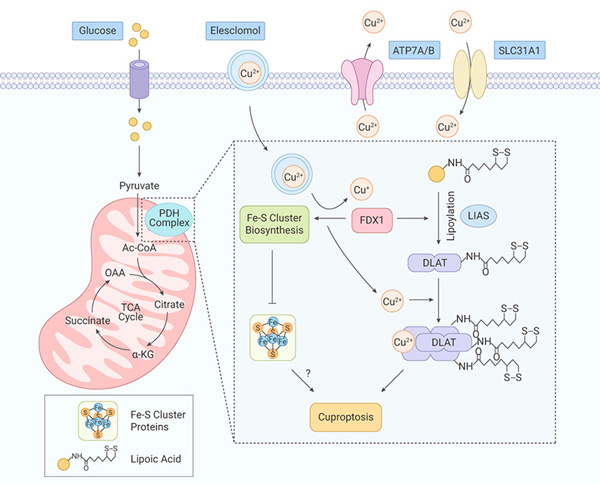
Briefly, initiated by the excessive accumulation of copper through ionophores and transporters, copper directly binds to lipoylated DLAT in cells that are dependent on mitochondrial respiration, subsequently induces aberrant oligomerization of DLAT and the formation of DLAT foci. The resulted increase of insoluble DLAT level leads to proteotoxicity and cell death [Fig. 1].
Ferrodoxin-1 (FDX1), a substrate of elesclomol, is an upstream regulator of protein lipoylation and is required for DLAT lipoylation. Additionally, as a reductase, FDX1 is known to reduce Cu (II) ions to the more toxic Cu(I) ions, subsequently leading to the inhibition of Fe-S cluster synthesis and reduction of Fe-S cluster proteins.
Copper homeostasis dysregulation
Copper homeostasis is mainly regulated by copper importer SLC31A1 and the copper exporters ATP7A and ATP7B.
In the copper dysregulation syndromes Menke’s disease and Wilson’s disease, the genes encoding these transporters are mutated. In the steady state of copper, ATP7A and ATP7B play essential roles in copper homeostasis, including intracellular copper delivery for inclusion in metalloproteins, membrane trafficking, and export of excess copper from cells. Cell death caused by dysregulation of copper homeostasis is comparable to cytotoxic effect caused by copper shuttling into the cell via copper ionophores (the copper-binding small molecules).
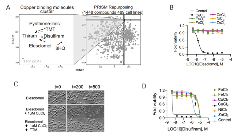
In this study, the cytotoxic effects of 1,448 copper ionophores with distinct structures were evaluated in 489 different cell lines [Fig. 2A]. As a highly lipophilic Cu (II) carrier, Elesclomol alone does not affect cell growth. But adding copper significantly increases sensitivity to Elesclomol, while supplementation with other metals, including iron, cobalt, zinc, and nickel, did not increase cell death [Fig 2B]. Notably, the addition of the copper chelator TTM abolished the cell growth inhibition activity by combination of Elesclomol and copper [Fig. 2C], confirming that copper ionophore-induced cell death is mainly dependent on the accumulation of intracellular copper. Treatment of cells with other copper ionophores such as NSC-319726 and Disulfiram showed the same results as elesclomol [Fig 3D-E].
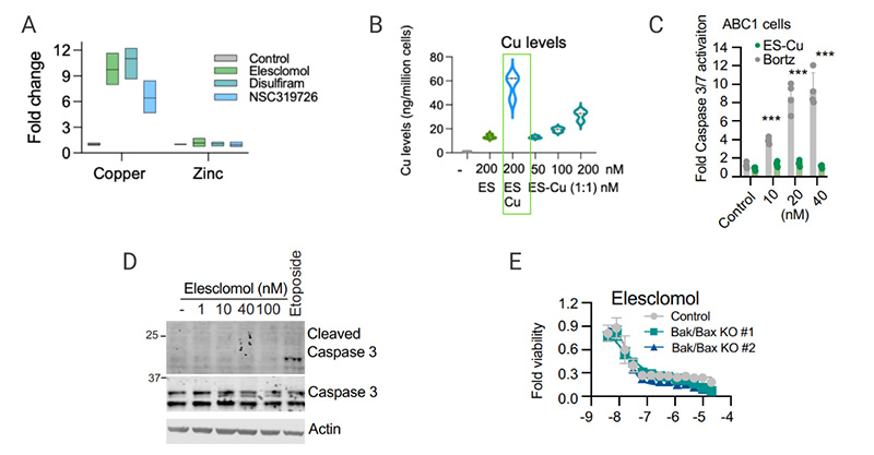
No cleavage or activation of caspase 3 activity was observed in elesclomol induced-cell death. [Fig. 3D] When key effectors of apoptosis BAX and BAK1 were knocked out or when cells were co-treated with pan-caspase inhibitors (Z-VAD-FMK and Boc-D-FMK), the inhibition activity of elecsclomol remained intact, [Fig. 3E], suggesting that the copper-induced cell death is not through the apoptotic pathway. Moreover, pre-treatments with inhibitors of ferroptosis (Ferrostatin-1), Necroptosis (Necrostatin-1), and oxidative stress (N-acetyl cysteine) did not affect copper ionophore-induced cell death, [Fig 3C (Fig 1G from the original article)], indicating the existence of a distinct cell death pathway.
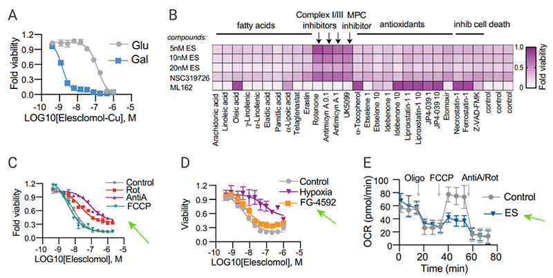
Cells that rely on mitochondrial respiration are more sensitive to copper ionophores than cells undergoing glycolysis [Fig. 4A]. In cell viability assays, cells pretreated with the ferroptosis inducer ML162 responded differently to variouis agents affecting mitochondrial functions compared to copper ionophores [Fig 4B].
Copper toxicity to the cells remained unchanged when cells were pretreated with the mitochondrial uncoupler FCCP, indicating that mitochondrial respiration is required for copper-induced cell death [Fig. 4C]. Although copper toxicity declined under hypoxic conditions, addition of the HIF prolyl hydroxylase inhibitor FG-4592 showed no effect on copper ionophore induced-cell death under normoxic conditions [Fig 4D] . It was observed that copper ionophores significantly reduced the spare capacity of respiration [Fig 4E]. These results support that copper ionophore induced-cell death is regulated by mitochondrial respiration.
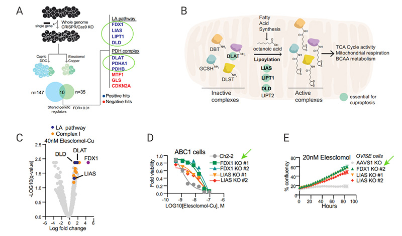
Using a genome-wide CRISPR-Cas9 positive selection screening, seven key genes were identified that play a role in copper-induced cell death, including FDX1 (encoding a direct target of elesclomol), and LIPT1, LIAS, DLD (three genes encoding lipoic acid pathway), or DLAT, PDHA1, and PDHB (encoding protein targets of lipoylation) [Fig 5A-C]. Individual gene knockout studies further confirmed that FDX1 and protein lipoylation are key regulators of copper ionophore-induced cell death [Fig.5D-E]. Therefore, Tsvetkov et al. thought that FDX1 was hypothesized to be an upstream regulator of protein thioctyl modification.
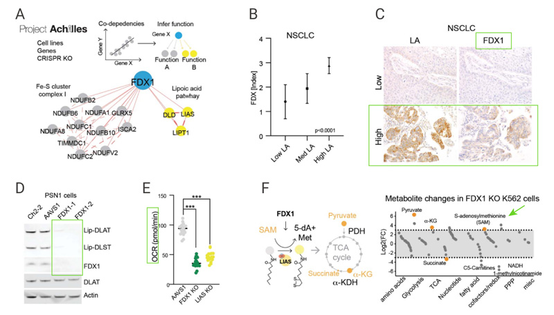
Correlation analysis of gene dependencies from the Cancer Dependency Map indicated that the FDX1 and components of the lipoic acid pathways were highly correlated across the panel of cell lines [Fig. 6A]. Immunohistochemistry staining results further confirmed this significant correlation [Fig. 6B-6C]. FDX1 knockout abolished protein lipoylation and resulted in a significant decrease in cellular respiration [Fig. 6D-E]. Furthermore, accumulation of pyruvate and α-ketoglutarate and depletion of succinate were observed followed deletion of FDX1 [Fig. 6F]. These results suggest that FDX1 is an upstream regulator of protein lipoylation.
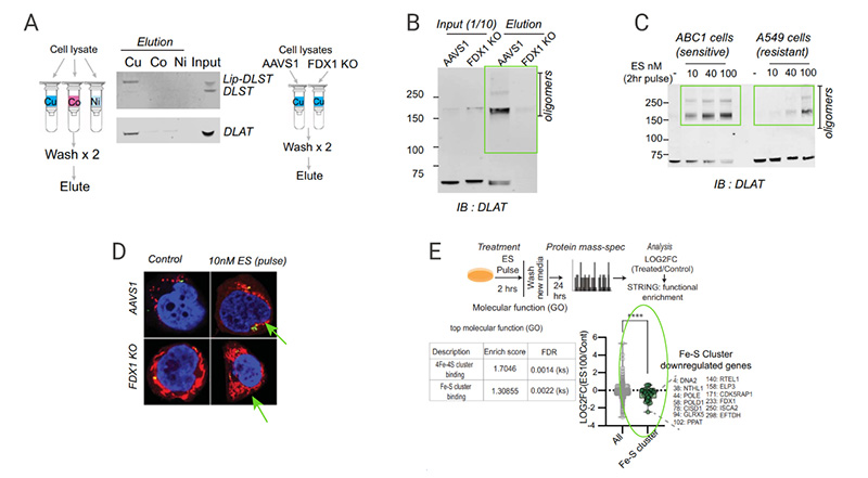
Some studies have reported that the dissociation constants of copper ions and free fatty acids are 10-17, which indicates that copper ions may bind directly to thiocylated proteins. DLAT and DLST proteins purified from cell lysates bound to copper-charged resin but not to cobalt or nickel resins [Fig. 7A]. FDX1 knockout abolished protein lipoylation and the resulted naked DLAT and DLST no longer bound copper [Fig. 7B-C], lipoylation is thus a prerequisite for copper binding. Immunofluorescence imaging results support that copper binding leads to the toxic aggregation of lipoylated DLAT [Fig 7D]. These results also suggested that the toxicity of thioacylated proteins after copper ionophore treatment is mediated by their abnormal oligomerization.
Proteomic analysis of control and elesclomol treatment showed the downregulation of Fe-S cluster genes [Fig 7E] and loss of Fe-S cluster proteins by copper ionophore treatment (Data not shown). These findings indicate that copper can destabilize Fe-S-containing proteins.
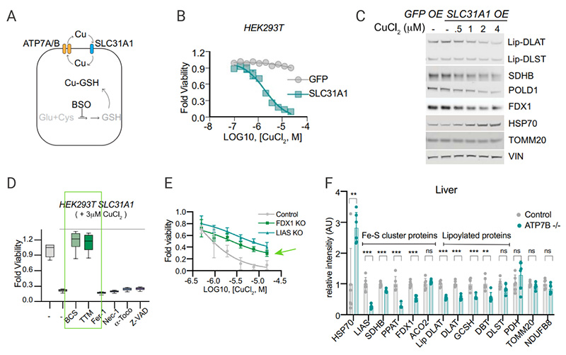
The copper importer SLC31A1 (CTR1) and copper exporters ATP7A and ATP78 regulate homeostatic state of copper and normally keep intracellular copper concentration low. Overexpression of SLC1A1 in HEK293T and ABC1 cells was found to significantly increase sensitivity to physiological copper concentrations. [Fig 8B] Treatment of SLC31A1 overexpressed cells with copper resulted in the reduction of protein lipoylation and Fe-S cluster protein level, as well as increase of HSP70 [Fig. 8C].
The use of ferrodeath, necrotizing apoptosis, and inhibitors of apoptosis in cells overexpressing SLC31A1 did not affect copper-induced cell death, but copper chelators alleviated the cell-killing effect produced by copper ionophore. Whereas copper chelators, FDX1 KO and LIAS KO each partially rescued cells from copper-induced cell death [Fig 8D-E]. Tsvetkov et al. demonstrated this same mechanism of copper-induced cell death in vivo. In Menke’s disease-associated Atpb7b−/− mice, it showed that the Fe-S cluster and lipoylated proteins were significantly reduced and Hsp70 protein was significantly increased compared with those in wild-type mice, further illustrating that excessive intracellular copper accumulation leads to cell death in vivo.These animal model results are in line with the copper ionophore induced cellular effects.
|
Related products |
|
ML162 is a covalent glutathione peroxidase 4 (GPX4) inhibitor. ML162 has a selective lethal effect on mutant RAS oncogene-expressing cell lines |
|
Ferrostatin-1, a selective ferroptosis inhibitor, suppresses Erastin-induced ferroptosis. |
|
FCCP is an uncoupler of oxidative phosphorylation (OXPHOS) in mitochondria. |
|
DL-Buthionine-(S,R)-sulfoximine DL-Buthionine-(S,R)-sulfoximine is a potent inhibitor of glutamylcysteine synthetase biosynthesis. |
|
DPQ is a potent PARP-1 inhibitor, which can reduce the N-methyl-d-aspartate (NMDA)-induced PARP activation. |
|
Elesclomol (STA-4783) is an oxidative stress inducer that can induce apoptosis in cancer cells. Elesclomol is also a highly lipophilic Cu2+ -binding molecule that can be used in the study of Menkes and hereditary copper deficiency related diseases. |
|
Zinc Pyrithione is an antifungal and antibacterial agent disrupting membrane transport by blocking the proton pump. Zinc Pyrithione is also a copper ionophore that delivers copper into cells and is a useful tool for studying cuproptosis. |
|
Boc-D-FMK is a cell-permeable, irreversible and broad spectrum caspase inhibitor. Boc-D-FMK inhibits apoptosis stimulated by TNF-α. |
|
Roxadustat (FG-4592) is a hypoxia-inducible factor prolyl hydroxylase (HIF-PHI) inhibitor. |
|
Etoposide inhibits topoisomerase II (topoisomerase-II), induces cell cycle arrest, induces apoptosis and autophagy. |
|
UK-5099 (PF-1005023) is a potent inhibitor of mitochondrial pyruvate transporter (MPC), inhibiting pyruvate-dependent O2 consumption. |
|
Necrostatin-1 (Nec-1) is a potent necroptosis inhibitor. |
|
Z-VAD-FMK is a pan-caspase (Caspase) inhibitor. |
|
NSC319726 is a mutant p53R175 reactivator that inhibits the proliferation of p53R175-expressing fibroblasts, but not wild-type p53 cells. |
|
8-Hydroxyquinoline (8-HQ) is a monoprotic bidentate chelating agent that acts as a preservative, disinfectant and insecticide, as a transcription inhibitor. |
[2]. Yongqiang Wang, Long Zhang, Fangfang Zhou. Cuproptosis: a new form of programmed cell death. Cell Mol Immunol. 2022 Apr 22.
[3]. Li SR, Bu LL, Cai L. Cuproptosis: lipoylated TCA cycle proteins-mediated novel cell death pathway. Signal Transduct Target Ther. 2022;7(1):158.
[4]. Tang D, Chen X, Kroemer G. Cuproptosis: a copper-triggered modality of mitochondrial cell death. Cell Res. 2022;32(5):417-418.
 LinkedIn
 Researchgate
 Instagram
 X
X Facebook
 WhatsApp
 Reddit
 Mail
Mail