



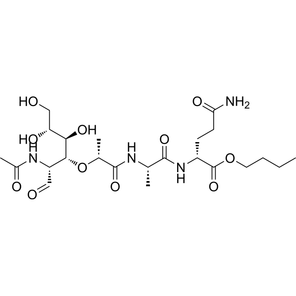
Murabutide
Murabutide is an immunomodulator and also a NOD2 receptor agonist. Murabutide enhances the signal transduction of ATR and NOD2, mediates the activation of the DDR pathway, and thereby repairs radiation-induced DNA double-strand breaks. Murabutide inhibits radiation-induced cell apoptosis and protects cells and mice from radiation-induced toxic damage. Murabutide induces the expression and DNA-binding activity of Oct-1 in macrophages, thereby inhibiting the transcription and replication of HIV-1. Murabutide reduces the expression levels of CD4 and CCR5 on the surface of macrophages, and induces the secretion of β-chemokines, TNF-α and IL-6. Murabutide enhances non-specific viral resistance and targets reticuloendothelial cells. Murabutide can be used in research related to radiation-induced damage and human immunodeficiency virus type 1 infection.
Nur für Forschungszwecke. Wir verkaufen nicht an Patienten.
- CAS. Nr.: 74817-61-1
- Formel: C23H40N4O11
- Molecular Weight:548.58
-
Speicherung:
Please store the product under the recommended conditions in the Certificate of Analysis.

To place orders, for customer services and technical support, please contact: MedChemExpress USA
Tel: 609-228-6898 E-mail: [email protected] [email protected]


-
 Biologische Aktivität
-
Chemical Information
- Protokoll
- Reinheit & Dokumentation
- Verweise
-
 Help & FAQs


Alle TNF Receptor Isoform-spezifische Produkte anzeigen
More

Biologische Aktivität
Beschreibung
In Vitro
Murabutide (1-5 μg/mL; pretreated for 2-12 h prior to 8 Gy IR, harvested 0.5-12 h post-IR) activates NOD2, enhances the phosphorylation levels of key DDR pathway proteins DNAPKcs, ATR and CHK1 in HIEC cells at 0.5 h post-irradiation, upregulates the level of anti-apoptotic protein Bcl2, downregulates the levels of pro-apoptotic protein Bax and activated caspase3, and reduces IR-induced γ-H2AX levels in HIEC cells at 0.5 h and 12 h post-irradiation[1].
Murabutide (10 μg/mL; 24 h) significantly regulates the expression of 28 genes in HIV-1-infected MDMs, among which 18 functionally characterized genes are upregulated (including Oct-1, MT-II and IL-13 receptor α-1 chain), and 4 functionally characterized genes are downregulated (including EGR2 and ferritin L chain)[2].
Murabutide (10 μg/mL; 24 h) upregulates the mRNA expression of Oct-1, MT-II and cytochrome b in HIV-1-infected MDM, and downregulates the mRNA expression of EGR2[2].
Murabutide (0.1-10 μg/mL; ≥8-20 days) inhibits the replication of M-tropic and T-tropic HIV-1 strains in human monocyte-derived dendritic cells; detection at 19 days post-infection shows that the average inhibition rate reaches 77% at the concentration of 10 μg/mL, with no alteration of cell proliferation[3].
Murabutide (10 μg/mL; 6-48 h) significantly reduces the expression of CD4 and CCR5 receptors on the surface of human monocyte-derived macrophages and dendritic cells after 24 h or 48 h of treatment, without altering the expression of CD14, HLA-DR or CXCR4[3].
MedChemExpress (MCE) has not independently confirmed the accuracy of these methods. They are for reference only.
-
Cell Line:normal human intestinal epithelial crypt (HIEC) cells
-
Concentration:0.25, 0.5, 1, 5 μg/mL
-
Incubation Time:2 h pretreatment before 8 Gy IR, measured 24 h post-IR
-
Result:Significantly promoted HIEC cell viability after 8 Gy IR, with the most pronounced effect observed at 1 μg/mL; cell viability was significantly higher in all murabutide-treated groups compared to the IR-only group.
-
Cell Line:normal human intestinal epithelial crypt (HIEC) cells
-
Concentration:1 μg/mL
-
Incubation Time:2 h pretreatment before 10 Gy IR, measured 24 h post-IR
-
Result:Significantly reduced IR-induced apoptosis in HIEC cells, lowering the apoptosis rate from ~14% in the IR-only group to ~8% in the murabutide-pretreated group.\nSignificantly reduced IR-induced apoptosis in HIEC cells, but this inhibitory effect was eliminated by co-treatment with VE-821, restoring apoptosis rates to levels comparable to the IR-only group.\nSignificantly reduced IR-induced apoptosis in HIEC cells, and co-treatment with KU-55933 did not reverse this anti-apoptotic effect.
-
Cell Line:normal human intestinal epithelial crypt (HIEC) cells
-
Concentration:1-5 μg/mL
-
Incubation Time:2 h pretreatment before 8 Gy IR, harvested 0.5 h post-IR
-
Result:Increased the expression of anti-apoptotic Bcl2 and decreased the expression of pro-apoptotic Bax and cleaved caspase3 in HIEC cells after 8 Gy IR, compared to the IR-only group.\nEnhanced NOD2 activation, increased the phosphorylation of DNAPKcs, ATR, and CHK1, and did not alter the phosphorylation of ATM or CHK2 in HIEC cells after 8 Gy IR, compared to the IR-only group.
-
Cell Line:normal human intestinal epithelial crypt (HIEC) cells
-
Concentration:1 μg/mL
-
Incubation Time:2 h or 12 h pretreatment before 8 Gy IR, harvested 0.5 h or 12 h post-IR
-
Result:Significantly reduced the IR-induced increase in γ-H2AX levels in HIEC cells at both 0.5 and 12 hours post-irradiation, compared to the IR-only group.
-
Cell Line:normal human intestinal epithelial crypt (HIEC) cells
-
Concentration:1 μg/mL
-
Incubation Time:2 h pretreatment before 8 Gy IR, analyzed 0.5, 8, 12, 24 h post-IR
-
Result:Significantly reduced the number of γ-H2AX foci in HIEC cells at all tested time points (0.5, 8, 12, 24 hours) post-8 Gy IR, compared to the IR-only group.
-
Cell Line:normal human intestinal epithelial crypt (HIEC) cells
-
Concentration:1 μg/mL
-
Incubation Time:2 h pretreatment before 8 Gy IR, harvested 0, 0.5, 2, 12 h post-IR
-
Result:Increased the phosphorylation of ATR, CHK1, and RPA2 in HIEC cells post-IR, but co-treatment with VE-821 diminished this enhanced phosphorylation, reducing levels to those comparable to the IR-only group.
-
Cell Line:normal human intestinal epithelial crypt (HIEC) cells
-
Concentration:1 μg/mL
-
Incubation Time:2 h pretreatment before 8 Gy IR, analyzed 24 h post-IR
-
Result:Significantly reduced the number of γ-H2AX foci in HIEC cells 24 hours post-IR, but co-treatment with VE-821 reversed this effect, increasing foci numbers to levels comparable to the IR-only group.
-
Cell Line:HIV-1-infected human monocyte-derived macrophages (MDMs)
-
Concentration:10 μg/mL
-
Incubation Time:24 h, 48 h, 72 h
-
Result:Up-regulated Oct-1 protein levels at 48-hour and 72-hour time points.
Showed no significant change in Oct-1 protein levels at 24-hour time point.
In Vivo
MedChemExpress (MCE) has not independently confirmed the accuracy of these methods. They are for reference only.
-
Animal Model:C57BL/6 (male, 8-week old)[1]
-
Dosage:300 μg/mouse
-
Administration:i.p.; single dose (2 hours before irradiation)
-
Result:Increased 30-day survival rate to 20% in mice exposed to 8.5 Gy γ-irradiation, compared to 0% in untreated controls.
Increased 30-day survival rate to 50% in mice exposed to 7 Gy γ-irradiation, and extended average survival time compared to untreated controls.
Maintained higher average body weight, better overall health, and prevented diarrhea in mice exposed to 7 Gy γ-irradiation, compared to untreated controls.
Increased the ratio of bone marrow nucleated cells on Day 1 and Day 3 post-7 Gy γ-irradiation, compared to untreated controls.
Increased intestinal villi length and intestinal crypts ratio on Day 1 and Day 3 post-7 Gy γ-irradiation, compared to untreated controls.
Increased white pulp area in the spleen on Day 1 and Day 3 post-7 Gy γ-irradiation, compared to untreated controls.
Reduced the number of TUNEL-labeled apoptotic cells in the small intestine, testis, and spleen on Day 1 post-7 Gy γ-irradiation, compared to untreated controls.


Chemical Information
-
CAS. Nr. 74817-61-1
-
Molecular Weight 548.58
-
Formel C23H40N4O11
-
SMILES
CC(N[C@@H](C=O)[C@H]([C@H](O)[C@H](O)CO)O[C@H](C)C(N[C@@H](C)C(N[C@H](CCC(N)=O)C(OCCCC)=O)=O)=O)=O
-
Versand
Room temperature in continental US; may vary elsewhere.
-
Speicherung
Please store the product under the recommended conditions in the Certificate of Analysis.


Reinheit & Dokumentation
Verweise
[1]. Liu L, et al. NOD2 agonist murabutide alleviates radiation-induced injury through DNA damage response pathway mediated by ATR. Journal of cellular physiology. 2019 Nov;234(11):21294-21306. [Content Brief]
[2]. Truong MJ, et al. Differentially expressed genes in HIV-1-infected macrophages following treatment with the virus-suppressive immunomodulator murabutide. Virus research. 2004 Jan;99(1):25-33. [Content Brief]
[3]. Darcissac EC, et al. The synthetic immunomodulator murabutide controls human immunodeficiency virus type 1 replication at multiple levels in macrophages and dendritic cells. Journal of virology. 2000 Sep;74(17):7794-802. [Content Brief]

Calculators
Konzentration (Stammlösung) × Volumen (Stammlösung) = Konzentration (Ziellösung) × Volumen (Ziellösung)


Murabutide Related Classifications
HY-106055 Related Classifications
 Powered by Bioz