



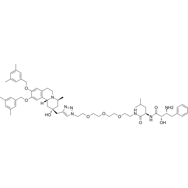
PROTAC GDI2 Degrader-1
PROTAC GDI2 Degrader-1 is a GDI2 PROTAC degrader with a DC50 value of 1.48 μM and a Kd value of 23.9 μM. PROTAC GDI2 Degrader-1 triggers paraptosis in tumor cells via endoplasmic reticulum expansion and fusion, endoplasmic reticulum stress, unfolded protein response, and cytoplasmic vacuolization. PROTAC GDI2 Degrader-1 exhibits significant in vivo anti-tumor activity in pancreatic cancer xenograft models. PROTAC GDI2 Degrader-1 can be used for research related to pancreatic cancer.
(Pink: GDI2 ligand (HY-156242); Blue: cIAP1 ligand (HY-175690); Black: linker).
For research use only. We do not sell to patients.
- Formula: C59H81N7O9
- Molecular Weight:1032.32
-
Storage:
Please store the product under the recommended conditions in the Certificate of Analysis.

To place orders, for customer services and technical support, please contact: MedChemExpress USA
Tel: 609-228-6898 E-mail: [email protected] [email protected]


-
 Biological Activity
-
Chemical Information
- Purity & Documentation
- References
-
 Help & FAQs


All PROTACs Isoforms
More

Biological Activity
Description
In Vitro
PROTAC GDI2 Degrader-1 (Compound 21) (0.63-10 μM; 24 h; 5 μM; 0-24 h) degrades GDI2 in AsPC-1 pancreatic cancer cells in a dose-dependent manner with a DC50 of 1.48 μM and a Dmax of 84.2%, and in a time-dependent manner with > 70% depletion observed by 9 h[1].
PROTAC GDI2 Degrader-1 (2 μM; 24 h) reduces GDI2 protein levels to 61% of control in PC-3 prostate cancer cells[1].
MedChemExpress (MCE) has not independently confirmed the accuracy of these methods. They are for reference only.
-
Cell Line:AsPC-1 pancreatic cancer cells
-
Concentration:0.63, 1.25, 2.5, 5, 10 μM (dose-response); 5 μM (time-course)
-
Incubation Time:24 h (dose-response); 0, 3, 6, 9, 12, 24 h (time-course)
-
Result:Induced dose-dependent degradation of GDI2 with a DC50 of 1.48 μM and a maximum degradation (Dmax) of 84.2% at 10 μM after 24 h.
Achieved > 70% depletion of GDI2 after 9 h of treatment with 5 μM.
-
Cell Line:PC-3 prostate cancer cells
-
Concentration:2 μM
-
Incubation Time:24 h
-
Result:Reduced GDI2 protein levels to 61% of DMSO control levels after 24 h.
In Vivo
PROTAC GDI2 Degrader-1 (15 mg/kg; i.p.; every 3 days; 18 days) significantly suppresses growth of patient-derived pancreatic adenocarcinoma tumors in nude mice via GDI2 depletion and induction of paraptosis[1].
MedChemExpress (MCE) has not independently confirmed the accuracy of these methods. They are for reference only.
-
Animal Model:BALB/c athymic nude mice (male, 6-8 weeks old, subcutaneous inoculation of 5 × 10⁶ AsPC-1 cells)[1]
-
Dosage:15 mg/kg
-
Administration:i.p.; every 3 days; 18 days
-
Result:Significantly attenuated tumor growth compared to the vehicle group.
Achieved over 90% GDI2 depletion that was not restored 72 hours post final injection.
Elevated protein levels of ER stress/UPR markers GRP78, CHOP, and p-eIF2α in tumor tissues.
Induced extensive cytoplasmic vacuolization indicating paraptosis in tumor tissues.
Caused no significant body weight loss or histopathological injury to major organs (heart, lung, liver, spleen, kidney).
-
Animal Model:BALB/c athymic nude mice (male, 6-8 weeks old, subcutaneous inoculation of ~3 mm3 human PDAC tumor fragments)[1]
-
Dosage:15 mg/kg
-
Administration:i.p.; every 3 days; 18 days
-
Result:Significantly suppressed PDAC tumor growth to levels similar to the gemcitabine positive control group.
Achieved excellent GDI2 depletion in PDAC tumors.
Elevated protein levels of ER stress/UPR markers GRP78, CHOP, and p-eIF2α in tumor tissues.
Induced cytoplasmic vacuolization in tumor tissues.
Caused no significant systemic toxicity.


Chemical Information
-
Molecular Weight 1032.32
-
Formula C59H81N7O9
-
SMILES
CC1=CC(C)=CC(COC2=CC3=C([C@](C[C@](O)(CC4=CN(CCOCCOCCOCCNC([C@@H](CC(C)C)NC([C@@H](O)[C@H](N)CC5=CC=CC=C5)=O)=O)N=N4)C[C@@H]6C)([H])N6CC3)C=C2OCC7=CC(C)=CC(C)=C7)=C1
-
Shipping
Room temperature in continental US; may vary elsewhere.
-
Storage
Please store the product under the recommended conditions in the Certificate of Analysis.


Purity & Documentation
References

Calculators
Concentration (start) × Volume (start) = Concentration (final) × Volume (final)


PROTAC GDI2 Degrader-1 Related Classifications
HY-156244 Related Classifications
 Powered by Bioz