



PD-1 Protein, Mouse (HEK293, His)

Based on 1 Customer Validation
PD-1 Protein is an important immune regulatory molecule expressed on the surface of activated immune cells. By binding to its ligands (PD-L1/PD-L2), the PD-1 Protein exerts immunosuppressive effects. PD-1 Protein can be used in the research of tumors, autoimmune diseases, and other conditions. PD-1 Protein, Mouse (HEK293, His) is a recombinant PD-1 protein expressed by HEK293 cells and carries a C-6*His tag and glycosylation modification.
- Species: Mouse
- Source: HEK293
-
Storage:Stored at -20°C for 2 years from date of receipt. After reconstitution, it is stable at 4°C for 1 week or -20°C for longer (with carrier protein). It is recommended to freeze aliquots at -20°C or -80°C for extended storage.


To place orders, for customer services and technical support, please contact: MedChemExpress USA
Tel: 609-228-6898 E-mail: [email protected] [email protected]


-
 Biological Activity
-
Technical Parameters
-
Product Properties
- Documentation
- References
- Help & FAQs




Biological Activity
Description
PD-1 Protein is an important immune regulatory molecule expressed on the surface of activated immune cells. By binding to its ligands (PD-L1/PD-L2), the PD-1 Protein exerts immunosuppressive effects. PD-1 Protein can be used in the research of tumors, autoimmune diseases, and other conditions. PD-1 Protein, Mouse (HEK293, His) is a recombinant PD-1 protein expressed by HEK293 cells and carries a C-6*His tag and glycosylation modification[1][2].
Background
PD-1, also known as CD279, is a 55 kDa transmembrane protein consisting of 288 amino acids, including a signal peptide, extracellular domain, transmembrane domain, and intracellular domain. As an immune checkpoint molecule, PD-1 is mainly expressed on the surface of activated immune cells such as T cells and B cells. Notably, PD-1 is highly expressed on tumor-specific T cells. PD-1 acts as an inhibitor of adaptive and innate immune responses. Under normal conditions, PD-1 maintains immune tolerance by binding to its ligands PD-L1/PD-L2 to avoid autoimmune damage. However, when PD-1 binds to PD-L1/PD-L2 on tumor cells, it leads to T cell functional exhaustion, inhibits cytokine secretion, and facilitates tumor immune escape. Currently, PD-1 serves as a core target for cancer immunotherapy, and its inhibitors can restore the tumor-killing ability of T cells by blocking this binding[1][2].
Verified Bioactivity
1.Determined by its ability to prevent plate adhesion of PHA-stimulated Jurkat cells in the presence of 625 ng/mL of bound hPD-L1. The ED50 for this effect is <1 μg/mL.
2.Immobilized Mouse PD-1 at 1 μg/ml (100 μl/well) can bind Biotinylated Mouse PD-L1, Fc (HY-P70633). The ED50 for this effect is <1 μg/mL.
3.Immobilized Mouse PD-1 at 1 μg/mL (100 μL/well) can bind Biotinylated Mouse PD-L1, His (HY-P70632). The ED50 for this effect is <1 μg/mL.
MCE Validation Data
-
Purity - SDS-PAGE
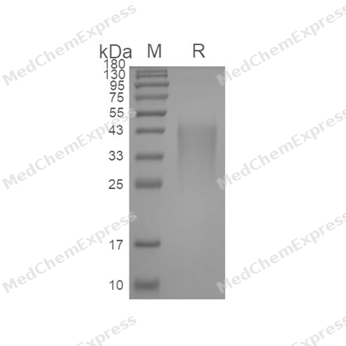
Purity - SDS-PAGE
-
Bioactivity - Cell-Based Assay
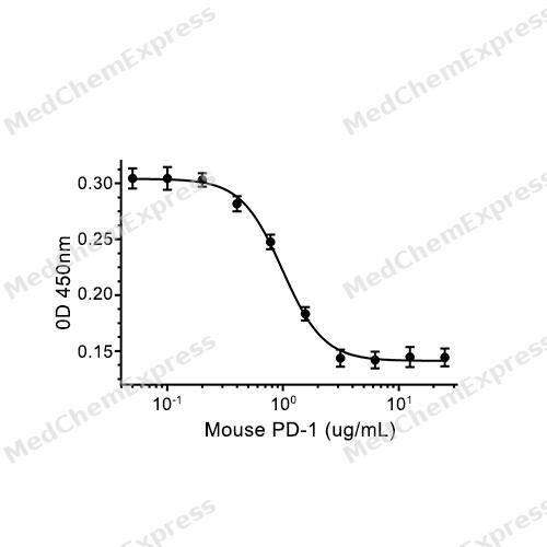
Bioactivity - Cell-Based Assay


Technical Parameters
-
Species Mouse
-
Source HEK293
-
Tag C-6*His
-
Accession
Q02242 (L25-Q167)
-
Molecular Construction
-
N-term
-
PD-1 (L25-Q167)
Accession # Q02242 -
6*His
-
C-term
-
-
Protein Length
Extracellular Domain
-
Synonyms
PDCD1; Systemic Lupus Erythematosus Susceptibility 2; Prev. SLEB2; HPD-1; PD1; Programmed Cell Death 1 Protein; Programmed Cell Death Protein 1; CD279 Antigen; CD279; ADMIO4; HSLE1; AIMTBS; PD-1; HPD-L; Programmed Cell Death 1; Protein PD-1
-
AA Sequence
LEVPNGPWRSLTFYPAWLTVSEGANATFTCSLSNWSEDLMLNWNRLSPSNQTEKQAAFCNGLSQPVQDARFQIIQLPNRHDFHMNILDTRRNDSGIYLCGAISLHPKAKIEESPGAELVVTERILETSTRYPSPSPKPEGRFQ
-
Molecular Weight
Approximately 33-40 kDa, based on SDS-PAGE under reducing conditions, due to the glycosylation.
-
Glycosylation
Yes
-
Purity
≥ 95%, as determined by reducing SDS-PAGE.


Product Properties
Lyophilized powder
1.Lyophilized from a 0.22 μm filtered solution of 20 mM Tris-HCl, 150 mM NaCl, pH 8.0.
2.Lyophilized from a 0.22 μm filtered solution of 20 mM PB, 150 mM NaCl, pH 7.4.
3.Lyophilized from a 0.22 μm filtered solution of PBS, pH 7.4.
Please refer to the lot-specific COA for specific buffer information.
Note: For SPR assay, please replace the buffer. Primary amine components (e.g., Tris, imidazole) can affect protein-coupled chips.
<1 EU/μg, determined by LAL method.
It is not recommended to reconstitute to a concentration less than 100 μg/mL in ddH2O. For long term storage it is recommended to add a carrier protein (0.1% BSA, 5% HSA, 10% FBS or 5% Trehalose).
Stored at -20°C for 2 years from date of receipt. After reconstitution, it is stable at 4°C for 1 week or -20°C for longer (with carrier protein). It is recommended to freeze aliquots at -20°C or -80°C for extended storage.
Room temperature in continental US; may vary elsewhere.


Documentation
-
Data Sheet (263 KB)
-
SDS (252 KB)
- English - EN (252 KB)
- Français - FR (252 KB)
- Deutsch - DE (252 KB)
- Norwegian - NO (252 KB)
- Español - ES (252 KB)
- Swedish - SV (252 KB)
- Italian - IT (252 KB)
- Korean - KR (252 KB)
- Portuguese - PT (252 KB)
-
Handling Instructions (2659 KB)


References

Calculators
Concentration (start) × Volume (start) = Concentration (final) × Volume (final)


 Powered by Bioz

