



Dryocrassin ABBA
Based on 2 publication(s) in Google Scholar
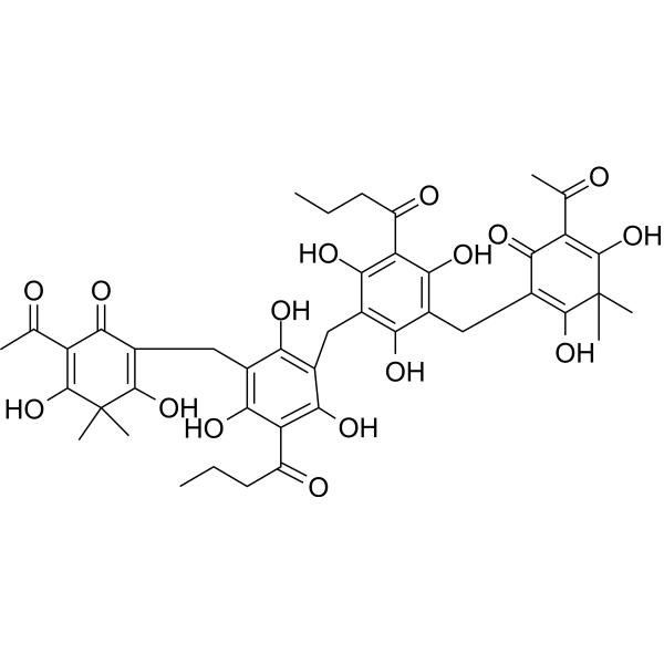
Dryocrassin ABBA (Dryocrassin) is an orally active phloroglucinol derivative that can be extracted from Phyllopteris officinalis. Dryocrassin ABBA has antitumor and antiviral activity. Dryocrassin ABBA induced apoptosis of human hepatocellular carcinoma cells through mitochondrial pathway mediated by Caspase.
For research use only. We do not sell to patients.
- CAS No.: 12777-70-7
- Formula: C43H48O16
- Molecular Weight:820.83
-
Storage:
-20°C, protect from light
* In solvent : -80°C, 6 months; -20°C, 1 month (protect from light)

To place orders, for customer services and technical support, please contact: MedChemExpress USA
Tel: 609-228-6898 E-mail: [email protected] [email protected]


-
 Biological Activity
-
Chemical Information
-
Solvent & Solubility
- Protocols
- Purity & Documentation
- References
-
 Help & FAQs


Publications Citing Use of MedChemExpress (MCE) Dryocrassin ABBA
More

Biological Activity
Description
Cellular Effect
|
Cell Line

|
Type | Value | Description | References |
|---|---|---|---|---|
| HEK293 | IC50 |
25.5 μM
Compound: Dry. ABBA
|
Inhibition of Influenza A virus (A/California/04/2009 (H1N1) neuraminidase expressed by HEK293 cells incubated for 1 hr using 4-MU-NANA substrate by spectrofluorometric assay
Inhibition of Influenza A virus (A/California/04/2009 (H1N1) neuraminidase expressed by HEK293 cells incubated for 1 hr using 4-MU-NANA substrate by spectrofluorometric assay
|
[PMID: 31324565] |
| HEK293 | IC50 |
279.6 μM
Compound: Dry. ABBA
|
Inhibition of Influenza A virus (A/Shanghai/1/2013 (H7N9)) neuraminidase R294K mutant expressed by HEK293 cells incubated for 1 hr using 4-MU-NANA substrate by spectrofluorometric assay1
Inhibition of Influenza A virus (A/Shanghai/1/2013 (H7N9)) neuraminidase R294K mutant expressed by HEK293 cells incubated for 1 hr using 4-MU-NANA substrate by spectrofluorometric assay1
|
[PMID: 31324565] |
| HEK293 | IC50 |
3.6 μM
Compound: Dry. ABBA
|
Inhibition of Influenza A virus (A/Anhui/1/2013 (H7N9) neuraminidase expressed by HEK293 cells incubated for 1 hr using 4-MU-NANA substrate by spectrofluorometric assay
Inhibition of Influenza A virus (A/Anhui/1/2013 (H7N9) neuraminidase expressed by HEK293 cells incubated for 1 hr using 4-MU-NANA substrate by spectrofluorometric assay
|
[PMID: 31324565] |
| HEK293 | IC50 |
36.1 μM
Compound: Dry. ABBA
|
Inhibition of Influenza A virus (A/Babol/36/2005 (H3N2) neuraminidase expressed by HEK293 cells incubated for 1 hr using 4-MU-NANA substrate by spectrofluorometric assay
Inhibition of Influenza A virus (A/Babol/36/2005 (H3N2) neuraminidase expressed by HEK293 cells incubated for 1 hr using 4-MU-NANA substrate by spectrofluorometric assay
|
[PMID: 31324565] |
| HEK293 | IC50 |
61.8 μM
Compound: Dry. ABBA
|
Inhibition of Influenza A virus (A/Anhui/1/2005(H5N1)) neuraminidase expressed by HEK293 cells incubated for 1 hr using 4-MU-NANA substrate by spectrofluorometric assay
Inhibition of Influenza A virus (A/Anhui/1/2005(H5N1)) neuraminidase expressed by HEK293 cells incubated for 1 hr using 4-MU-NANA substrate by spectrofluorometric assay
|
[PMID: 31324565] |
| MDCK | CC50 |
>400 μM
Compound: 8
|
Cytotoxicity against MDCK cells assessed as reduction in cell viability measured after 72 hrs by CCK8 assay
Cytotoxicity against MDCK cells assessed as reduction in cell viability measured after 72 hrs by CCK8 assay
|
[PMID: 38283223] |
In Vitro
Dryocrassin ABBA (Dryocrassin) inhibits the activity of S. aureus SrtA (sortase A) with an IC50 of 24.17 μM[2].
MedChemExpress (MCE) has not independently confirmed the accuracy of these methods. They are for reference only.
In Vivo
Dryocrassin ABBA (100 mg/kg; s.c.; every 12 h for 96 h) can reduce infection and prevent mice from contracting S. aureus pneumonia[3].
MedChemExpress (MCE) has not independently confirmed the accuracy of these methods. They are for reference only.
-
Animal Model:SPF BALB/C female mice inoculated intra-nasally with 104.5ELD50 H5N1 viruses in 100 μl saline[1]
-
Dosage:33, 18, and 12.5 mg/kg
-
Administration:Oral gavage, daily, 7 days from day 2 to day 8
-
Result:Significantly reduced mortality, prolonged survival rate, and improved survival time throughout the infection period. Increased body weight and reduced lung lesions and virus loads. Significantly increased MCP-1 and IL-10 while significantly decreased IL-12, IL-6, IFN-γ and TNF-α.
-
Animal Model:Eight-week-old C57BL/6J mice infected with 30 μL of S. aureus Newman (2 × 10⁸ CFU/10 μL) by intranasal administration[3]
-
Dosage:100 mg/kg
-
Administration:Subcutaneous injection, every 12 h for 96 h
-
Result:Resulted a significant increase in survival.


Chemical Information
-
CAS No. 12777-70-7
-
Appearance Solid
-
Molecular Weight 820.83
-
Formula C43H48O16
-
Color Light yellow to green yellow
-
SMILES
OC1=C(C(O)=C(C(CCC)=O)C(O)=C1CC(C(C(C(C)=O)=C(O)C2(C)C)=O)=C2O)CC(C(O)=C(C(CCC)=O)C(O)=C3CC(C(C(C(C)=O)=C(O)C4(C)C)=O)=C4O)=C3O
-
Synonyms
Dryocrassin
-
Structure Classification
-
Initial Source
-
Shipping
Room temperature in continental US; may vary elsewhere.
-
Storage
-20°C, protect from light
* In solvent : -80°C, 6 months; -20°C, 1 month (protect from light)


Publications (2)
-
Journal Impact Factor
-
Most Recent
-
PLoS One
2024 Dec 13;19(12):e0309624. PMID: 39671442 -


Solvent & Solubility
In Vitro:
DMSO : 16.67 mg/mL (20.31 mM; Need ultrasonic; Hygroscopic DMSO has a significant impact on the solubility of product, please use newly opened DMSO)
Please refer to the solubility information to select the appropriate solvent. Once prepared, please aliquot and store the solution to prevent product inactivation from repeated freeze-thaw cycles.
Storage method and period of stock solution: -80°C, 6 months; -20°C, 1 month (protect from light). When stored at -80°C, please use it within 6 months. When stored at -20°C, please use it within 1 month.
Please refer to the solubility information to select the appropriate solvent. Once prepared, please aliquot and store the solution to prevent product inactivation from repeated freeze-thaw cycles.
Storage method and period of stock solution: -80°C, 6 months; -20°C, 1 month (protect from light). When stored at -80°C, please use it within 6 months. When stored at -20°C, please use it within 1 month.
Concentration (start) × Volume (start) = Concentration (final) × Volume (final)
In Vivo:
Select the appropriate dissolution method based on your experimental animal and administration route.
- For the following dissolution methods, please ensure to first prepare a clear stock solution using an In Vitro approach and then sequentially add co-solvents:
- To ensure reliable experimental results, the clarified stock solution can be appropriately stored based on storage conditions. As for the working solution for In Vivo experiments, it is recommended to prepare freshly and use it on the same day.
- The percentages shown for the solvents indicate their volumetric ratio in the final prepared solution. If precipitation or phase separation occurs during preparation, heat and/or sonication can be used to aid dissolution.
Add each solvent one by one: 10% DMSO 40% PEG300 5% Tween-80 45% Saline
Solubility: ≥ 1.67 mg/mL (2.03 mM); Clear solution
This protocol yields a clear solution of ≥ 1.67 mg/mL (saturation unknown).
Taking 1 mL working solution as an example, add 100 μL DMSO stock solution (16.7 mg/mL) to 400 μL PEG300, and mix evenly; then add 50 μL Tween-80 and mix evenly; then add 450 μL Saline to adjust the volume to 1 mL.
Preparation of Saline: Dissolve 0.9 g sodium chloride in ddH₂O and dilute to 100 mL to obtain a clear Saline solution.
In Vivo Dissolution Calculator
Please enter the basic information of animal experiments:
-
-
-
-
Recommended: Prepare an additional quantity of animals to account for potential losses during experiments.
Please enter your animal formula composition:
-
%DMSO +
Recommended: Keep the proportion of DMSO in working solution below 2% if your animal is weak.
-
%+
-
+%Tween-80 + +
-
%Saline +
The co-solvents required include: DMSO, . All of co-solvents are available by MedChemExpress (MCE). , Tween 80. All of co-solvents are available by MedChemExpress (MCE).
Working solution concentration: 0.22 mg/mL
Method for preparing stock solution: mg drug dissolved in μL DMSO. Stock solution concentration: mg/mL. * In solvent : -80°C, 6 months; -20°C, 1 month (protect from light)
1. Take μL DMSO stock solution;
2. Add μL .
μL , mix evenly;
3. Then add μL Tween 80, mix evenly;
4. Then add μL
Please ensure that the stock solution in the first step is dissolved to a clear state, and add co-solvents in sequence. You can use ultrasonic heating (ultrasonic cleaner, recommended frequency 20-40 kHz), vortexing, etc. to assist dissolution.


Purity & Documentation
-
Data Sheet (279 KB)
-
SDS (393 KB)
- English - EN (393 KB)
- Français - FR (393 KB)
- Deutsch - DE (393 KB)
- Norwegian - NO (393 KB)
- Español - ES (393 KB)
- Swedish - SV (393 KB)
- Italian - IT (393 KB)
- Korean - KR (393 KB)
- Portuguese - PT (393 KB)
-
Handling Instructions (2659 KB)
References
[1]. Ou C, et al. Dryocrassin ABBA, a novel active substance for use against amantadine-resistant H5N1 avian influenza virus. Front Microbiol. 2015 Jun 16;6:592. [Content Brief]
[2]. Ou C, et al. Dryocrassin ABBA, a novel active substance for use against amantadine-resistant H5N1 avian influenza virus. Front Microbiol. 2015 Jun 16;6:592. [Content Brief]
[3]. Jin Z, et al. Dryocrassin ABBA Induces Apoptosis in Human Hepatocellular Carcinoma HepG2 Cells Through a Caspase-Dependent Mitochondrial Pathway. Asian Pac J Cancer Prev. 2016;17(4):1823-8. [Content Brief]
[4]. Zhang B, et al. Molecular Mechanism of the Flavonoid Natural Product Dryocrassin ABBA against Staphylococcus aureus Sortase A. Molecules. 2016 Oct 26;21(11). [Content Brief]
[5]. Li B, et al. An Inhibitory Effect of Dryocrassin ABBA on Staphylococcus aureus vWbp That Protects Mice From Pneumonia. Front Microbiol. 2019 Jan 23;10:7. [Content Brief]
[6]. Fu RH, et al. Dryocrassin suppresses immunostimulatory function of dendritic cells and prolongs skin allograft survival. Cell Transplant. 2014;23(4-5):641-56. [Content Brief]


Complete Stock Solution Preparation Table
Please refer to the solubility information to select the appropriate solvent. Once prepared, please aliquot and store the solution to prevent product inactivation from repeated freeze-thaw cycles.
Storage method and period of stock solution: -80°C, 6 months; -20°C, 1 month (protect from light). When stored at -80°C, please use it within 6 months. When stored at -20°C, please use it within 1 month.
| Optional Solvent | Concentration Solvent Mass | 1 mg | 5 mg | 10 mg | 25 mg |
|---|---|---|---|---|---|
| DMSO | 1 mM | 1.2183 mL | 6.0914 mL | 12.1828 mL | 30.4570 mL |
| 5 mM | 0.2437 mL | 1.2183 mL | 2.4366 mL | 6.0914 mL | |
| 10 mM | 0.1218 mL | 0.6091 mL | 1.2183 mL | 3.0457 mL | |
| 15 mM | 0.0812 mL | 0.4061 mL | 0.8122 mL | 2.0305 mL | |
| 20 mM | 0.0609 mL | 0.3046 mL | 0.6091 mL | 1.5228 mL |


Dryocrassin ABBA Related Classifications
HY-N0530 Related Classifications
 Powered by Bioz