



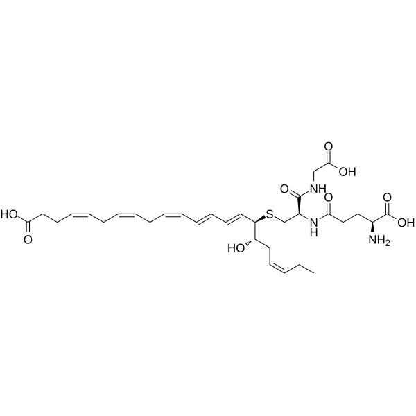
(7S,8R,17S)-RCTR1
(7S,8R,17S)-RCTR1 is a specialized pro-resolving mediator, that exhibits anti-inflammatory and anti-infective activities. (7S,8R,17S)-RCTR1 promotes pathogen clearance, wound healing, tissue repair and regeneration.
For research use only. We do not sell to patients.
- CAS No.: 2095607-49-9
- Formula: C32H47N3O10S
- Molecular Weight:665.79
-
Storage:
Please store the product under the recommended conditions in the Certificate of Analysis.


To place orders, for customer services and technical support, please contact: MedChemExpress USA
Tel: 609-228-6898 E-mail: [email protected] [email protected]


-
 Biological Activity
-
Chemical Information
- Protocols
- Purity & Documentation
- References
-
 Help & FAQs


-
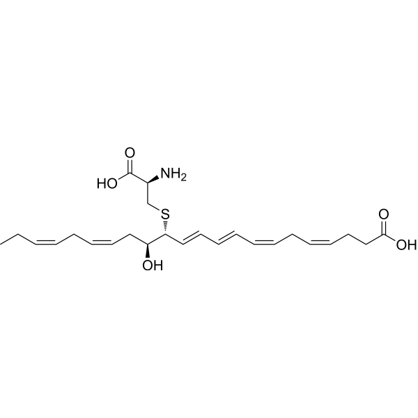
HY-125516
MCTR3Pro-resolving Mediator -
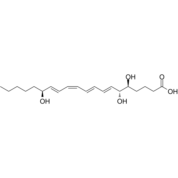
HY-113509
Lipoxin A4Anti-inflammatory Agent -
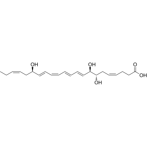
HY-125527A
17(R)-Resolvin D1TRPV3 Inhibitor -
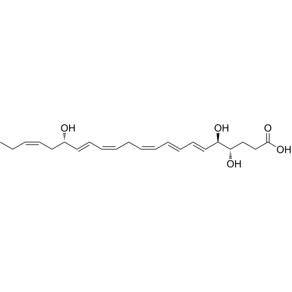
HY-113533
Resolvin D4Proresolving Mediator -
HY-125445
PCTR1Pro-resolving Mediator -
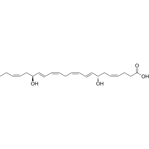
HY-145491
Resolvin D5Inflammation Inhibitor -
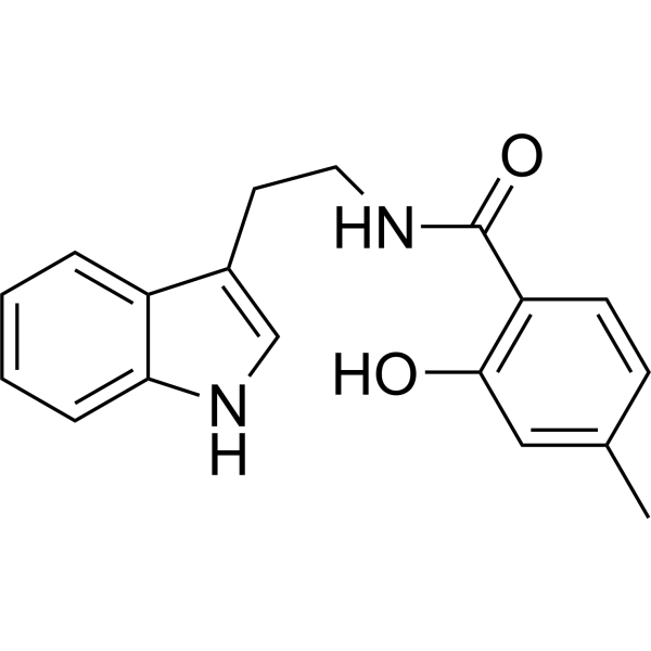
HY-155126
LZWL02003Anti-Neuroinflammatory Agent -

HY-125408
Maresin conjugates in tissue regeneration 1cytokine -

HY-130322
Maresin Conjugates in Tissue Regeneration 2Specialized Pro-resolving Mediator -

HY-158792
GK241 -

HY-170645
Neuroprotectin D1 methyl esterProtectin D1 Intermediate -
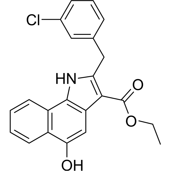
HY-120502
5-LOX-IN-65-LO Inhibitor


Biological Activity
Description


Chemical Information
-
CAS No. 2095607-49-9
-
Molecular Weight 665.79
-
Formula C32H47N3O10S
-
SMILES
OC(CC/C=C\C[C@H](O)[C@@H](/C=C/C=C/C=C\C=C\[C@@H](O)C/C=C\CC)SC[C@@H](C(NCC(O)=O)=O)NC(CC[C@H](N)C(O)=O)=O)=O
-
Shipping
Room temperature in continental US; may vary elsewhere.
-
Storage
Please store the product under the recommended conditions in the Certificate of Analysis.

Protocols
-
Research Protocol for Infectious Diseases
Infectious-disease experiments test how pathogens interact with host barriers, innate immune receptors, inflammatory signaling, pathogen replication, and tissue injury; pattern-recognition receptors such as TLRs, RIG-I-like receptors, NOD-like receptors, and inflammasomes detect microbial molecules and activate NF-κB, interferon, and cytokine responses. The central hypothesis is that infection severity reflects the balance between pathogen burden and host response: protective inflammation restricts pathogen growth, whereas excessive or mislocalized inflammation contributes to tissue damage and disease phenotype. Unresolved questions include which host pathways are protective versus pathogenic, why some infection models fail to translate to human disease, and which combined readouts best predict clinically relevant infection outcomes.
-
Cell migration
Cell migration is a method that plays an important role in wound healing, cell differentiation, embryonic development, etc.
-
Protocol for Pharmacokinetic Study
Pharmacokinetic studies quantify how an organism handles a drug over time through absorption, distribution, metabolism, and excretion, and the core experimental readout is the concentration-time profile of parent drug and, when relevant, metabolites in biological matrices such as plasma, whole blood, urine, bile, or tissue. Pharmacokinetic analysis links dose, route, exposure, clearance, half-life, distribution, bioavailability, and systemic exposure to drug efficacy and toxicity hypotheses rather than measuring a signaling pathway directly. The literature links pharmacokinetics to drug-development phenotypes by showing that drug metabolism and pharmacokinetics influence compound progression, exposure-response interpretation, safety margins, dosing strategy, and failure risk during discovery and development. DMPK science contributes to compound optimization by integrating physicochemical properties, in vitro metabolism, transporter behavior, in vivo exposure, and pharmacodynamic contex
-
Research Protocol for Inflammation-related Diseases
The NLRP3 inflammasome is a cytosolic innate immune signaling platform that integrates priming signals and danger-signal activation to promote caspase-1 activation, maturation of IL-1β and IL-18, and gasdermin D-mediated pyroptotic cell death. The core experimental logic is to determine whether inflammatory disease phenotypes are driven by increased NLRP3 expression, ASC-containing inflammasome assembly, caspase-1 cleavage, GSDMD cleavage, and extracellular release of IL-1β/IL-18 rather than by nonspecific cell injury alone. The pathway is strongly linked to inflammation-related disease phenotypes because monosodium urate crystals activate NALP3/NLRP3 inflammasome signaling in gout-like crystal inflammation, cholesterol crystals activate NLRP3 inflammasomes in atherogenesis models, and DSS-induced intestinal inflammation has been reported to involve NLRP3 inflammasome activity. However, experimental colitis studies also show context-dependent protective effects of NLRP3 inflammasome co
-
Cell-Exclusion Zone Migration Assay
The Cell-Exclusion Zone (CEZ) migration assay is an in vitro 2D cell migration method in which a defined cell-free area is created using removable physical barriers such as silicone stoppers, allowing cells to be seeded around the barrier and subsequently migrate into the cleared zone after barrier removal. This approach enables quantification of collective cell migration by monitoring repopulation of the initially cell-free region over time using microscopy-based imaging. Compared with scratch-based wound healing assays, barrier-based exclusion methods are designed to avoid mechanical damage to the extracellular matrix and reduce injury-induced effects on boundary cells, thereby improving interpretability of migration behavior in vitro. The assay readout is typically the progressive reduction in the cell-free area or the number of cells invading the exclusion zone, reflecting coordinated cell motility relevant to physiological processes such as wound healing, epithelial repair, and ca


Purity & Documentation
References

Calculators
Concentration (start) × Volume (start) = Concentration (final) × Volume (final)


(7S,8R,17S)-RCTR1 Related Classifications
HY-155286 Related Classifications
 Powered by Bioz