



Fluorescent Dye
Fluorescence is a phenomenon of photoluminescence in luminescence. When an atom is irradiated by light, some electrons around the nucleus jump from their original orbitals to higher energy orbitals under the influence of light energy, i.e., from the ground state to the first or second excited single-line state, etc. Due to the instability of the first or second excited singlet state, the electron returns from the excited singlet state to the ground state accompanied by released energy, resulting in fluorescence.
Fluorescent dyes can be covalently bound or physically adsorbed in the structure of the substance. The fluorescence properties allow for the characterization, localization and quantitative analysis of the labeled object. Fluorescent dyes have shown great potential for the detection of proteins, nucleic acids, cells, and immunoassays, attributed to the advantages in non-radioactive contamination, simple experimental operation and easy observation.
Fluorescent dyes have penetrated many fields such as pharmacology, physiology, environmental science, and information science, as well as functional studies of proteins and drug screening.
All Product Categories
-
Fluorescent Dye Related Products (4888)
Related Products (4888)

-
Antibodies (4)
-
Photoacoustic contrast agent-1
 0 ImagesCat. No.: HY-D1291CAS No.: 2255329-06-5Photoacoustic contrast agent-1 (Compound F-1) is a photo/sono-sensitizer that can produce a significant cell killing effect under illuminated conditions. Photoacoustic contrast agent-1 can be used in the research of tumor diagnosis and treatment. Photoacoustic contrast agent-1 is a multifunctional dye. Dyes are important tools in biological experiments. They can help researchers observe and analyze cell structures, track biomolecules, evaluate cell functions, distinguish cell types, detect biomolecules, study tissue pathology and monitor microorganisms. Their applications range from basic scientific research to clinical A wide range of diagnostics. Dyes are also widely used in traditional fields such as textile dyeing, as well as in emerging fields such as functional textile processing, food pigments and dye-sensitized solar cells.
-
Anticancer agent 327
 0 ImagesCat. No.: HY-183786Anticancer agent 327, a fluorescent Andrographolide (HY-N0191) derivative, is an NF-κB p50 inhibitor. Anticancer agent 327 covalently binds to the p50 subunit of NF-κB. Anticancer agent 327 reduces levels of multiple oncogenic p53 proteins via the autophagy/lysosome pathway. Anticancer agent 327 can be used for the research of pancreatic ductal adenocarcinoma (Ex/Em = 488/515 nm)[1].
-
Cy3 labled Elsunersen sodium
 0 ImagesCat. No.: HY-177629DCy3 labled Elsunersen sodium is a Cy3 labled Elsunersen sodium.
-
AG2
 0 ImagesCat. No.: HY-D3196CAS No.: 1198157-60-6AG2 is a glucose uptake tracer and two-photon fluorescent probe. AG2 is taken up by cells via glucose-specific transport systems, rather than passive diffusion; it preferentially accumulates in cancer cells and colon cancer tissues compared with normal cells and tissues; it mainly localizes to mitochondria, with a small amount also distributed in the cytoplasm and cell membrane. AG2 can be used for colon cancer research.


-
FAM-Danvatirsen
 0 ImagesCat. No.: HY-D145729FFAM-Danvatirsen is a FAM-labeled Danvatirsen (HY-145729). Danvatirsen is an antisense oligonucleotide targeting STAT3 that can be used in the study of cancer.
-
Nile Blue Methacrylamide
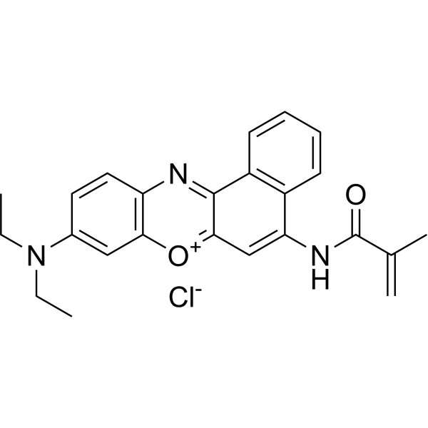 0 ImagesCat. No.: HY-D1463CAS No.: 699018-10-5Nile Blue Methacrylamide is a pH-responsive probe. Nile Blue Methacrylamide exhibits pH-sensitive absorption and fluorescence emission in the physiological pH range, with spectral shifts occurring between acidic and near-physiological pH conditions. (Ex/Em = 590/670-700 nm)
-
3-Methyl-8-(2'-deoxy-β-D-ribofuranosyl)isoxanthopterin
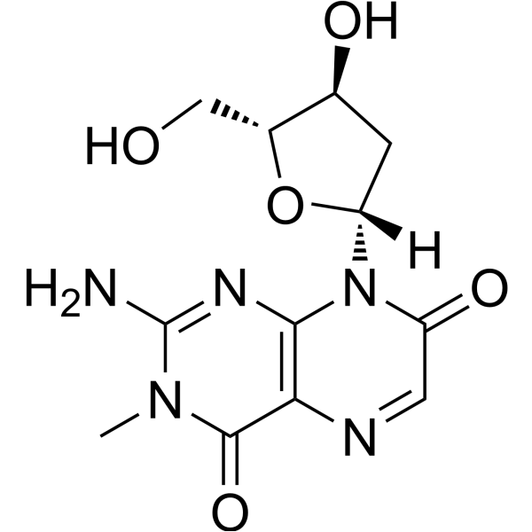 0 ImagesCat. No.: HY-147114CAS No.: 170379-51-83-Methyl-8-(2'-deoxy-β-D-ribofuranosyl)isoxanthopterin is a DNA uptake tracer, a pteridine-based fluorescent guanosine analogue. 3-Methyl-8-(2'-deoxy-β-D-ribofuranosyl)isoxanthopterin is widely used in studies of DNA binding and dynamics, with an absorbance maximum at 350 nm and an emission maximum at 430 nm.
-
ATTO 495 NHS ester
 0 ImagesCat. No.: HY-D3494CAS No.: 948041-99-4ATTO 495 NHS ester features an NHS ester reactive group used for conjugation and labeling via primary amine groups. It is primarily used for labeling antibodies and proteins (Ex/Em = 495/520 nm).
-
Zinc(II) probe-1
 0 ImagesCat. No.: HY-D3226CAS No.: 2417345-13-0Zinc (II) probe-1 (Compound DNP) is a dual-color Fluorescent probe that can simultaneously monitor Zn2+ and H+. Upon interaction with Zn2+, Zinc (II) probe-1 produces bright blue fluorescence (excitation wavelength: 405 nm; blue channel wavelength: 420-500 nm). Upon interaction with H+, Zinc (II) probe-1 exhibits red fluorescence (excitation wavelength: 561 nm; red channel emission wavelength: 630-730 nm). Zinc (II) probe-1 can be used in studies related to depression.
-
CY5-β-Amyloid (1-42), (rat/mouse)
 0 ImagesCat. No.: HY-P1388F1Synonyms: CY5-Ahx-Amyloid β-peptide (1-42) (rat/mouse)CY5-β-Amyloid (1-42), (rat/mouse) (CY5-Ahx-Amyloid β-peptide (1-42) (rat/mouse)) is a far-red fluorescent dye Cy5-labeled β-Amyloid (1-42), (rat/mouse) (HY-P1388) (Ex/Em ≈ 651 nm/670 nm). CY5-β-Amyloid (1-42), (rat/mouse) can be used in the basic research of Alzheimer's disease (AD) and related neurodegenerative diseases to monitor the aggregation, distribution and interaction of Aβ peptide segments in real time.
-
CGTDP
 0 ImagesCat. No.: HY-D3102CAS No.: 1015251-87-2CGTDP is a fluorescent probe used for selective labeling of active-site arginine residues, detection of local polarity, and monitoring of conformational changes in enzyme active sites. CGTDP contains a neutral red moiety serving as a polarity-sensitive fluorophore, and a phenylglyoxal unit acting as an arginine-specific labeling group; it undergoes a selective reaction via its α-dicarbonyl moiety with the guanidyl group of arginine residues in enzyme active sites to form a covalent linkage, thereby enabling the reporting of the local microenvironment at the labeled site. The excitation/emission wavelengths are Ex/Em = 398/607 nm and Ex/Em = 507/607 nm; these wavelengths blue-shift in organic solvents, such as Ex/Em = 375/569 nm and Ex/Em = 469/569 nm in THF, and Ex/Em = 375/587 nm and Ex/Em = 470/587 nm in acetonitrile. When labeled onto creatine kinase, its excitation/emission wavelengths are Ex/Em = 493/590 nm.
-
PerCP Maleimide
 0 ImagesCat. No.: HY-D2094PerCP Maleimide is a fluorescent dye that reacts with free sulfhydryl groups on proteins. PerCP is a red fluorescence albuminous dye for immunostaining and Maleimide can be leveraged for the preparation of fluorogenic probe, which is mainly used for the specific detection of thiol analytes.
-
Ratio-pHCL-1
 0 ImagesCat. No.: HY-D3129Ratio-pHCL-1 is a fluorescent probe for quantitative ratiometric pH imaging in biological systems. Ratio-pHCL-1 consists of an acrylamide 1,2-dioxetane chemiluminescent scaffold and a linked pH-sensitive carbon fluorescein fluorophore, and functions via chemically initiated electron exchange luminescence (CIEEL) decomposition and chemiluminescence resonance energy transfer (CRET). Ratio-pHCL-1 can cross cell membranes and accumulate intracellularly, enabling pH measurement within the range of 6.8-8.4.


-
C-TPA
 0 ImagesCat. No.: HY-D3084C-TPA is a fluorescent probe used for hypochlorite detection and bioimaging of lipid droplet polarity in cancer cells. For hypochlorite detection, when its derivative C-TPA-S is exposed to hypochlorite, an oxidative desulfurization reaction converts the thiocarbonyl group of C-TPA-S to the carbonyl group of C-TPA, thus restoring bright fluorescence from the almost non-fluorescent C-TPA-S. For bioimaging of lipid droplet polarity, the intramolecular charge transfer process between its Triphenylamine (HY-W011998) donor group and Coumarin (HY-N0709) acceptor group endows C-TPA with solvatochromism-it exhibits stronger fluorescence in less polar environments (such as lipid droplets in cancer cells) and weaker fluorescence in more polar environments (such as lipid droplets in normal cells), enabling the differentiation of cancer cells and cancer tissues from normal cells and normal tissues. The excitation wavelength of C-TPA is 405 nm, and the emission wavelengths used for cell and tissue imaging cover 425-475 nm and 500-550 nm; in solvents, its emission peak shifts from 494 nm in non-polar toluene to 528 nm in polar water.




