



Caspase-8 Antibody (YA554)
(Synonyms: MCH5, CASP8, Caspase-8, CASP-8, Apoptotic cysteine protease, Apoptotic protease Mch-5, CAP4, FADD-homologous ICE/ced-3-like protease, FADD-like ICE, ICE-like apoptotic protease 5, MORT1-associated ced-3 homolog, FLICE, MACH)Based on 1 Customer Validation
Caspase-8 Antibody (YA554) is a Rabbit-derived and non-conjugated IgG monoclonal antibody, targeting to Caspase-8.
-
Host:
Rabbit
-
Isotype:
IgG
-
Application:
WB, ICC/IF, IHC-P
-
Reactivity :
Human
-
Formulation:
Supplied in 1*TBS (pH7.4), 0.05% BSA and 40% Glycerol. Preservative: 0.05% Sodium Azide.
-
Conjugation:
Non-conjugated


To place orders, for customer services and technical support, please contact: MedChemExpress USA
Tel: 609-228-6898 E-mail: [email protected] [email protected]



-

HY-P80046
Caspase-3 Antibody (YA557) -
HY-P80624
Cleaved-Caspase 8 Antibody (YA792) -
HY-P80028
Bax Antibody (YA591) -

HY-K0010
Protease Inhibitor Cocktail (EDTA-Free, 100× in DMSO) -
HY-P80050
Caspase-9 Antibody (YA553) -
HY-P80450
GPX4 Antibody (YA399) -
HY-P80468
Phospho-MLKL (Ser345) Antibody (YA174) -
HY-P80137
GAPDH Antibody (YA418) -
HY-P80623
Cleaved-Caspase 3 p12 Antibody (YA499) -
HY-P80029
Bcl-2 Antibody (YA589) -
HY-P80589
Caspase-7 Antibody (YA561)


Applications
| Application |
WB

WB: Western Blot
|
ICC/IF
ICC/IF: Immunocytochemistry/
Immunofluorescence |
IHC-P
IHC-P: Immunohistochemistry-Paraffin
|
|---|---|---|---|
| Dilution Ratio | 1:1000-1:2000 | 1:50-1:200 | 1:50-1:200 |


Product Details
Caspase-8 Antibody (YA554) is a Rabbit-derived and non-conjugated IgG monoclonal antibody, targeting to Caspase-8.
-
Host Rabbit

-
Clonality Recombinant,Monoclonal

-
Species ReactivityHuman
-
Observed Molecular WeightObserved band size: 55 kDa
Note: Due to possible protein modifications or aggregation, the molecular weight should be confirmed by actual measurement, and the predicted value is for reference only.

-
Calculated Molecular Weight Predicted band size: 55 kDa

Synthetic peptide corresponding to Human Caspase-8.AA range:200-249.
Endogenous
Protein A affinity purified.
Non-conjugated
Unmodified
IgG


Product Properties
-
Appearance
Solution
-
Formulation
Supplied in 1*TBS (pH7.4), 0.05% BSA and 40% Glycerol. Preservative: 0.05% Sodium Azide.
-
Concentration
Batch-dependent, Please check the COA for the concentration of each lot. Check Lot Concentration
-
Storage & Stability
Stored at -20°C for 1 year. Avoid repeated freeze / thaw cycles.
-
Shipping
Shipping with blue ice.


Verification Images
-
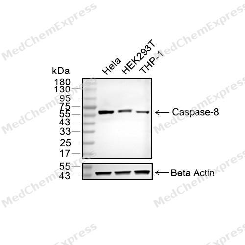 Western blot analysis of extracts from Hela (lane 2(20μg), HEK293T (lane 3(20μg) and THP-1(lane 4(20μg) using Caspase-8 (HY-P80049) Rabbit mAb. Proteins were transferred to a PVDF membrane and blocked with 5% non-fat milk in TBST for 2 hour at room temperature. The primary antibody (1/1000) and Loading control antibody (Beta Actin, HY-P80438, 1/10000) was used in 5% non-fat milk in TBST at 4°C overnight. Goat Anti-Mouse/Rabbit IgG-HRP Secondary Antibody (1/10000) was used for 1 hour at room temperature.
 -
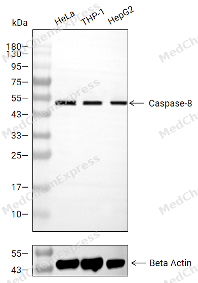 Western blot analysis of extracts from HeLa (lane 2(20μg), THP-1 (lane 3(20μg), HepG2 (lane 4(20μg),using Caspase-8 Antibody. Proteins were transferred to a PVDF membrane and blocked with 5% BSA in TBST for 2 hour at room temperature. The primary antibody and Loading control antibody (Beta Actin, HY-P80438, 1/3000) was used in 5% BSA in TBST at 4°C overnight. Goat Anti-Mouse/Rabbit IgG-HRP Secondary Antibody (HY-P8004/HY-P8001, 1/10,000) was used for 1 hour at room temperature.
 -
 Immunohistochemical analysis of paraffin-embedded human pancreas using Caspase-8 antibody. The section was pre-treated using heat mediated antigen retrieval with EDTA (pH 9.0) for 14 minutes. The tissues were blocked in 5% BSA for 30 minutes at room temperature, washed with TBST, and then probed with the primary antibody (HY-P80049, 1/200) for 1 hour at room temperature. The detection was performed using Polymer HRP-conjugated Goat Anti-Mouse/Rabbit lgG(H&L) secondary antibody (HY-P83652). DAB was used as the chromogen. Tissues were counterstained with hematoxylin and mounted with DPX.
 -
 Immunohistochemical analysis of paraffin-embedded human liver cancer using Caspase-8 antibody. The section was pre-treated using heat mediated antigen retrieval with EDTA (pH 9.0) for 14 minutes. The tissues were blocked in 5% BSA for 30 minutes at room temperature, washed with TBST, and then probed with the primary antibody (HY-P80049, 1/200) for 1 hour at room temperature. The detection was performed using Polymer HRP-conjugated Goat Anti-Mouse/Rabbit lgG(H&L) secondary antibody (HY-P83652). DAB was used as the chromogen. Tissues were counterstained with hematoxylin and mounted with DPX.
 -
 Immunohistochemical analysis of paraffin-embedded human breast cancer using Caspase-8 antibody. The section was pre-treated using heat mediated antigen retrieval with EDTA (pH 9.0) for 14 minutes. The tissues were blocked in 5% BSA for 30 minutes at room temperature, washed with TBST, and then probed with the primary antibody (HY-P80049, 1/200) for 1 hour at room temperature. The detection was performed using Polymer HRP-conjugated Goat Anti-Mouse/Rabbit lgG(H&L) secondary antibody (HY-P83652). DAB was used as the chromogen. Tissues were counterstained with hematoxylin and mounted with DPX.
 -
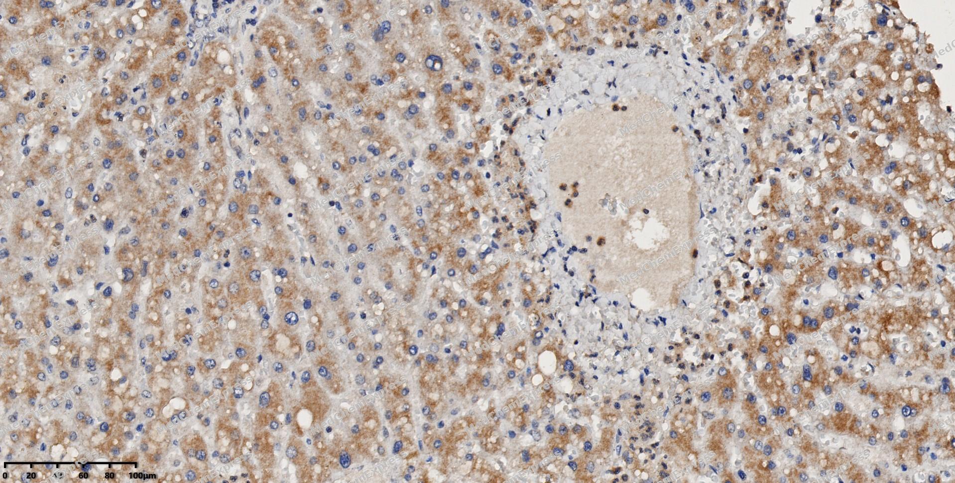 Immunohistochemical analysis of paraffin-embedded human liver using Caspase-8 antibody. The section was pre-treated using heat mediated antigen retrieval with EDTA (pH 9.0) for 14 minutes. The tissues were blocked in 5% BSA for 30 minutes at room temperature, washed with TBST, and then probed with the primary antibody (HY-P80049, 1/200) for 1 hour at room temperature. The detection was performed using Polymer HRP-conjugated Goat Anti-Mouse/Rabbit lgG(H&L) secondary antibody (HY-P83652). DAB was used as the chromogen. Tissues were counterstained with hematoxylin and mounted with DPX.
 -
 Immunohistochemical analysis of paraffin-embedded human pancreatic cancer using Caspase-8 antibody. The section was pre-treated using heat mediated antigen retrieval with EDTA (pH 9.0) for 14 minutes. The tissues were blocked in 5% BSA for 30 minutes at room temperature, washed with TBST, and then probed with the primary antibody (HY-P80049, 1/200) for 1 hour at room temperature. The detection was performed using Polymer HRP-conjugated Goat Anti-Mouse/Rabbit lgG(H&L) secondary antibody (HY-P83652). DAB was used as the chromogen. Tissues were counterstained with hematoxylin and mounted with DPX.
 -
 Immunohistochemical analysis of paraffin-embedded human breast cancer using Caspase-8 antibody. The section was pre-treated using heat mediated antigen retrieval with EDTA (pH 9.0) for 14 minutes. The tissues were blocked in 5% BSA for 30 minutes at room temperature, washed with TBST, and then probed with the primary antibody (HY-P80049, 1/200) for 1 hour at room temperature. The detection was performed using Polymer HRP-conjugated Goat Anti-Mouse/Rabbit lgG(H&L) secondary antibody (HY-P83652). DAB was used as the chromogen. Tissues were counterstained with hematoxylin and mounted with DPX.
 -
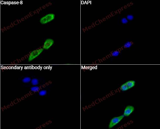 Immunocytochemistry analysis of Hela cells labeling Caspase-8 with Caspase-8 Antibody (HY-P80049)at 1/50 dilution. Cells were fixed in 4% paraformaldehyde for 15 minutes at room temperature, permeabilized with 0.1% Triton X-100 for 10 minutes at room temperature, then blocked with QuickBlock™ Blocking Buffer for Immunol Staining for 10 min at room temperature. Cells were then incubated with Caspase-8 Antibody (HY-P80049) at 1/50 dilution in QuickBlock™ Blocking Buffer for Immunol Staining at 4 ℃. Alexa Fluor® 488-conjugated AffiniPure Goat Anti-Rabbit IgG H&L(HY-P8002, Green) was used as the secondary antibody at 1/1,000 dilution. PBS instead of the primary antibody was used as the secondary antibody only control. The Nuclear counterstain was DAPI (Blue).
 -
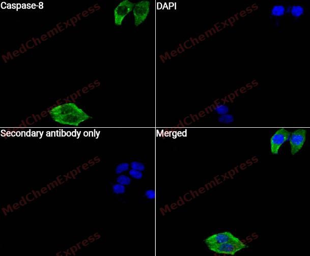 Immunocytochemistry analysis of Hela cells labeling Caspase-8 with Caspase-8 Antibody (HY-P80049) at 1/100 dilution. Cells were fixed in 4% paraformaldehyde for 15 minutes at room temperature, permeabilized with 0.1% Triton X-100 for 10 minutes at room temperature, then blocked with QuickBlock™ Blocking Buffer for Immunol Staining for 10 min at room temperature. Cells were then incubated with Caspase-8 Antibody (HY-P80049) at 1/100 dilution in QuickBlock™ Blocking Buffer for Immunol Staining at 4 ℃. Alexa Fluor® 488-conjugated AffiniPure Goat Anti-Rabbit IgG H&L(HY-P8002, Green) was used as the secondary antibody at 1/1,000 dilution. PBS instead of the primary antibody was used as the secondary antibody only control. The Nuclear counterstain was DAPI (Blue).


Background
-
Function
Caspase 8 is an initiator protease of death receptor-mediated extrinsic apoptosis and also supports cell survival by restraining RIP-family kinase-dependent necroptosis[1]. Mechanistically, caspase 8 works with cFLIP to regulate the switch among survival, apoptosis, and necroptosis, while cFLIP is structurally related to caspase 8 but lacks proteolytic activity[1][2]. Therefore, caspase 8 differs from inflammatory caspases such as caspase 1, 4, 5, and 12, which participate in inflammasome-related innate immune responses[3]. In intestinal disease, caspase 8 controls Paneth cell necroptosis and may contribute to epithelial cell death and mucosal inflammation in Crohn’s disease[3]. In severe SARS-CoV-2 mouse models, non-apoptotic caspase 8 promoted cytokine release, IL-1β elevation, NF-κB-linked inflammation, and disease pathology[4]. In ovarian cancer, low caspase 8 expression correlated with shorter overall survival, while caspase 8 loss enhanced non-apoptotic necroptotic death under NF-κB blockade[5]. For experimental applications, studies use genetic caspase 8 loss, viral caspase 8 inhibitors, and necroptosis pathway modulation to separate apoptosis, inflammation, and RIPK3-MLKL-dependent cell death[6].
-
Subcellular Localization
Cytoplasm; Nucleus; Cell projection, lamellipodium
-
Expression
Tissue_specificity:Isomers 1, 5, and 7 are expressed in a variety of tissues. They are most highly expressed in peripheral blood leukocytes, spleen, thymus, and liver. They are almost undetectable in the brain, testes, and skeletal muscle. -
Isoforms & Post-Translational Modification
Q14790 has 9 isomers: Q14790-1: 55391 Da (predicted); Q14790-2: 53768 Da (predicted); Q14790-3: 45929 Da (predicted); Q14790-4: 57701 Da (predicted); Q14790-5: 27484 Da (predicted); Q14790-6: 25862 Da (predicted); Q14790-7: 32330 Da (predicted); Q14790-8: 30707 Da (predicted); Q14790-9: 61836 Da (predicted).
Generation of the p10 and p18 subunits requires association with the death-inducing signaling complex (DISC), whereas additional processing is likely due to the autocatalytic activity of the activated protease. GZMB and CASP10 can be involved in these processing events;Phosphorylation on Ser-387 during mitosis by CDK1 inhibits activation by proteolysis and prevents apoptosis (PubMed:20937773). Phosphorylation on Tyr-380 by SRC is mediated by interaction with the SRC SH2 domain and does not affect dimerization or recruitment to the death-inducing signaling complex (DISC) but negatively regulates DISC-mediated processing and activation of CASP8, down-regulating its proapoptotic function (PubMed:16619028, PubMed:27109099). Phosphorylation on Tyr-380 also enhances localization to lamellipodia in migrating cells (PubMed:18216014);(Microbial infection) ADP-riboxanation by C.violaceum CopC blocks CASP8 processing, preventing CASP8 activation and ability to mediate extrinsic apoptosis;(Microbial infection) Proteolytically cleaved by the cowpox virus CRMA death inhibitory protein -
Subunit
Heterotetramer that consists of two anti-parallel arranged heterodimers, each one formed by a 18 kDa (p18) and a 10 kDa (p10) subunit (PubMed:10508784). Component of the death-induced signaling complex (DISC) composed of cell surface receptor FAS/CD95 or TNFRSF1A, adapter protein FADD and the CASP8 protease; recruitment of CASP8 to the complex is required for processing of CASP8 into the p18 and p10 subunits (PubMed:8681376, PubMed:8681377, PubMed:9184224). Component of the AIM2 PANoptosome complex, a multiprotein complex that drives inflammatory cell death (PANoptosis) (By similarity). Interacts with CFLAR and PEA15 (PubMed:10442631). Interacts with TNFAIP8L2 (By similarity). Interacts with CASP8AP2 (PubMed:16378960). Interacts with RFFL and RNF34; negatively regulate CASP8 through proteasomal degradation (PubMed:15069192). Interacts with NOL3; decreases CASP8 activity in a mitochondria localization- and phosphorylation-dependent manner and this interaction is dissociated by calcium (PubMed:15509781). Interacts with UBR2ca (PubMed:28602583). Interacts with RIPK1 (By similarity). Interacts with stimulated TNFRSF10B; this interaction is followed by CASP8 proteolytic cleavage and activation (PubMed:18846110). Interacts (phosphorylated on Tyr-380) with PIK3R1 (PubMed:27109099)
-
SwissProt ID
-
Synonyms
MCH5, CASP8, Caspase-8, CASP-8, Apoptotic cysteine protease, Apoptotic protease Mch-5, CAP4, FADD-homologous ICE/ced-3-like protease, FADD-like ICE, ICE-like apoptotic protease 5, MORT1-associated ced-3 homolog, FLICE, MACH
-
Research Field
Cell Biology


Documentation
-
Data Sheet (262 KB)
-
SDS (252 KB)
- English - EN (252 KB)
- Français - FR (252 KB)
- Deutsch - DE (252 KB)
- Norwegian - NO (252 KB)
- Español - ES (252 KB)
- Swedish - SV (252 KB)
- Italian - IT (252 KB)
- Korean - KR (252 KB)
- Portuguese - PT (252 KB)
-
User Guide for Antibodies (1077 KB)
References
[1]. Salvesen GS, et al. Functions of caspase 8: the identified and the mysterious. Semin Immunol. 2014 Jun;26(3):246-52. [Content Brief]
[2]. Tsuchiya Y, et al. FLIP the Switch: Regulation of Apoptosis and Necroptosis by cFLIP. Int J Mol Sci. 2015 Dec 18;16(12):30321-41. [Content Brief]
[3]. Becker C, et al. Complex roles of caspases in the pathogenesis of inflammatory bowel disease. Gastroenterology. 2013 Feb;144(2):283-293. [Content Brief]
[4]. M Bader S, et al. Non-apoptotic caspase-8 is critical for orchestrating exaggerated inflammation during severe SARS-CoV-2 infection. Nat Commun. 2025 Nov 13;16(1):9822. [Content Brief]
[5]. Kim M, et al. Caspase 8 expression may determine the survival of women with ovarian cancer. Cell Death Dis. 2016 Jan 14;7(1):e2045. [Content Brief]
[6]. Daley-Bauer LP, et al. Mouse cytomegalovirus M36 and M45 death suppressors cooperate to prevent inflammation resulting from antiviral programmed cell death pathways. Proc Natl Acad Sci U S A. 2017 Mar 28;114(13):E2786-E2795. [Content Brief]

 Powered by Bioz
