



Caspase-9 Antibody (YA553)
(Synonyms: MCH6, CASP9, Caspase-9, CASP-9, Apoptotic protease Mch-6, Apoptotic protease-activating factor 3, ICE-like apoptotic protease 6, APAF-3, ICE-LAP6)Based on 2 publication(s) in Google Scholar
Caspase-9 Antibody (YA553) is a Rabbit-derived and non-conjugated IgG monoclonal antibody, targeting to Caspase-9.
-
Host:
Rabbit
-
Isotype:
IgG
-
Application:
WB, IHC-P, ICC/IF, IP, FC
-
Reactivity :
Human, Mouse
-
Formulation:
Supplied in 1*TBS (pH7.4), 0.05% BSA and 40% Glycerol. Preservative: 0.05% Sodium Azide.
-
Conjugation:
Non-conjugated


To place orders, for customer services and technical support, please contact: MedChemExpress USA
Tel: 609-228-6898 E-mail: [email protected] [email protected]



-

HY-P80046
Caspase-3 Antibody (YA557) -
HY-P80029
Bcl-2 Antibody (YA589) -
HY-P80028
Bax Antibody (YA591) -
HY-P80964
Cleaved-Caspase 9 Antibody (YA852) -
HY-P80623
Cleaved-Caspase 3 p12 Antibody (YA499) -
HY-P80566
Bcl-2 Antibody (YA590) -
HY-P8001
HRP-conjugated Goat Anti-Rabbit IgG H&L -
HY-P80049
Caspase-8 Antibody (YA554) -
HY-P80450
GPX4 Antibody (YA399) -
HY-P80102
Cytochrome C Antibody (YA479) -
HY-P80344
STAT3 Antibody (YA056) -
HY-P80448
Cleaved-PARP1 Antibody (YA500)
Publications Citing Use of MedChemExpress (MCE) Caspase-9 Antibody (YA553)
More

Applications
| Application |
WB

WB: Western Blot
|
ICC/IF
ICC/IF: Immunocytochemistry/
Immunofluorescence |
IHC-P
IHC-P: Immunohistochemistry-Paraffin
|
IP
IP: Immunoprecipitation
|
FC
FC: Flow Cytometry
|
|---|---|---|---|---|---|
| Dilution Ratio | 1:500-1:5000 | 1:100-1:500 | 1:50-1:200 | Use at an assay dependent concentration | 1:1000 |


Product Details
Caspase-9 Antibody (YA553) is a Rabbit-derived and non-conjugated IgG monoclonal antibody, targeting to Caspase-9.
-
Host Rabbit

-
Clonality Recombinant,Monoclonal

-
Species ReactivityHuman, Mouse
-
Observed Molecular WeightObserved band size: 46 kDa
Note: Due to possible protein modifications or aggregation, the molecular weight should be confirmed by actual measurement, and the predicted value is for reference only.

-
Calculated Molecular Weight Predicted band size: 46 kDa

Synthetic peptide corresponding to Human Caspase-9.AA range:289-338.
Endogenous
Protein A affinity purified.
Non-conjugated
Unmodified
IgG


Product Properties
-
Appearance
Solution
-
Formulation
Supplied in 1*TBS (pH7.4), 0.05% BSA and 40% Glycerol. Preservative: 0.05% Sodium Azide.
-
Concentration
Batch-dependent, Please check the COA for the concentration of each lot. Check Lot Concentration
-
Storage & Stability
Stored at -20°C for 1 year. Avoid repeated freeze / thaw cycles.
-
Shipping
Shipping with blue ice.


Publications (2)
-
Journal Impact Factor
-
Most Recent
-
Naunyn Schmiedebergs Arch Pharmacol
Dehydrocostus lactone induces apoptosis and mitophagy in gastric cancer cells through the ROS-mediated mitochondrial pathway. [Abstract]2025 Oct 3. PMID: 41039061


Verification Images
-
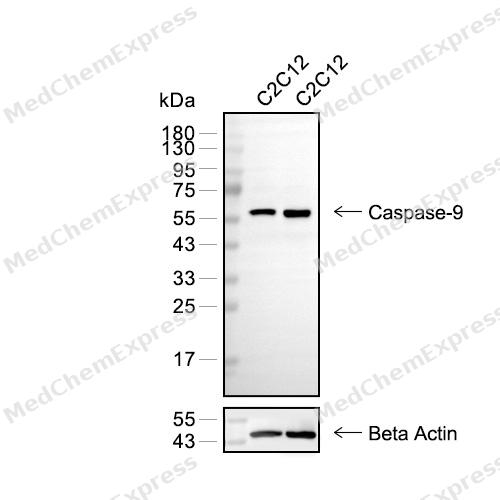 Western blot analysis of extracts from C2C12 (lane 2(20μg) and C2C12 (lane 3(40μg) using Caspase-9 (HY-P80050) Rabbit mAb. Proteins were transferred to a PVDF membrane and blocked with 5% defatted milk powder in TBST for 2 hour at room temperature. The primary antibody (HY-P80050, 1/1000) and Loading control antibody (Beta Actin, HY-P80438, 1/3000) was used in 5% defatted milk powder in TBST at 4°C overnight. Goat Anti-Mouse/Rabbit IgG-HRP Secondary Antibody (HY-P8004/HY-P8001, 1/10,000) was used for 1 hour at room temperature.
 -
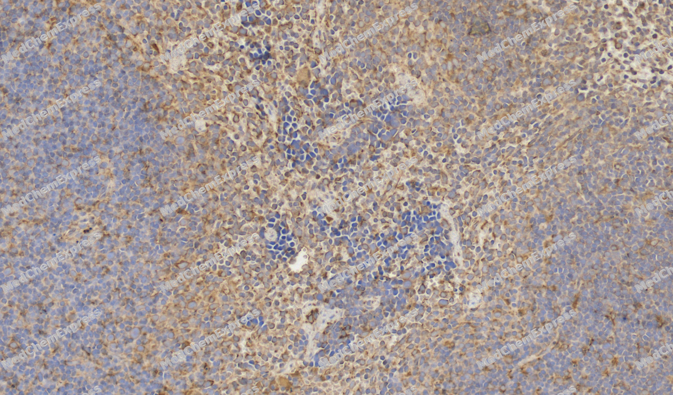 Immunohistochemical analysis of paraffin-embedded Mouse spleen tissue using Caspase-9 Antibody. The section was pre-treated using heat mediated antigen retrieval with sodium citrate buffer (pH 6.0) for 8 minutes. The tissues were blocked in QuickBlock for 20 minutes at room temperature, washed with ddH2O and PBS, and then probed with the primary antibody (HY-P80050, 1/100) in 4℃ overnight. The detection was performed using an HRP conjugated compact polymer system. DAB was used as the chromogen. Tissues were counterstained with hematoxylin and mounted with DPX.
 -
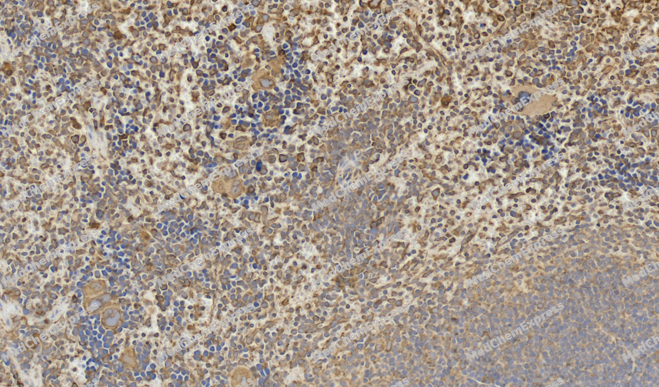 Immunohistochemical analysis of paraffin-embedded Mouse spleen tissue using Caspase-9 Antibody. The section was pre-treated using heat mediated antigen retrieval with sodium citrate buffer (pH 6.0) for 8 minutes. The tissues were blocked in QuickBlock for 20 minutes at room temperature, washed with ddH2O and PBS, and then probed with the primary antibody (HY-P80050, 1/100) in 4℃ overnight. The detection was performed using an HRP conjugated compact polymer system. DAB was used as the chromogen. Tissues were counterstained with hematoxylin and mounted with DPX.
 -
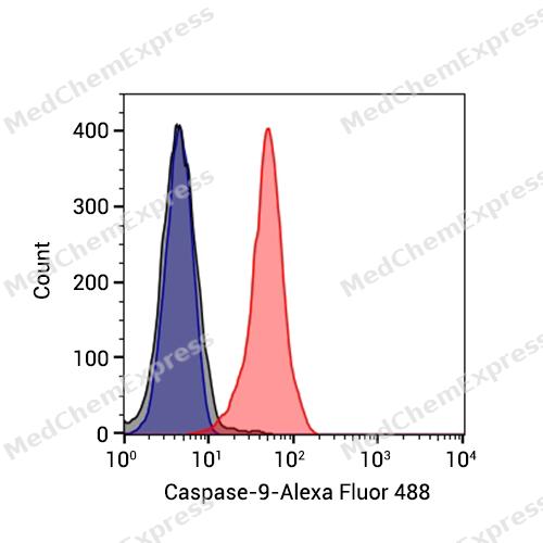 Flow cytometric analysis of 1X10^6 NIH3T3 cells labeling Caspase-9 Antibody(HY-P80050, red). Cells were fixed with 4% paraformaldehyde and permeabilised with 90% methanol. Then stained with the primary antibody at 1μg/mL dilution for an hour at 4℃. Alexa Fluor® 488-conjugated AffiniPure Goat Anti-Rabbit IgG H&L (HY-P8002) was used as the secondary antibody at 1/1,000 dilution for 30 minutes at 4℃. Rabbit IgG Isotype Control (HY-P80879, blue) was used as the isotype control, cells without incubation with primary antibody were used as the unlabeled control (black).
 -
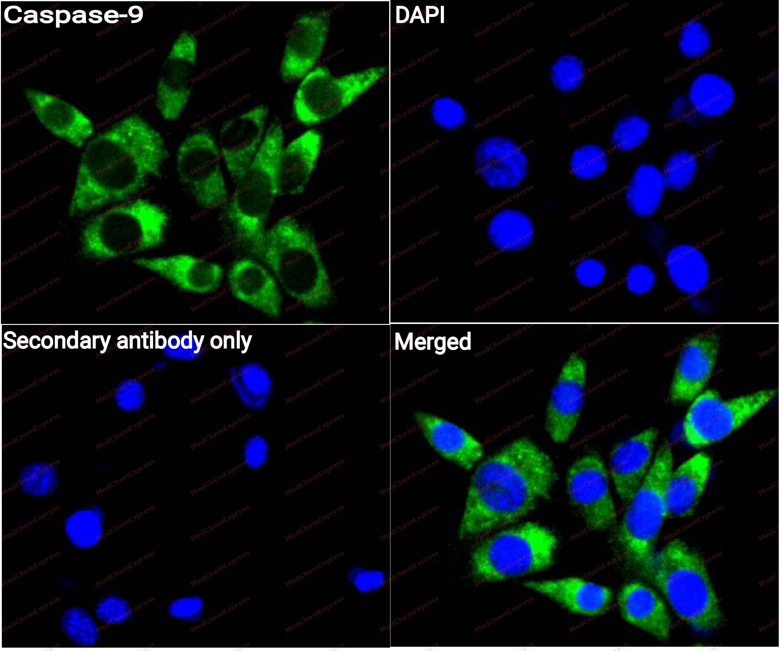 Immunocytochemistry analysis of NIH3T3 cells labeling Caspase-9 with Caspase-9 antibody (HY-P80050) at 1/100 dilution. Cells were fixed in 4% paraformaldehyde for 15 minutes at room temperature, permeabilized with 0.1% Triton X-100 for 10 minutes at room temperature, then blocked with QuickBlock™ Blocking Buffer for Immunol Staining for 10 min at room temperature. Cells were then incubated with Caspase-9 antibody (HY-P80050) at 1/100 dilution in QuickBlock™ Blocking Buffer for Immunol Staining at 4 ℃. Alexa Fluor® 488-conjugated AffiniPure Goat Anti-Rabbit IgG H&L(HY-P8002, Green) was used as the secondary antibody at 1/1,000 dilution. PBS instead of the primary antibody was used as the secondary antibody only control. The Nuclear counterstain was DAPI (Blue).
 -
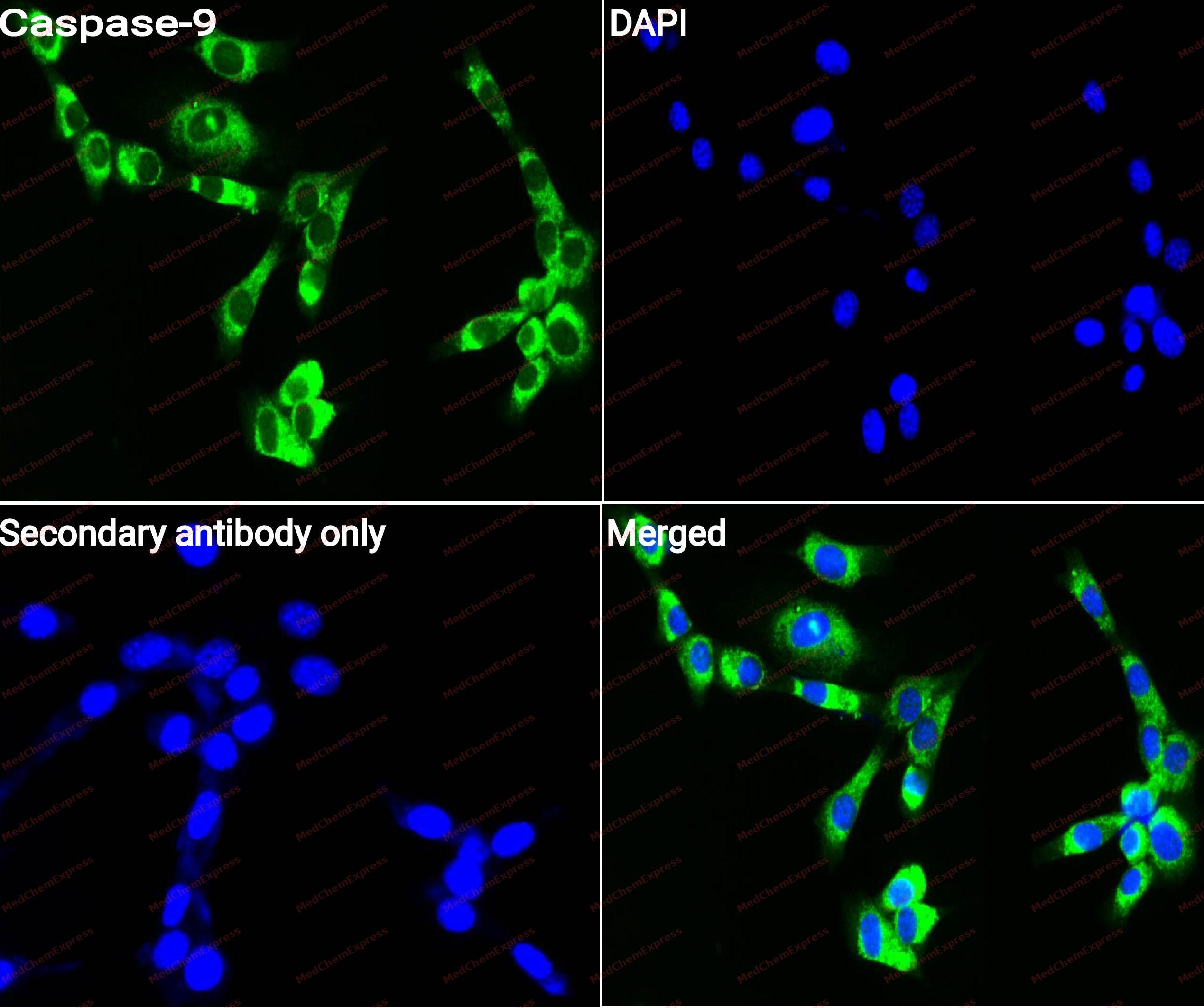 Immunocytochemistry analysis of NIH3T3 cells labeling Caspase-9 with Caspase-9 antibody (HY-P80050) at 1/250 dilution. Cells were fixed in 4% paraformaldehyde for 15 minutes at room temperature, permeabilized with 0.1% Triton X-100 for 10 minutes at room temperature, then blocked with QuickBlock™ Blocking Buffer for Immunol Staining for 10 min at room temperature. Cells were then incubated with Caspase-9 antibody (HY-P80050) at 1/100 dilution in QuickBlock™ Blocking Buffer for Immunol Staining at 4 ℃. Alexa Fluor® 488-conjugated AffiniPure Goat Anti-Rabbit IgG H&L(HY-P8002, Green) was used as the secondary antibody at 1/1,000 dilution. PBS instead of the primary antibody was used as the secondary antibody only control. The Nuclear counterstain was DAPI (Blue).


Background
-
Function
Caspase 9 plays a central role in the mitochondrial, or intrinsic, apoptotic pathway, where cytochrome c release promotes apoptosome formation with Apaf-1 and procaspase-9[1]. Mechanistically, activated caspase 9 cleaves and activates effector caspases 3 and 7, linking mitochondrial dysfunction to proteolytic cell death execution[1][2]. In disease models, diminished Apaf-1 activity reduced cytochrome c-dependent caspase activation in ovarian cancer cell lines, while ischemic hippocampal neurons showed caspase 9 activation before neuronal death[2][3]. Compared with caspase 8 in the extrinsic pathway, caspase 9 primarily mediates the intrinsic route to caspase 3 activation in ischemic neurons and TNFR-mediated hepatocyte apoptosis[3][4]. For experimental applications, Apaf-1 ligands inhibited apoptosome-mediated procaspase-9 activation and reduced apoptotic phenotypes in mitochondrial apoptosis models[5].
-
Expression
Tissue_specificity:It is widely distributed throughout the body, with the highest expression level in the heart, moderate expression levels in the liver, skeletal muscle, and pancreas, and low expression levels in all other tissues. In the heart, it is specifically expressed in cardiomyocytes. -
Subunit
Heterotetramer that consists of two anti-parallel arranged heterodimers, each one formed by a 35 kDa (p35) and a 10 kDa (p10) subunit. Caspase-9 and APAF1 bind to each other via their respective NH2-terminal CED-3 homologous domains in the presence of cytochrome C and ATP. Interacts (inactive form) with EFHD2. Interacts with HAX1. Interacts with BIRC2/c-IAP1, XIAP/BIRC4, BIRC5/survivin, BIRC6/bruce and BIRC7/livin. Interacts with ABL1 (via SH3 domain); the interaction is direct and increases in the response of cells to genotoxic stress and ABL1/c-Abl activation. Interacts with BCL2L10 (PubMed:19255499). Interacts with NleF from pathogenic E.coli
-
SwissProt ID
-
Synonyms
MCH6, CASP9, Caspase-9, CASP-9, Apoptotic protease Mch-6, Apoptotic protease-activating factor 3, ICE-like apoptotic protease 6, APAF-3, ICE-LAP6
-
Research Field
Cell Biology
Documentation
-
Data Sheet (262 KB)
-
SDS (252 KB)
- English - EN (252 KB)
- Français - FR (252 KB)
- Deutsch - DE (252 KB)
- Norwegian - NO (252 KB)
- Español - ES (252 KB)
- Swedish - SV (252 KB)
- Italian - IT (252 KB)
- Korean - KR (252 KB)
- Portuguese - PT (252 KB)
-
User Guide for Antibodies (1077 KB)
References
[1]. Allan LA, et al. Apoptosis and autophagy: Regulation of caspase-9 by phosphorylation. FEBS J. 2009 Nov;276(21):6063-73. [Content Brief]
[2]. Wolf BB, et al. Defective cytochrome c-dependent caspase activation in ovarian cancer cell lines due to diminished or absent apoptotic protease activating factor-1 activity. J Biol Chem. 2001 Sep 7;276(36):34244-51. [Content Brief]
[3]. Cao G, et al. Cloning and characterization of rat caspase-9: implications for a role in mediating caspase-3 activation and hippocampal cell death after transient cerebral ischemia. J Cereb Blood Flow Metab. 2002 May;22(5):534-46. [Content Brief]
[4]. Imao M, et al. Differential caspase-9-dependent signaling pathway between tumor necrosis factor receptor- and Fas-mediated hepatocyte apoptosis in mice. Liver Int. 2006 Feb;26(1):137-46. [Content Brief]
[5]. Malet G, et al. Small molecule inhibitors of Apaf-1-related caspase- 3/-9 activation that control mitochondrial-dependent apoptosis. Cell Death Differ. 2006 Sep;13(9):1523-32. [Content Brief]

 Powered by Bioz
