



Caspase-7 Antibody (YA561)
(Synonyms: Mch3; CMH-1; mCASP-7; AI314680; ICE-IAP3; caspase-7)
Based on 1 Customer Validation
Caspase-7 Antibody (YA561) is a Rabbit-derived and non-conjugated IgG monoclonal antibody, targeting to Caspase-7.
-
Host:
Rabbit
-
Isotype:
IgG
-
Application:
WB, ICC/IF, IP
-
Reactivity :
Mouse, Rat
-
Formulation:
Supplied in 50 mM Tris-Glycine (pH 7.4), 0.15 M NaCl, 40% Glycerol and 0.05% BSA. Preservative: 0.01% Sodium azide
-
Conjugation:
Non-conjugated


To place orders, for customer services and technical support, please contact: MedChemExpress USA
Tel: 609-228-6898 E-mail: [email protected] [email protected]





Applications
| Application |
WB

WB: Western Blot
|
ICC/IF
ICC/IF: Immunocytochemistry/
Immunofluorescence |
IP
IP: Immunoprecipitation
|
|---|---|---|---|
| Dilution Ratio | 1:500-1:1000 | 1:50-1:200 | 1:20 |


Product Details
Caspase-7 Antibody (YA561) is a Rabbit-derived and non-conjugated IgG monoclonal antibody, targeting to Caspase-7.
-
Host Rabbit

-
Clonality Recombinant,Monoclonal

-
Species ReactivityMouse, Rat
-
Observed Molecular WeightObserved band size: 34 kDa
Note: Due to possible protein modifications or aggregation, the molecular weight should be confirmed by actual measurement, and the predicted value is for reference only.

-
Calculated Molecular Weight Predicted band size: 34 kDa

Entrez Gene: 840 Human ; 12369 Mouse ; 64026 Rat
SwissProt: P55210 Human ; P97864 Mouse ;
OMIM: 601761 Human
Synthetic peptide corresponding to Mouse Caspase-7.The exact sequence is proprietary to MCE.
Endogenous
affinity purified
Non-conjugated
Unmodified
IgG


Product Properties
-
Appearance
Solution
-
Formulation
Supplied in 50 mM Tris-Glycine (pH 7.4), 0.15 M NaCl, 40% Glycerol and 0.05% BSA. Preservative: 0.01% Sodium azide
-
Concentration
Batch-dependent, Please check the COA for the concentration of each lot. Check Lot Concentration
-
Storage & Stability
Stored at -20°C for 1 year. Avoid repeated freeze / thaw cycles.
-
Shipping
Shipping with blue ice.


Verification Images
-
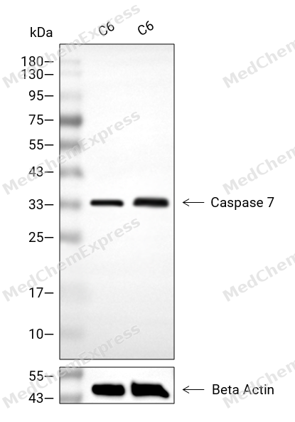 Western blot analysis of extracts from C6 (lane 2(20μg), C6 (lane 3(40μg), using Caspase 7 Antibody. Proteins were transferred to a PVDF membrane and blocked with 5% BSA in TBST for 2 hour at room temperature. The primary antibody and Loading control antibody (Beta Actin, HY-P80438, 1/3000) was used in 5% BSA in TBST at 4°C overnight. Goat Anti-Mouse/Rabbit IgG-HRP Secondary Antibody (HY-P8004/HY-P8001, 1/10,000) was used for 1 hour at room temperature.
 -
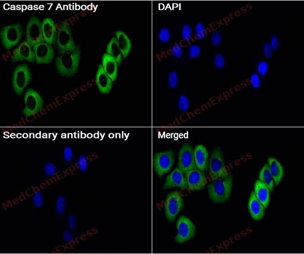 Immunocytochemistry analysis of HeLa cells labeling Caspase 7 with Caspase 7 antibody (HY-P80589) at 1/50 dilution. Cells were fixed in 4% paraformaldehyde for 15 minutes at room temperature, permeabilized with 0.1% Triton X-100 for 10 minutes at room temperature, then blocked with BSA for Immunol Staining for 10 min at room temperature. Cells were then incubated with Caspase 7 antibody (HY-P80589) at 1/50 dilution in BSA for Immunol Staining at 4 ℃,Stay overnight. AF488-conjugated AffiniPure Goat Anti-Rabbit IgG H&L(HY-P8002, Green) was used as the secondary antibody at 1/1,000 dilution. PBS instead of the primary antibody was used as the secondary antibody only control. The Nuclear counterstain was DAPI (Blue).
 -
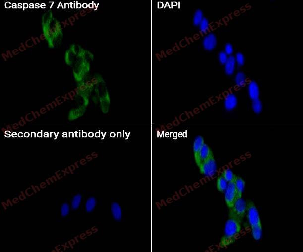 Immunocytochemistry analysis of HepG2 cells labeling Caspase 7 with Caspase 7 antibody (HY-P80589) at 1/50 dilution. Cells were fixed in 4% paraformaldehyde for 15 minutes at room temperature, permeabilized with 0.1% Triton X-100 for 10 minutes at room temperature, then blocked with BSA for Immunol Staining for 10 min at room temperature. Cells were then incubated with Caspase 7 antibody (HY-P80589) at 1/50 dilution in BSA for Immunol Staining at 4℃,Stay overnight. AF488-conjugated AffiniPure Goat Anti-Rabbit IgG H&L(HY-P8002, Green) was used as the secondary antibody at 1/1,000 dilution. PBS instead of the primary antibody was used as the secondary antibody only control. The Nuclear counterstain was DAPI (Blue).


Background
-
Function
Caspase 7 is an executioner cysteine protease that cleaves after aspartate residues and participates in programmed cell death and inflammation[1][2]. Mechanistically, death receptor and DNA-damage pathways activate caspase 7 through caspase 8 and caspase 9, placing it within apoptosis signaling cascades[1]. In Fas-stimulated Jurkat cells, caspase 8 activates caspase 3 and caspase 7, supporting a branched protease cascade during apoptotic execution[3]. In inflammatory models, caspase 1 inflammasomes activate caspase 7, and caspase 7 can mediate caspase 1-induced apoptosis upstream of caspase 3[1][4]. Disease and model evidence links caspase 7 to endotoxemia resistance in deficient mice, Salmonella-infected macrophages, neuropathic rat spinal cord apoptosis, and CHO-cell viability studies[1][4][5][6]. Compared with caspase 3, caspase 7 shares substrate recognition and some endogenous substrates, but specific substrates, caspase 1-dependent activation, and endotoxemia phenotypes distinguish the isoforms[1]. For experimental applications, isatin sulfonamides inhibit caspases 3 and 7 and serve as PET or SPECT apoptosis-imaging recognition units after radiolabeling[7].
-
Subcellular Localization
Cytoplasm, cytosol; Nucleus; Secreted, extracellular space
-
Expression
Tissue_specificity:It is highly expressed in the heart, lungs, liver, and kidneys. It is expressed at low levels in the spleen, skeletal muscle, and testes. It is not expressed in the brain. -
Subunit
Heterotetramer that consists of two anti-parallel arranged heterodimers, each one formed by a 20 kDa (p20) and a 11 kDa (p11) subunit (By similarity). Interacts with XIAP (via its second BIR domain); inhibiting CASP7 activity (By similarity). Interacts with BIRC6/bruce. Interacts with ATXN3 (short isoform 1) (By similarity). Interacts with HSPA5 (By similarity)
-
SwissProt ID
-
Synonyms
Mch3; CMH-1; mCASP-7; AI314680; ICE-IAP3; caspase-7
-
Research Field
Cell Biology


Documentation
-
Data Sheet (261 KB)
-
SDS (251 KB)
- English - EN (251 KB)
- Français - FR (251 KB)
- Deutsch - DE (251 KB)
- Norwegian - NO (251 KB)
- Español - ES (251 KB)
- Swedish - SV (251 KB)
- Italian - IT (251 KB)
- Korean - KR (251 KB)
- Portuguese - PT (251 KB)
-
User Guide for Antibodies (1077 KB)
References
[1]. Lamkanfi M, et al. Caspase-7: a protease involved in apoptosis and inflammation. Int J Biochem Cell Biol. 2010 Jan;42(1):21-4. [Content Brief]
[2]. Denault JB, et al. Caspases. Curr Protoc Protein Sci. 2002 Feb;Chapter 21:21.8.1-21.8.16. [Content Brief]
[3]. Hirata H, et al. Caspases are activated in a branched protease cascade and control distinct downstream processes in Fas-induced apoptosis. J Exp Med. 1998 Feb 16;187(4):587-600. [Content Brief]
[4]. Mahib MR, et al. Caspase-7 mediates caspase-1-induced apoptosis independently of Bid. Microbiol Immunol. 2020 Feb;64(2):143-152. [Content Brief]
[5]. Siniscalco D, et al. Involvement of subtype 1 metabotropic glutamate receptors in apoptosis and caspase-7 over-expression in spinal cord of neuropathic rats. Pharmacol Res. 2008 Mar;57(3):223-33. [Content Brief]
[6]. Safari F, et al. Caspase-7 deficiency in Chinese hamster ovary cells reduces cell proliferation and viability. Biol Res. 2020 Nov 13;53(1):52. [Content Brief]
[7]. Limpachayaporn P, et al. Isatin sulfonamides: potent caspases-3 and -7 inhibitors, and promising PET and SPECT radiotracers for apoptosis imaging. Future Med Chem. 2015;7(9):1173-96. [Content Brief]

 Powered by Bioz
