



PGK1 Antibody
(Synonyms: PGKA; MIG10; HEL-S-68p)Based on 1 Customer Validation
PGK1 Antibody is a Rabbit-derived and non-conjugated IgG polyclonal antibody, targeting to PGK1.
-
Host:
Rabbit
-
Isotype:
IgG
-
Application:
WB, ICC/IF, FC
-
Reactivity :
Human, Mouse, Rat
-
Formulation:
Supplied in 50mM Tris-Glycine(pH 7.4), 0.15M NaCl, 40%Glycerol, 0.01% sodium azide and 0.05% BSA.
-
Conjugation:
Non-conjugated


To place orders, for customer services and technical support, please contact: MedChemExpress USA
Tel: 609-228-6898 E-mail: [email protected] [email protected]





Applications
| Application |
WB

WB: Western Blot
|
ICC/IF
ICC/IF: Immunocytochemistry/
Immunofluorescence |
FC
FC: Flow Cytometry
|
|---|---|---|---|
| Dilution Ratio | 1:500-1:1000 | 1:50-1:200 | 1:50-1:100 |


Product Details
PGK1 Antibody is a Rabbit-derived and non-conjugated IgG polyclonal antibody, targeting to PGK1.
-
Host Rabbit

-
Clonality Polyclonal

-
Species ReactivityHuman, Mouse, Rat
-
Observed Molecular WeightObserved band size: 45 kDa
Note: Due to possible protein modifications or aggregation, the molecular weight should be confirmed by actual measurement, and the predicted value is for reference only.

-
Calculated Molecular Weight Predicted band size: 45 kDa

Entrez Gene: 5230 Human ; 18655 Mouse ; 24644 Rat
SwissProt: P00558 Human ; P09411 Mouse ; P16617 Rat
OMIM: 300653 Human
Synthetic peptide corresponding to Human PGK1.The exact sequence is proprietary to MCE.
Endogenous
affinity purified
Non-conjugated
Unmodified
IgG


Product Properties
-
Appearance
Solution
-
Formulation
Supplied in 50mM Tris-Glycine(pH 7.4), 0.15M NaCl, 40%Glycerol, 0.01% sodium azide and 0.05% BSA.
-
Concentration
Batch-dependent, Please check the COA for the concentration of each lot. Check Lot Concentration
-
Storage & Stability
Stored at -20°C for 1 year. Avoid repeated freeze / thaw cycles.
-
Shipping
Shipping with blue ice.


Verification Images
-
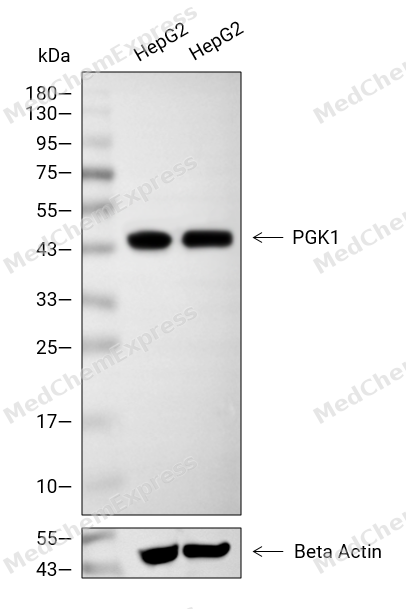 Western blot analysis of extracts from HepG2 (lane 2(20μg), HepG2 (lane 3(40μg), using PGK1 Antibody. Proteins were transferred to a PVDF membrane and blocked with 5% BSA in TBST for 2 hour at room temperature. The primary antibody and Loading control antibody (Beta Actin, HY-P80438, 1/3000) was used in 5% BSA in TBST at 4°C overnight. Goat Anti-Mouse/Rabbit IgG-HRP Secondary Antibody (HY-P8004/HY-P8001, 1/10,000) was used for 1 hour at room temperature.
 -
 Western blot analysis of extracts from HepG2 (lane 1(20μg)) 、HEK293 (lane 2(20μg)) 、NIH-3T3 (lane 3(20μg)) and C6 (lane 4(20μg)) using PGK1 Antibody (HY-P80784) . Proteins were transferred to a PVDF membrane and blocked with 5% nonfat dry milk in TBST for 1.5 hour at room temperature. The primary antibody (1/1000) and Loading control antibody (Hsp90, 1/10000) was used in 5% nonfat dry milk in TBST at 4℃ overnight. Goat Anti-Rabbit IgG-HRP Secondary Antibody (1/10,000) was used for 1 hour at room temperature.
 -
 Western blot analysis of extracts from WT (lane 1(20μg)) and KD (lane 2(20μg)) using PGK1 Antibody (HY-P80784) . Proteins were transferred to a PVDF membrane and blocked with 5% nonfat dry milk in TBST for 1.5 hour at room temperature. The primary antibody (1/1000) and Loading control antibody (Hsp90, 1/10000) was used in 5% nonfat dry milk in TBST at 4℃ overnight. Goat Anti-Rabbit IgG-HRP Secondary Antibody (1/10,000) was used for 1 hour at room temperature.
 -
 Flow cytometric analysis of 1X10⁶ HeLa cells labeling PGK1 Antibody (HY-P80784, red). Cells were fixed with 4% paraformaldehyde and permeabilised with 90% methanol. Then stained with the primary antibody at 1/100 dilution for an hour at 4℃. AF488-conjugated Goat Anti-Rabbit IgG H&L (HY-P8002) was used as the secondary antibody at 1/1,000 dilution for 30 minutes at 4℃. Rabbit IgG Isotype Control (HY-P80879, blue) was used as the isotype control, cells without incubation with primary antibody were used as the unlabeled control (black).
 -
 Immunocytochemistry analysis of Hela cells labeling PGK1 with PGK1 Antibody (HY-P80784) at 1/50 dilution. Cells were fixed in 4% paraformaldehyde for 15 minutes at room temperature, permeabilized with 0.1% Triton X-100 for 10 minutes at room temperature, then blocked with QuickBlock™ Blocking Buffer for Immunol Staining for 10 min at room temperature. Cells were then incubated with PGK1 Antibody (HY-P80784) at 1/50 dilution in QuickBlock™ Blocking Buffer for Immunol Staining at 4 ℃. Alexa Fluor® 488-conjugated AffiniPure Goat Anti-Rabbit IgG H&L(HY-P8002, Green) was used as the secondary antibody at 1/1,000 dilution. PBS instead of the primary antibody was used as the secondary antibody only control. The Nuclear counterstain was DAPI (Blue).
 -
 Immunocytochemistry analysis of Hela cells labeling PGK1 with PGK1 Antibody (HY-P80784) at 1/100 dilution. Cells were fixed in 4% paraformaldehyde for 15 minutes at room temperature, permeabilized with 0.1% Triton X-100 for 10 minutes at room temperature, then blocked with QuickBlock™ Blocking Buffer for Immunol Staining for 10 min at room temperature. Cells were then incubated with PGK1 Antibody (HY-P80784)at 1/100 dilution in QuickBlock™ Blocking Buffer for Immunol Staining at 4 ℃. Alexa Fluor® 488-conjugated AffiniPure Goat Anti-Rabbit IgG H&L(HY-P8002,Green) was used as the secondary antibody at 1/1,000 dilution. PBS instead of the primary antibody was used as the secondary antibody only control. The Nuclear counterstain was DAPI (Blue).


Background
-
Function
PGK1 catalyzes an ATP-producing step in glycolysis and supports tumor-cell metabolic fitness through glucose metabolism[1]. Mechanistically, mitochondrial PGK1 phosphorylates PDHK1 Thr338, activates PDHK1, inhibits the PDH complex, reduces mitochondrial pyruvate utilization, increases lactate production, and promotes brain tumorigenesis[2]. Under hypoxia or glutamine deprivation, PGK1 phosphorylates Beclin1 Ser30, enhances VPS34-Beclin1 activity, and drives stress-induced autophagy linked to glioblastoma prognosis[3]. In liver cancer models, PGK1 depletion reduced proliferation and tumorigenesis, while PGK1 acetylation promoted liver cancer progression[4]. Compared with PGK2, PGK1 represents the cytoplasmic isoform, whereas PGK2 is sperm-specific; PGK2 keeps an essentially identical active site but differs in C-terminal regions away from the active site[5]. For experimental applications, NG52 inhibited PGK1 kinase activity, restored PDH activity, reversed the Warburg effect, and suppressed glioma xenograft growth[1]. Ilicicolin H acted as a non-ATP-competitive PGK1 inhibitor that reduced glucose uptake, lactate production, and hepatocellular carcinoma proliferation[6]. Structure-based screening also identified CHR-6494 and Z57346765 as PGK1-binding inhibitors that reduced PGK1 metabolic enzyme activity and inhibited kidney renal clear cell carcinoma growth[7].- PGK1 connects glycolytic ATP production with PDHK1-PDH control and tumor metabolic remodeling[1][2]. - PGK1 differs from sperm-specific PGK2 mainly by expression context and distal structural regions[5]. - PGK1 inhibitors support mechanistic studies of glycolysis, autophagy, and cancer-cell proliferation[1][6][7].
-
Subcellular Localization
Cytoplasm, cytosol; Mitochondrion matrix
-
Expression
Tissue_specificity:It is primarily expressed in spermatogonia. It is located in the main sperm segment (protein level) . Its expression is significantly reduced in the testes of older men. -
Isoforms & Post-Translational Modification
P00558 has 2 isomers: P00558-1: 44615 Da (predicted); P00558-2: 41429 Da (predicted).
Phosphorylated at Ser-203 by MAPK1/ERK2 under hypoxic conditions, which promotes its mitochondrial targeting -
Subunit
Monomer (PubMed:18463139). Interacts with kinase MAPK1/ERK2; the interaction is direct, occurs under hypoxic conditions, and promotes its interaction with PIN1 (PubMed:26942675). Interacts with peptidyl-prolyl cis-trans isomerase PIN1; the interaction is direct, occurs under hypoxic conditions, and targets the protein to the mitochondrion by promoting interactions with the TOM complex (PubMed:26942675). Interacts with mitochondrial circRNA mcPGK1 (via its 2nd stem-loop); the interaction is direct and targets the protein to the mitochondrion by promoting interactions with the TOM complex (PubMed:36849569). Interacts with pyruvate dehydrogenase kinase PDK1; the interaction is direct, occurs under hypoxic conditions and leads to PDK1-mediated inhibition of pyruvate dehydrogenase complex activity (PubMed:26942675)
-
SwissProt ID
-
Synonyms
PGKA; MIG10; HEL-S-68p
-
Research Field
Cardiovascular


Documentation
-
Data Sheet (262 KB)
-
SDS (251 KB)
- English - EN (251 KB)
- Français - FR (251 KB)
- Deutsch - DE (251 KB)
- Norwegian - NO (251 KB)
- Español - ES (251 KB)
- Swedish - SV (251 KB)
- Italian - IT (251 KB)
- Korean - KR (251 KB)
- Portuguese - PT (251 KB)
-
User Guide for Antibodies (1077 KB)
References
[1]. Wang WL, et al. Pharmacologically inhibiting phosphoglycerate kinase 1 for glioma with NG52. Acta Pharmacol Sin. 2021 Apr;42(4):633-640. [Content Brief]
[2]. Li X, et al. Mitochondria-Translocated PGK1 Functions as a Protein Kinase to Coordinate Glycolysis and the TCA Cycle in Tumorigenesis. Mol Cell. 2016 Mar 3;61(5):705-719. [Content Brief]
[3]. Qian X, et al. Phosphoglycerate Kinase 1 Phosphorylates Beclin1 to Induce Autophagy. Mol Cell. 2017 Mar 2;65(5):917-931.e6. [Content Brief]
[4]. Hu H, et al. Acetylation of PGK1 promotes liver cancer cell proliferation and tumorigenesis. Hepatology. 2017 Feb;65(2):515-528. [Content Brief]
[5]. Sawyer GM, et al. X-ray analysis of phosphoglycerate kinase 2, a sperm-specific isoform from Mus musculus. Proteins. 2008 May 15;71(3):1134-44. [Content Brief]
[6]. Li M, et al. A novel inhibitor of PGK1 suppresses the aerobic glycolysis and proliferation of hepatocellular carcinoma. Biomed Pharmacother. 2023 Feb;158:114115. [Content Brief]
[7]. He Y, et al. Novel inhibitors targeting the PGK1 metabolic enzyme in glycolysis exhibit effective antitumor activity against kidney renal clear cell carcinoma in vitro and in vivo. Eur J Med Chem. 2024 Mar 5;267:116209. [Content Brief]

 Powered by Bioz
