



S100 A8 Antibody (YA1170)
(Synonyms: Calgranulin-A antibody; Calprotectin L1L subunit antibody; CFAG antibody; Cystic fibrosis antigen antibody; Leukocyte L1 complex light chain antibody; Migration inhibitory factor-related protein 8 antibody; MRP-8 antibody; N-terminally processed antibody; p8 antibody; Protein S100-A8 antibody; S100 calcium-binding protein A8 antibody; S100A8 antibody; S10A8_HUMAN antibody; Urinary stone protein band A antibody)Based on 1 Customer Validation
S100 A8 Antibody (YA1170) is a Rabbit-derived and non-conjugated IgG monoclonal antibody, targeting to S100 A8.
-
Host:
Rabbit
-
Isotype:
IgG
-
Application:
WB, IHC-P, ICC/IF, IP, FC, IF-Tissue
-
Reactivity :
Human
-
Formulation:
Supplied in PBS (pH7.4), 0.1% BSA, 40% Glycerol. Preservative: 0.05% Sodium Azide.
-
Conjugation:
Non-conjugated


To place orders, for customer services and technical support, please contact: MedChemExpress USA
Tel: 609-228-6898 E-mail: [email protected] [email protected]





Applications
| Application |
WB

WB: Western Blot
|
IHC-P
IHC-P: Immunohistochemistry-Paraffin
|
ICC/IF
ICC/IF: Immunocytochemistry/
Immunofluorescence |
FC
FC: Flow Cytometry
|
IF-Tissue
IF-Tissue: Immunofluorescence-Tissue
|
IP
IP: Immunoprecipitation
|
|---|---|---|---|---|---|---|
| Dilution Ratio | 1:2000-1:5000 | 1:10000 | 1:50000 | 1:1000 | 1:1000-1:3000 | 1-2μg/sample |


Product Details
S100 A8 Antibody (YA1170) is a Rabbit-derived and non-conjugated IgG monoclonal antibody, targeting to S100 A8.
-
Host Rabbit

-
Clonality Recombinant,Monoclonal

-
Species ReactivityHuman
-
Observed Molecular WeightObserved band size: 14 kDa
Note: Due to possible protein modifications or aggregation, the molecular weight should be confirmed by actual measurement, and the predicted value is for reference only.

-
Calculated Molecular Weight Predicted band size: 11 kDa

Recombinant protein within human S100A8 aa 1-93/93.
Endogenous
Affinity Purified
Non-conjugated
Unmodified
IgG


Product Properties
-
Appearance
Solution
-
Formulation
Supplied in PBS (pH7.4), 0.1% BSA, 40% Glycerol. Preservative: 0.05% Sodium Azide.
-
Concentration
Batch-dependent, Please check the COA for the concentration of each lot. Check Lot Concentration
-
Storage & Stability
Stored at -20°C for 1 year. Avoid repeated freeze / thaw cycles.
-
Shipping
Shipping with blue ice.


Verification Images
-
 Western blot analysis of extracts from SK-BR-3(lane 2(20ug) and SK-BR-3(lane 3(40ug) using S100 A8 Antibody (HY-P81425) Rabbit mAb. Proteins were transferred to a PVDF membrane and blocked with 5% non-fat milk in TBST for 2 hour at room temperature. The primary antibody (1/1000) and Loading control antibody (Beta Actin, HY-P83730, 1/10000) was used in 5% non-fat milk in TBST at 4°C overnight. Goat Anti-Mouse/Rabbit IgG-HRP Secondary Antibody (1/10000) was used for 1 hour at room temperature.
 -
 Immunohistochemical analysis of paraffin-embedded human oral cancer tissue using S100 A8 Antibody (HY-P81425, 1/10000). The section was pretreated using heat mediated antigen retrieval with sodium citrate buffer (pH 6.0) for 8 minutes. The tissues were blocked with quick block buffer for 0.5 hours at room temperature, washed with PBS and PBST, and then incubated with the primary antibody overnight at 4℃. The detection was performed using an HRP conjugated compact polymer system. DAB was used as the chromogen. Tissues were counterstained with hematoxylin and mounted with DPX.
 -
 Immunohistochemical analysis of paraffin-embedded human throat cancer tissue using S100 A8 Antibody (HY-P81425, 1/10000). The section was pretreated using heat mediated antigen retrieval with sodium citrate buffer (pH 6.0) for 8 minutes. The tissues were blocked with quick block buffer for 0.5 hours at room temperature, washed with PBS and PBST, and then incubated with the primary antibody overnight at 4℃. The detection was performed using an HRP conjugated compact polymer system. DAB was used as the chromogen. Tissues were counterstained with hematoxylin and mounted with DPX.
 -
 Immunohistochemical analysis of paraffin-embedded human lung cancer tissue using S100 A8 Antibody (HY-P81425, 1/10000). The section was pretreated using heat mediated antigen retrieval with sodium citrate buffer (pH 6.0) for 8 minutes. The tissues were blocked with quick block buffer for 0.5 hours at room temperature, washed with PBS and PBST, and then incubated with the primary antibody overnight at 4℃. The detection was performed using an HRP conjugated compact polymer system. DAB was used as the chromogen. Tissues were counterstained with hematoxylin and mounted with DPX.
 -
 Immunohistochemical analysis of paraffin-embedded human endometrial cancer tissue using S100 A8 Antibody (HY-P81425, 1/10000). The section was pretreated using heat mediated antigen retrieval with sodium citrate buffer (pH 6.0) for 8 minutes. The tissues were blocked with quick block buffer for 0.5 hours at room temperature, washed with PBS and PBST, and then incubated with the primary antibody overnight at 4℃. The detection was performed using an HRP conjugated compact polymer system. DAB was used as the chromogen. Tissues were counterstained with hematoxylin and mounted with DPX.
 -
 Immunohistochemical analysis of paraffin-embedded human breast cancer tissue using S100 A8 Antibody (HY-P81425, 1/10000). The section was pretreated using heat mediated antigen retrieval with sodium citrate buffer (pH 6.0) for 8 minutes. The tissues were blocked with quick block buffer for 0.5 hours at room temperature, washed with PBS and PBST, and then incubated with the primary antibody overnight at 4℃. The detection was performed using an HRP conjugated compact polymer system. DAB was used as the chromogen. Tissues were counterstained with hematoxylin and mounted with DPX.
 -
 Immunohistochemical analysis of paraffin-embedded human thyroid cancer tissue using S100 A8 Antibody (HY-P81425, 1/10000). The section was pretreated using heat mediated antigen retrieval with sodium citrate buffer (pH 6.0) for 8 minutes. The tissues were blocked with quick block buffer for 0.5 hours at room temperature, washed with PBS and PBST, and then incubated with the primary antibody overnight at 4℃. The detection was performed using an HRP conjugated compact polymer system. DAB was used as the chromogen. Tissues were counterstained with hematoxylin and mounted with DPX.
 -
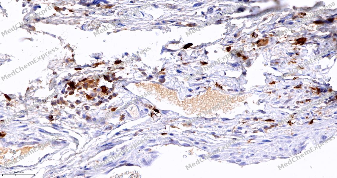 Immunohistochemical analysis of paraffin-embedded human lymphoma tissue using S100 A8 Antibody (HY-P81425, 1/10000). The section was pretreated using heat mediated antigen retrieval with sodium citrate buffer (pH 6.0) for 8 minutes. The tissues were blocked with quick block buffer for 0.5 hours at room temperature, washed with PBS and PBST, and then incubated with the primary antibody overnight at 4℃. The detection was performed using an HRP conjugated compact polymer system. DAB was used as the chromogen. Tissues were counterstained with hematoxylin and mounted with DPX.
 -
 Immunohistochemical analysis of paraffin-embedded human cervical cancer tissue using S100 A8 Antibody (HY-P81425, 1/10000). The section was pretreated using heat mediated antigen retrieval with sodium citrate buffer (pH 6.0) for 8 minutes. The tissues were blocked with quick block buffer for 0.5 hours at room temperature, washed with PBS and PBST, and then incubated with the primary antibody overnight at 4℃. The detection was performed using an HRP conjugated compact polymer system. DAB was used as the chromogen. Tissues were counterstained with hematoxylin and mounted with DPX.
 -
 Immunohistochemical analysis of paraffin-embedded human skin tissue using S100 A8 Antibody (HY-P81425, 1/10000). The section was pretreated using heat mediated antigen retrieval with sodium citrate buffer (pH 6.0) for 8 minutes. The tissues were blocked with quick block buffer for 0.5 hours at room temperature, washed with PBS and PBST, and then incubated with the primary antibody overnight at 4℃. The detection was performed using an HRP conjugated compact polymer system. DAB was used as the chromogen. Tissues were counterstained with hematoxylin and mounted with DPX.
 -
 Immunohistochemical analysis of paraffin-embedded human spleen tissue using S100 A8 Antibody (HY-P81425, 1/10000). The section was pretreated using heat mediated antigen retrieval with sodium citrate buffer (pH 6.0) for 8 minutes. The tissues were blocked with quick block buffer for 0.5 hours at room temperature, washed with PBS and PBST, and then incubated with the primary antibody overnight at 4℃. The detection was performed using an HRP conjugated compact polymer system. DAB was used as the chromogen. Tissues were counterstained with hematoxylin and mounted with DPX.
 -
 Flow cytometric analysis of 1X10⁶ THP-1 cells labeling S100 A8 Antibody (HY-P81425, red). Cells were fixed with 4% paraformaldehyde and permeabilised with 90% methanol. Then stained with the primary antibody at 1/1000 dilution for an hour at 4℃. AF488-conjugated Goat Anti-Rabbit IgG H&L (HY-P8002) was used as the secondary antibody at 1/1,000 dilution for 30 minutes at 4℃. Rabbit IgG Isotype Control (HY-P80879, blue) was used as the isotype control, cells without incubation with primary antibody were used as the unlabeled control (black).
 -
 Flow cytometric analysis of 1X10⁶ THP-1 cells labeling S100 A8 Antibody (HY-P81425, red). Cells were fixed with 4% paraformaldehyde and permeabilised with 90% methanol. Then stained with the primary antibody at 1/1000 dilution for an hour at 4℃. AF488-conjugated Goat Anti-Rabbit IgG H&L (HY-P8002) was used as the secondary antibody at 1/1,000 dilution for 30 minutes at 4℃. Rabbit IgG Isotype Control (HY-P80879, blue) was used as the isotype control, cells without incubation with primary antibody were used as the unlabeled control (black).


Background
-
Function
S100A8 is a calcium- and zinc-binding member of the S100 protein family that plays a prominent role in regulating inflammatory processes and innate immune responses, and it is most commonly present as the calprotectin complex together with S100A9[4]. Mechanistically, S100A8 functions as a damage-associated molecular pattern (DAMP) and participates in activation of TLR4- and AGER-dependent signaling, leading to downstream NF-κB and MAPK pathway activation that amplifies inflammatory responses. Through these pathways, S100A8 contributes to neutrophil chemotaxis, leukocyte adhesion, antimicrobial defense, and coordination of myeloid-cell-driven inflammation[4]. In disease settings, elevated S100A8/S100A9 signaling has been associated with inflammatory disorders, cardiovascular pathology, and tissue injury responses, supporting its value as a biomarker and mechanistic mediator of disease progression[1][2][3]. Experimental studies and clinical observations further indicate that circulating S100A8/A9 levels can provide prognostic information in cardiovascular disease, including heart failure following acute myocardial infarction[3]. Compared with the related isoform S100A9, S100A8 is frequently studied as a component of the S100A8/S100A9 heterodimer rather than as an isolated protein, reflecting the dominant biological activity of calprotectin in inflammatory microenvironments[4]. For experimental applications, inhibition of S100A8/A9 activity using small-molecule inhibitors or antibodies has improved pathological outcomes in murine models, making this signaling axis a useful target for mechanistic and translational research[2].
-
Subcellular Localization
Secreted; Cytoplasm; Cytoplasm, cytoskeleton; Cell membrane; Peripheral membrane protein
-
Expression
Tissue_specificity:Calprotectin (S100A8/9) is primarily expressed in myeloid cells. Except in inflammatory states, its expression is limited to specific stages of myeloid differentiation, as both proteins are expressed in circulating neutrophils and monocytes, but absent in normal tissue macrophages and lymphocytes. Calprotectin is also expressed in the epidermis in cH2O2ic inflammatory states, such as psoriasis and malignancies. Calprotectin concentrations are higher in serum at sites of inflammation or in patients with inflammatory diseases such as rheumatoid arthritis, cystic fibrosis, inflammatory bowel disease, Crohn's disease, giant cell arteritis, Sjgren's syndrome, systemic lupus erythematosus, and progressive systemic sclerosis. Calprotectin is involved in the formation and deposition of amyloid proteins (called amyloid inclusion bodies) in the aging prostate. It is significantly upregulated in a variety of tumors, including gastric cancer, esophageal cancer, colon cancer, pancreatic cancer, bladder cancer, ovarian cancer, thyroid cancer, breast cancer, and skin cancer.
Induction: (Microbial infection) Expression is highly induced in CD14 (+) monocytes, neutrophils, and developing neutrophils of infected by SARS-COV-2
Positive sample: SK-Br-3 cell lysate, human breast carcinoma tissue, human liver tissue, human spleen tissue, human tonsil tissue, SK-Br-3. -
Subunit
Homodimer. Preferentially exists as a heterodimer or heterotetramer with S100A9 known as calprotectin (S100A8/A9).
-
SwissProt ID
-
Synonyms
Calgranulin-A antibody; Calprotectin L1L subunit antibody; CFAG antibody; Cystic fibrosis antigen antibody; Leukocyte L1 complex light chain antibody; Migration inhibitory factor-related protein 8 antibody; MRP-8 antibody; N-terminally processed antibody; p8 antibody; Protein S100-A8 antibody; S100 calcium-binding protein A8 antibody; S100A8 antibody; S10A8_HUMAN antibody; Urinary stone protein band A antibody
-
Research Field
Immunology


Documentation
-
Data Sheet (262 KB)
-
SDS (254 KB)
- English - EN (254 KB)
- Français - FR (254 KB)
- Deutsch - DE (254 KB)
- Norwegian - NO (254 KB)
- Español - ES (254 KB)
- Swedish - SV (254 KB)
- Italian - IT (254 KB)
- Korean - KR (254 KB)
- Portuguese - PT (254 KB)
-
User Guide for Antibodies (1077 KB)
References
[1]. Averill MM, et al. S100A8 and S100A9 in cardiovascular biology and disease. Arterioscler Thromb Vasc Biol. 2012 Feb;32(2):223-9. [Content Brief]
[2]. Wang S, et al. S100A8/A9 in Inflammation. Front Immunol. 2018 Jun 11;9:1298. [Content Brief]
[3]. Ma J, et al. S100A8/A9 as a prognostic biomarker with causal effects for post-acute myocardial infarction heart failure. Nat Commun. 2024 Mar 27;15(1):2701. [Content Brief]
[4]. UCSC.edu. DATAbase.

 Powered by Bioz
