



SIRT1 Antibody (YA082)
(Synonyms: SIRT1; SIR2L1; NAD-dependent protein deacetylase sirtuin-1; hSIRT1; Regulatory protein SIR2 homolog 1; SIR2-like protein 1; hSIR2; Sirtuin 1)Based on 4 publication(s) in Google Scholar
SIRT1 Antibody (YA082) is a Rabbit-derived and non-conjugated IgG monoclonal antibody, targeting to SIRT1.
-
Host:
Rabbit
-
Isotype:
IgG
-
Application:
WB, IHC-P, ICC/IF, ChIP
-
Reactivity :
Human, Mouse
-
Formulation:
Supplied in 1*TBS (pH7.4), 0.05% BSA and 40% Glycerol. Preservative: 0.05% Sodium Azide.
-
Conjugation:
Non-conjugated


To place orders, for customer services and technical support, please contact: MedChemExpress USA
Tel: 609-228-6898 E-mail: [email protected] [email protected]



-

HY-P8001
HRP-conjugated Goat Anti-Rabbit IgG H&L -
HY-15452
Selisistat -
HY-P81051
Nrf2 Antibody(YA895) -
HY-P80245
NF-KB p65 Antibody (YA267) -
HY-K1048
AntiFade Reagent (with DAPI) -
HY-P80725
iNOS Antibody -
HY-P80541
AMPK alpha 2 Antibody (YA622) -
HY-P80871
PPAR alpha Antibody -
HY-P80769
NRF-1 Antibody (YA260) -
HY-P80155
Hes1 Antibody (YA387) -
HY-P80532
Acetyl-p53 (Lys370) Antibody (YA640)
Publications Citing Use of MedChemExpress (MCE) SIRT1 Antibody (YA082)
More

Applications
| Application |
WB

WB: Western Blot
|
ICC/IF
ICC/IF: Immunocytochemistry/
Immunofluorescence |
IHC-P
IHC-P: Immunohistochemistry-Paraffin
|
ChIP
ChIP: Chromatin Immunoprecipitation
|
|---|---|---|---|---|
| Dilution Ratio | 1:1000 | 1:50-1:200 | 1:50-1:1000 | Use 0.5~2 μg for 25 μg of chromatin. |


Product Details
SIRT1 Antibody (YA082) is a Rabbit-derived and non-conjugated IgG monoclonal antibody, targeting to SIRT1.
-
Host Rabbit

-
Clonality Recombinant,Monoclonal

-
Species ReactivityHuman, Mouse
-
Observed Molecular WeightObserved band size: 110-130 kDa
Note: Due to possible protein modifications or aggregation, the molecular weight should be confirmed by actual measurement, and the predicted value is for reference only.

-
Calculated Molecular Weight Predicted band size: 82 kDa

Entrez Gene: 23411 Human ; 93759 Mouse ; 309757 Rat
SwissProt: Q96EB6 Human ; Q923E4 Mouse ;
OMIM: 604479 Human
Synthetic peptide corresponding to Human SIRT1.AA range:698-747.
Endogenous
Protein A affinity purified.
Non-conjugated
Unmodified
IgG


Product Properties
-
Appearance
Solution
-
Formulation
Supplied in 1*TBS (pH7.4), 0.05% BSA and 40% Glycerol. Preservative: 0.05% Sodium Azide.
-
Concentration
Batch-dependent, Please check the COA for the concentration of each lot. Check Lot Concentration
-
Storage & Stability
Stored at -20°C for 1 year. Avoid repeated freeze / thaw cycles.
-
Shipping
Shipping with blue ice.


Publications (4)
-
Journal Impact Factor
-
Most Recent
-
Aging Cell
Pathogenic TDP-43 accelerates the generation of toxic exon1 HTT in Huntington's disease knock-in mice. [Abstract]2024 Dec;23(12):e14325. PMID: 39185703 -
J Inflamm Res
NAD-Dependent Protein Deacetylase Sirtuin-1 Mediated Mitophagy Regulates Early Brain Injury After Subarachnoid Hemorrhage. [Abstract]2024 Mar 28:17:1971-1981. PMID: 38562659 -
Naunyn Schmiedebergs Arch Pharmacol
L-theanine prevents myocardial injury in sleep‑deprived mice by suppressing ferroptosis through SIRT1. [Abstract]2025 May 6. PMID: 40328913


Verification Images
-
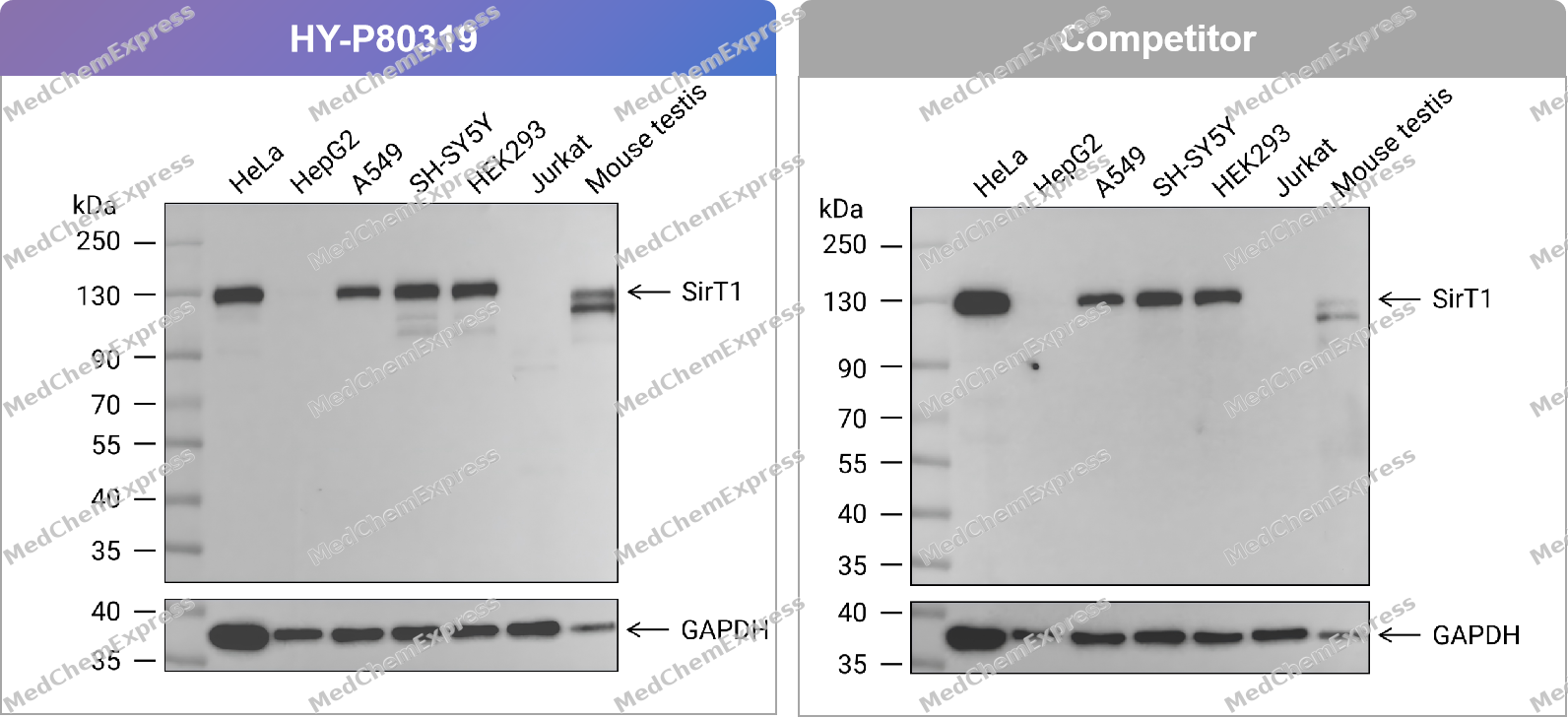 Western blot analysis of extracts from HeLa (lane 1(20μg)) 、HepG2 (lane 2(20μg)) 、A549 (lane 3(20μg)) 、SH-SY5Y (lane 4(20μg)) 、HEK293 (lane 5(20μg)) 、Jurkat (lane 6(20μg)) and Mouse testis (lane 7(20μg)) using SirT1 Antibody. Proteins were transferred to a PVDF membrane and blocked with 5% nonfat dry milk in TBST for 1.5 hour at room temperature. The primary antibody (HY-P80319, 1/1000) , competitor's antibody (1/1000) and Loading control antibody (GAPDH, 1/10000) was used in 5% nonfat dry milk in TBST at 4℃ overnight. Goat Anti-Rabbit IgG-HRP Secondary Antibody (1/10,000) was used for 1 hour at room temperature.
 -
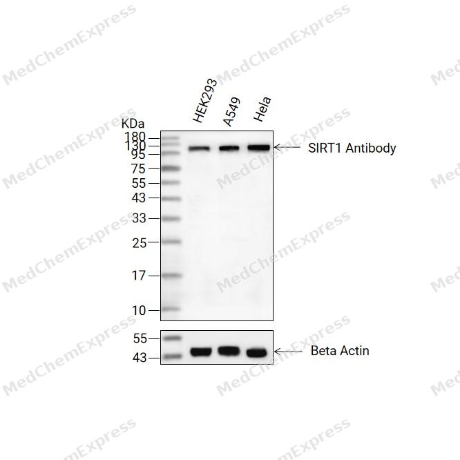 Western blot analysis of extracts from HEK293(lane 2(20ug) , A549(lane 3(20ug) and Hela(lane 4(20ug) using SIRT1 Antibody (HY-P80319) Rabbit mAb. Proteins were transferred to a PVDF membrane and blocked with 5% non-fat milk in TBST for 2 hour at room temperature. The primary antibody (1/1000) and Loading control antibody (Beta Actin, HY-P83730, 1/10000) was used in 5% non-fat milk in TBST at 4°C overnight. Goat Anti-Mouse/Rabbit IgG-HRP Secondary Antibody (1/10000) was used for 1 hour at room temperature.
 -
 Western blot analysis of extracts from HeLa (lane 1(20μg)) 、HepG2 (lane 2(20μg)) 、A549 (lane 3(20μg)) 、SH-SY5Y (lane 4(20μg)) 、HEK293 (lane 5(20μg)) 、Jurkat (lane 6(20μg)) and Mouse testis (lane 7(20μg)) using SirT1 Antibody (HY-P80319) . Proteins were transferred to a PVDF membrane and blocked with 5% nonfat dry milk in TBST for 1.5 hour at room temperature. The primary antibody (1/1000) and Loading control antibody (GAPDH, 1/10000) was used in 5% nonfat dry milk in TBST at 4℃ overnight. Goat Anti-Rabbit IgG-HRP Secondary Antibody (1/10,000) was used for 1 hour at room temperature.
 -
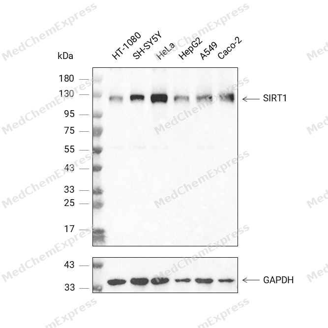 Western blot analysis was performed on protein extracts (30 μg) from HT-1080 (lane 2), SH-SY5Y (lane 3), HeLa (lane 4), HepG2 (lane 5), A549 (lane 6), and Caco-2 (lane 7) using SIRT1 antibody. Proteins were transferred onto a 0.45 μm PVDF membrane using the Trans-Blot® Turbo™ system for 13 min. The membrane was then blocked with 5% nonfat milk in TBST (HY-K1025) for 1 h at room temperature. Thhe primary antibody (1:1000) and loading control antibody GAPDH Antibody (HRP) (HY-P80954A) (1:2500) were diluted in 5% nonfat milk in TBST and incubated with the membrane overnight at 4°C. After washing, the membrane of primary antibody was incubated with HRP-conjugated goat anti-rabbit IgG (H&L) secondary antibody (HY-P8001) (1:5000) diluted in 5% nonfat milk in TBST for 1 h at room temperature. Protein bands were visualized using an Ultra High Sensitivity ECL detection kit (HY-K1005).
 -
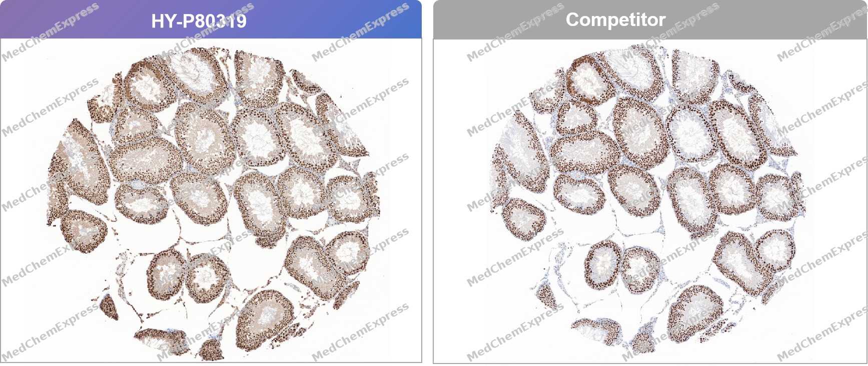 Immunohistochemical analysis of paraffin-embedded rat testis tissue using SIRT 1 Antibody. The section was pre-treated using heat mediated antigen retrieval with Tris/EDTA buffer (pH 9.0) for 20 minutes. The tissues were probed with the primary antibody (HY-P80319, 1/200) , competitor's antibody (1/200) overnight at 4℃. The detection was performed using an HRP conjugated compact polymer system. DAB was used as the chromogen. Tissues were counterstained with hematoxylin and mounted with neutral balsam.
 -
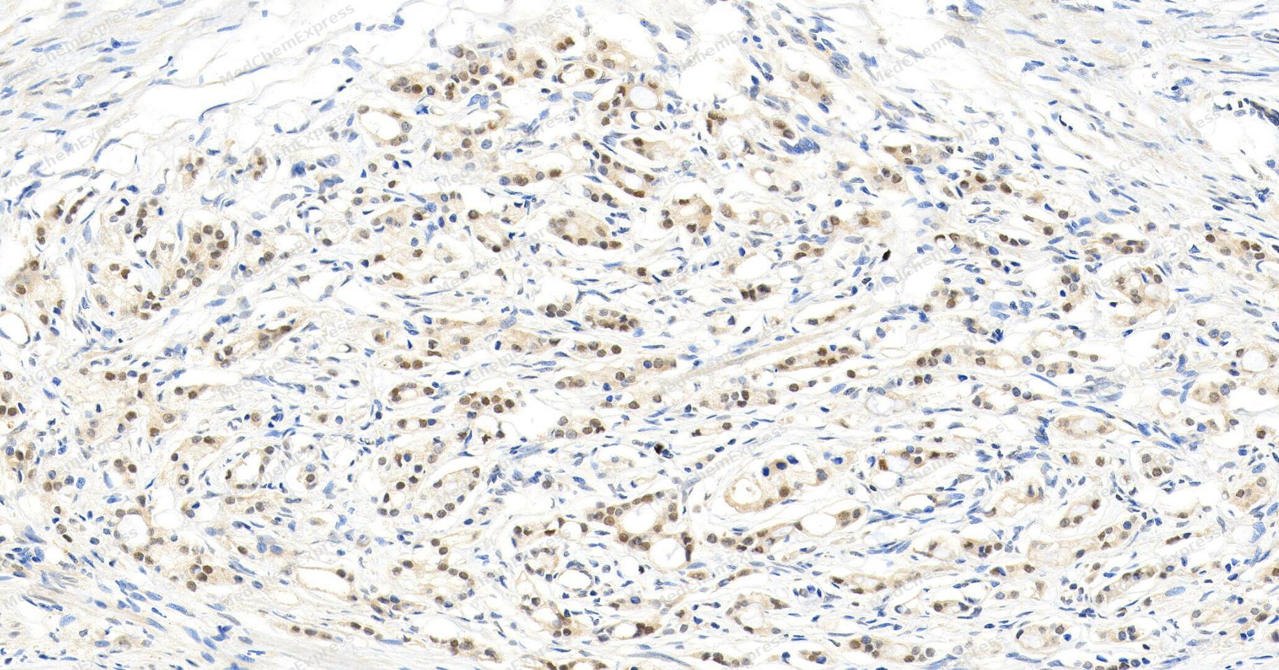 Immunohistochemical analysis of paraffin-embedded human prostate cancer tissue using SIRT1 antibody. The section was pre-treated using heat mediated antigen retrieval with Tris/EDTA buffer (pH 9.0) for 20 minutes. The tissues were probed with the primary antibody (HY-P80319, 1/100) overnight at 4℃. The detection was performed using an HRP conjugated compact polymer system. DAB was used as the chromogen. Tissues were counterstained with hematoxylin and mounted with neutral balsam.
 -
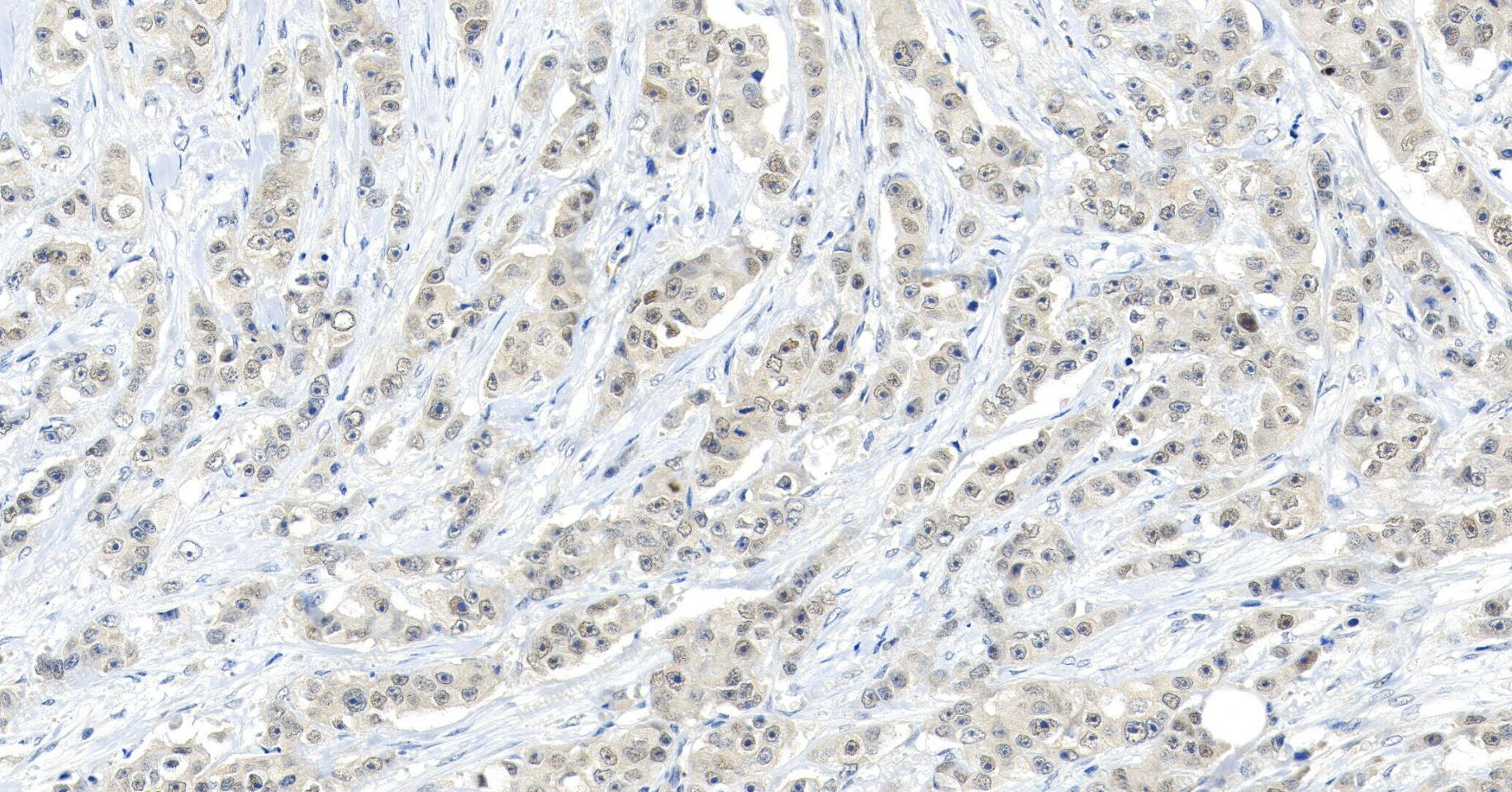 Immunohistochemical analysis of paraffin-embedded human breast cancer tissue using SIRT1 antibody. The section was pre-treated using heat mediated antigen retrieval with Tris/EDTA buffer (pH 9.0) for 20 minutes. The tissues were probed with the primary antibody (HY-P80319, 1/100) overnight at 4℃. The detection was performed using an HRP conjugated compact polymer system. DAB was used as the chromogen. Tissues were counterstained with hematoxylin and mounted with neutral balsam.
 -
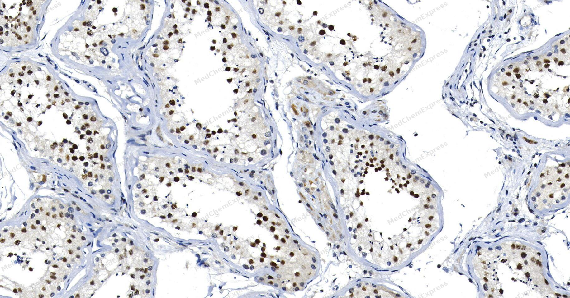 Immunohistochemical analysis of paraffin-embedded human testis tissue using SIRT1 antibody. The section was pre-treated using heat mediated antigen retrieval with Tris/EDTA buffer (pH 9.0) for 20 minutes. The tissues were probed with the primary antibody (HY-P80319, 1/100) overnight at 4℃. The detection was performed using an HRP conjugated compact polymer system. DAB was used as the chromogen. Tissues were counterstained with hematoxylin and mounted with neutral balsam.
 -
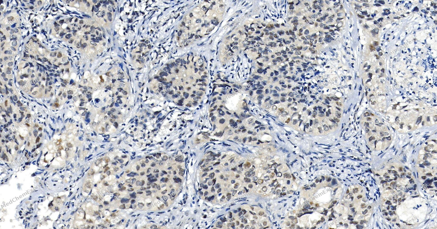 Immunohistochemical analysis of paraffin-embedded human lung cancer tissue using SIRT1 antibody. The section was pre-treated using heat mediated antigen retrieval with Tris/EDTA buffer (pH 9.0) for 20 minutes. The tissues were probed with the primary antibody (HY-P80319, 1/100) overnight at 4℃. The detection was performed using an HRP conjugated compact polymer system. DAB was used as the chromogen. Tissues were counterstained with hematoxylin and mounted with neutral balsam.
 -
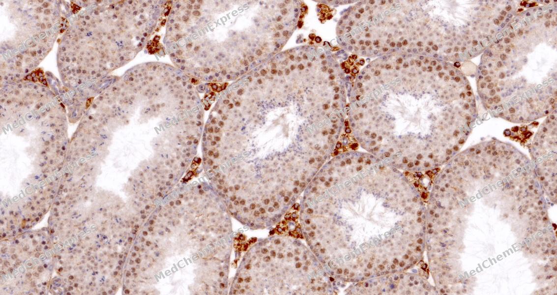 Immunohistochemical analysis of paraffin-embedded mouse testis tissue using SIRT1 Antibody. The section was pre-treated using heat mediated antigen retrieval with sodium citrate buffer (pH 6.0) for 8 minutes. The tissues were blocked in QuickBlock for 20 minutes at room temperature, washed with ddH2O and PBS, and then probed with the primary antibody at 1/100 dilution in 4℃ overnight. The detection was performed using an HRP conjugated compact polymer system. DAB was used as the chromogen. Tissues were counterstained with hematoxylin and mounted with DPX.
 -
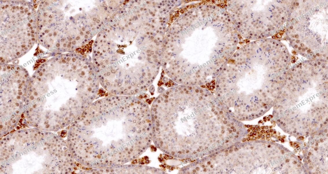 Immunohistochemical analysis of paraffin-embedded mouse testis tissue using SIRT1 Antibody. The section was pre-treated using heat mediated antigen retrieval with sodium citrate buffer (pH 6.0) for 8 minutes. The tissues were blocked in QuickBlock for 20 minutes at room temperature, washed with ddH2O and PBS, and then probed with the primary antibody at 1/100 dilution in 4℃ overnight. The detection was performed using an HRP conjugated compact polymer system. DAB was used as the chromogen. Tissues were counterstained with hematoxylin and mounted with DPX.
 -
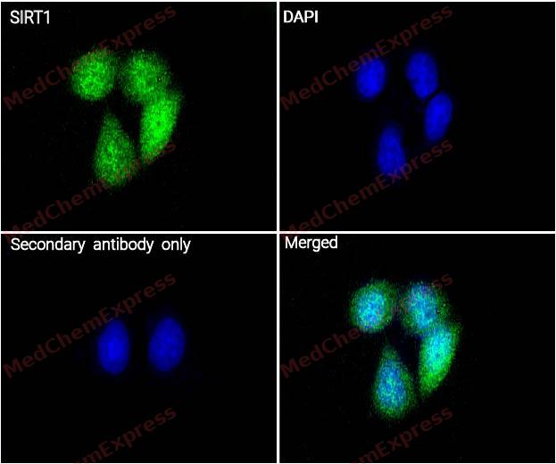 Immunocytochemistry analysis of Hela cells labeling SIRT1 with SIRT1 Antibody (HY-P80319) at 1/100 dilution . Cells were fixed in 4% paraformaldehyde for 15 minutes at room temperature, permeabilized with 0.1% Triton X-100 in PBS for 15 minutes at room temperature, then blocked with quick block buffer for 10 minutes at room temperature. Cells were then incubated with SIRT1 Antibody (HY-P80319) at 1/100 dilution in quick block buffer overnight at 4 ℃. AF488-conjugated Goat Anti-Rabbit IgG H&L(HY-P8002, Green) was used as the secondary antibody at 1/1,000 dilution. PBS instead of the primary antibody was used as the secondary antibody only control. The Nuclear counterstain was DAPI (Blue).
 -
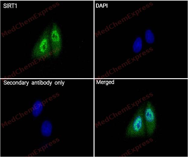 Immunocytochemistry analysis of Hela cells labeling SIRT1 with SIRT1 Antibody (HY-P80319) at 1/200 dilution . Cells were fixed in 4% paraformaldehyde for 15 minutes at room temperature, permeabilized with 0.1% Triton X-100 in PBS for 15 minutes at room temperature, then blocked with quick block buffer for 10 minutes at room temperature. Cells were then incubated with SIRT1 Antibody (HY-P80319) at 1/200 dilution in quick block buffer overnight at 4 ℃. AF488-conjugated Goat Anti-Rabbit IgG H&L(HY-P8002, Green) was used as the secondary antibody at 1/1,000 dilution. PBS instead of the primary antibody was used as the secondary antibody only control. The Nuclear counterstain was DAPI (Blue).


Background
-
Function
SIRT1 is a NAD\\+-dependent deacetylase that functions as a central regulator of cellular metabolism, stress responses, genomic stability, and aging through deacetylation of histones and multiple transcriptional regulators[1][2]. Mechanistically, SIRT1 integrates nutrient and energy signals with transcriptional programs that control mitochondrial biogenesis, fatty acid oxidation, glucose metabolism, autophagy, and cellular adaptation to metabolic stress[1][3]. Through coordinated interactions with metabolic signaling networks, SIRT1 contributes to maintenance of metabolic health across liver, skeletal muscle, adipose tissue, heart, and other organs[1]. In disease-related contexts, altered SIRT1 activity has been associated with aging, chronic inflammatory conditions, obesity, metabolic dysfunction, cardiovascular disorders, neurodegenerative diseases, and cancer-related processes, making it a widely used experimental target in translational research[2][4][5]. Compared with related sirtuin isoforms, SIRT1 is the most extensively characterized mammalian sirtuin and primarily mediates transcriptional regulation through deacetylation of nuclear histone and non-histone substrates, whereas other family members display distinct cellular localization and substrate preferences[1][2][4]. For experimental applications, SIRT1 activity can be modulated by small-molecule activators and inhibitors, and naturally occurring polyphenols such as resveratrol have been reported to activate SIRT1 in multiple model systems, providing useful tools for mechanistic studies of metabolism, inflammation, oxidative stress, and aging biology[3][4].
-
Subcellular Localization
Nucleus, PML body; Cytoplasm; Nucleus; Cytoplasm; Mitochondrion
-
Expression
Tissue_specificity:Broad expression
Induction:Up-regulated by methyl methanesulfonate (MMS) . In H293T cells by presence of rat calorie restriction (CR) serum
Positive sample: HeLa cell lysate, HEK-293 cell lysate, A549 cell lysate, F9 cell lysate, Mouse testis tissue lysate, Rat testis tissue lysate, Human testis tissue. -
Isoforms & Post-Translational Modification
Human (Q96EB6) has 2 isomers: Q96EB6-1: 81,681 Da (predicted); Q96EB6-2: 61,066 Da (predicted).
Methylated on multiple lysine residues.
Phosphorylated.
Proteolytically cleaved by cathepsin B upon TNF-alpha treatment to yield catalytic inactive but stable SirtT1 75 kDa fragment (75SirT1).
S-nitrosylated by GAPDH.
Acetylated at various Lys residues.
Ubiquitinated. -
Subunit
Interacts with XBP1 isoform 2 (PubMed:20955178). Found in a complex with PCAF and MYOD1. Interacts with FOXO1; the interaction deacetylates FOXO1, resulting in its nuclear retention and promotion of its transcriptional activity Component of the eNoSC complex, composed of SIRT1, SUV39H1 and RRP8. Interacts with HES1, HEY2 and PML. Interacts with RPS19BP1/AROS. Interacts with CCAR2 (via N-terminus); the interaction disrupts the interaction between SIRT1 and p53/TP53. Interacts with SETD7; the interaction induces the dissociation of SIRT1 from p53/TP53 and increases p53/TP53 activity. Interacts with MYCN, NR1I2, CREBZF, TSC2, TLE1, FOS, JUN, NR0B2, PPARG, NCOR, IRS1, IRS2 and NMNAT1. Interacts with HNF1A; the interaction occurs under nutrient restriction. Interacts with SUZ12; the interaction mediates the association with the PRC4 histone methylation complex which is specific as an association with PCR2 and PCR3 complex variants is not found. Interacts with BCL6; leads to a epigenetic repression of specific target genes. Interacts with CLOCK, BMAL1 and PER2 (By similarity). Interacts with PPARA; the interaction seems to be modulated by NAD(+) levels (PubMed:24043310). Interacts with NR1H3 and this interaction is inhibited in the presence of CCAR2. Interacts with CHEK2. Interacts with p53/TP53. Exhibits a preferential interaction with sumoylated CCAR2 over its unmodified form. Interacts with PACS2 (PubMed:29656858). Interacts with SIRT7 (By similarity). Interacts with PUS7 (PubMed:31451225). Interacts with TULP3 (PubMed:35397207). Interacts with MORN3; the interaction enhances the ubiquitination of p53/TP53 (PubMed:29681526)
-
SwissProt ID
-
Synonyms
SIRT1; SIR2L1; NAD-dependent protein deacetylase sirtuin-1; hSIRT1; Regulatory protein SIR2 homolog 1; SIR2-like protein 1; hSIR2; Sirtuin 1
-
Research Field
Cell Biology
Documentation
-
Data Sheet (262 KB)
-
SDS (252 KB)
- English - EN (252 KB)
- Français - FR (252 KB)
- Deutsch - DE (252 KB)
- Norwegian - NO (252 KB)
- Español - ES (252 KB)
- Swedish - SV (252 KB)
- Italian - IT (252 KB)
- Korean - KR (252 KB)
- Portuguese - PT (252 KB)
-
User Guide for Antibodies (1077 KB)
References
[1]. Dijk W, et al. Regulation of lipoprotein lipase by Angptl4. Trends Endocrinol Metab. 2014 Mar;25(3):146-55. [Content Brief]
[2]. Zhao L, et al. Sirtuins and their Biological Relevance in Aging and Age-Related Diseases. Aging Dis. 2020 Jul 23;11(4):927-945. [Content Brief]
[3]. Chung S, et al. Regulation of SIRT1 in cellular functions: role of polyphenols. Arch Biochem Biophys. 2010 Sep 1;501(1):79-90. [Content Brief]
[4]. Carafa V, et al. Sirtuins and disease: the road ahead. Front Pharmacol. 2012 Jan 31;3:4. [Content Brief]
[5]. Herskovits AZ, et al. Sirtuin deacetylases in neurodegenerative diseases of aging. Cell Res. 2013 Jun;23(6):746-58. [Content Brief]

 Powered by Bioz
