



Armepavine

Based on 1 Customer Validation
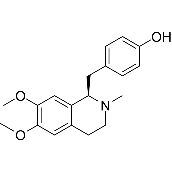
Armepavine, found in Nelumbo nucifera, is an orally active NF-κB inhibitor. Armepavine attenuates expression of p-p65, α-SMA, p-JNK1/2, p-ERK1/2, p-p38α stimulated by TNF-α and LPS. Armepavine suppresses NF-κB nuclear translocation, IκBα phosphorylation, and collagen deposition. Armepavine can be used for the research of hepatic fibrosis and leukemia.
For research use only. We do not sell to patients.
- Purity : 99.19%
- CAS No.: 524-20-9
- Formula: C19H23NO3
- Molecular Weight:313.39
-
Storage:
4°C, protect from light
* In solvent : -80°C, 6 months; -20°C, 1 month (protect from light)

To place orders, for customer services and technical support, please contact: MedChemExpress USA
Tel: 609-228-6898 E-mail: [email protected] [email protected]


-
 Biological Activity
-
Chemical Information
-
Solvent & Solubility
- Protocols
- Purity & Documentation
- References
-
 Help & FAQs




All AP-1 Isoforms
More

Biological Activity
Description
IC50 & Target
[1]|
p65 |
NF-κB |
p38 MAPK |
ERK1 |
ERK2 |
JNK |
In Vitro
Armepavine (1-10 μM; 24 h) does not exert cytotoxic effects on HSC-T6 rat hepatic stellate cells[1].
Armepavine (1-10 μM; 24 h) concentration-dependently inhibits TNF-α-induced collagen deposition in HSC-T6 rat hepatic stellate cells, with significant inhibition at 10 μM[1].
Armepavine (1-10 μM; 24 h) concentration-dependently inhibits TNF−α- and LPS-induced α-SMA protein expression in HSC-T6 rat hepatic stellate cells[1].
Armepavine (1-10 μM; 24 h) concentration-dependently inhibits TNF-α- and LPS-induced AP-1 transcriptional activity in HSC-T6 rat hepatic stellate cells, with significant effects at 10 μM for TNF-α and 3-10 μM for LPS[1].
Armepavine (1-10 μM; 6 h) concentration-dependently inhibits TNF-α-induced IκBα phosphorylation and NFκB p65 nuclear translocation in HSC-T6 rat hepatic stellate cells[1].
Armepavine (1-10 μM; 6 h) concentration-dependently inhibits TNF-α-induced NFκB transcriptional activity in HSC-T6 rat hepatic stellate cells, with significant effects at 1 and 10 μM[1].
Armepavine (10 μM; 15-60 min) inhibits TNF-α-induced phosphorylation of p38, ERK1/2, and JNK1/2 in HSC-T6 rat hepatic stellate cells[1].
Armepavine (1-10 μM; 24 h) concentration-dependently inhibits TNF-α-induced mRNA expression of iNOS, collagen 1α2, TIMP-1, and α-SMA in HSC-T6 rat hepatic stellate cells[1].
MedChemExpress (MCE) has not independently confirmed the accuracy of these methods. They are for reference only.
-
Cell Line:rat hepatic stellate cell line HSC-T6
-
Concentration:1 μM; 3 μM; 10 μM
-
Incubation Time:24 h
-
Result:Did not reduce HSC-T6 cell viability, with viability measured at 98% of untreated controls, showing no significant difference from controls.
Showed no cytotoxicity across the concentration range of 1-10 μM.
-
Cell Line:rat hepatic stellate cell line HSC-T6 stimulated with TNF-α or LPS
-
Concentration:1 μM; 3 μM; 10 μM
-
Incubation Time:24 h
-
Result:Reduced TNF-α-stimulated α-SMA expression to 160%, 127%, and 106% of control levels at 1, 3, and 10 μM respectively.
Reduced LPS-stimulated α-SMA expression in a concentration-dependent manner across 1-10 μM.
-
Cell Line:rat hepatic stellate cell line HSC-T6 stimulated with TNF-α
-
Concentration:1 μM; 3 μM; 10 μM
-
Incubation Time:6 h
-
Result:Reduced TNF-α-increased cytoplasmic IκBα phosphorylation and nuclear NFκB p65 protein levels in a concentration-dependent manner across 1-10 μM, with significant inhibition observed at 3 and 10 μM.
-
Cell Line:rat hepatic stellate cell line HSC-T6 stimulated with TNF-α
-
Concentration:10 μM
-
Incubation Time:15 min; 30 min; 60 min
-
Result:Significantly attenuated the TNF-α-induced phosphorylation of ERK1/2 at 15 minutes, p38 at 30 minutes, and JNK1/2 at 60 minutes.
-
Cell Line:rat hepatic stellate cell line HSC-T6 stimulated with TNF-α
-
Concentration:1 μM; 3 μM; 10 μM
-
Incubation Time:24 h
-
Result:Reduced TNF-α-stimulated mRNA expression of iNOS, collagen 1α2, TIMP-1, and α-SMA in a concentration-dependent manner across 1-10 μM, with significant inhibition observed at 3 and 10 μM for most genes.
In Vivo
MedChemExpress (MCE) has not independently confirmed the accuracy of these methods. They are for reference only.
-
Animal Model:Sprague-Dawley (male, 250~300 g, bile duct ligation-induced hepatic fibrosis)[1]
-
Dosage:3 mg/kg; 10 mg/kg
-
Administration:p.o.; twice daily; 3 weeks
-
Result:Increased body weight to 333 g, reduced liver weight to 27.5 g, decreased plasma ALT to 536 U/L and AST to 127 U/L, reduced fibrosis score to 1.85 (24% reduction from vehicle-treated BDL rats), reduced hepatocyte necrosis score to 0.38 (68% reduction from vehicle-treated BDL rats), reduced hepatic mRNA expression of col 1α2 to 7.45, TGF-β1 to 14.5, TIMP-1 to 4.36, ICAM-1 to 15.5, and IL-6 to 10.5 relative to GAPDH.
Increased body weight to 345 g, reduced liver weight to 26.3 g, decreased plasma ALT to 434 U/L and AST to 73 U/L, reduced hepatic collagen content to 4.38 mg/g liver, reduced α-SMA/α-tubulin ratio to 141%, reduced fibrosis score to 1.28 (47% reduction from vehicle-treated BDL rats), reduced hepatocyte necrosis score to 0.50 (57% reduction from vehicle-treated BDL rats), reduced hepatic mRNA expression of col 1α2 to 5.09, TGF-β1 to 7.09, TIMP-1 to 3.87, ICAM-1 to 7.03, iNOS to 3.09, and IL-6 to 4.09 relative to GAPDH; increased hepatic metallothionein mRNA expression to 0.57 relative to GAPDH.
- Kinase activity and phosphorylation assays
- Fibrosis/Collagen Morphometry
- RNA extraction experimental
- Cytoplasmic-Nuclear Fractionated Protein Extraction
- Western Blot
- Collagen: Sirius Red Staining
- Connective Tissue: Masson's Trichrome/Collagen Trichrome Staining
- Research Protocol for Inflammation-related Diseases
- Protocol for Kinase activity and phosphorylation assays


Chemical Information
-
CAS No. 524-20-9
-
Appearance Solid
-
Molecular Weight 313.39
-
Formula C19H23NO3
-
Color White to off-white
-
SMILES
OC(C=C1)=CC=C1C[C@@H]2C3=CC(OC)=C(OC)C=C3CCN2C
-
Structure Classification
-
Shipping
Room temperature in continental US; may vary elsewhere.
-
Storage
4°C, protect from light
* In solvent : -80°C, 6 months; -20°C, 1 month (protect from light)


Solvent & Solubility
In Vitro:
DMSO : 100 mg/mL (319.09 mM; Need ultrasonic; Hygroscopic DMSO has a significant impact on the solubility of product, please use newly opened DMSO)
Please refer to the solubility information to select the appropriate solvent. Once prepared, please aliquot and store the solution to prevent product inactivation from repeated freeze-thaw cycles.
Storage method and period of stock solution: -80°C, 6 months; -20°C, 1 month (protect from light). When stored at -80°C, please use it within 6 months. When stored at -20°C, please use it within 1 month.
Please refer to the solubility information to select the appropriate solvent. Once prepared, please aliquot and store the solution to prevent product inactivation from repeated freeze-thaw cycles.
Storage method and period of stock solution: -80°C, 6 months; -20°C, 1 month (protect from light). When stored at -80°C, please use it within 6 months. When stored at -20°C, please use it within 1 month.
Concentration (start) × Volume (start) = Concentration (final) × Volume (final)
In Vivo:
Select the appropriate dissolution method based on your experimental animal and administration route.
- For the following dissolution methods, please ensure to first prepare a clear stock solution using an In Vitro approach and then sequentially add co-solvents:
- To ensure reliable experimental results, the clarified stock solution can be appropriately stored based on storage conditions. As for the working solution for In Vivo experiments, it is recommended to prepare freshly and use it on the same day.
- The percentages shown for the solvents indicate their volumetric ratio in the final prepared solution. If precipitation or phase separation occurs during preparation, heat and/or sonication can be used to aid dissolution.
Add each solvent one by one: 10% DMSO 40% PEG300 5% Tween-80 45% Saline
Solubility: ≥ 2.5 mg/mL (7.98 mM); Clear solution
This protocol yields a clear solution of ≥ 2.5 mg/mL (saturation unknown).
Taking 1 mL working solution as an example, add 100 μL DMSO stock solution (25.0 mg/mL) to 400 μL PEG300, and mix evenly; then add 50 μL Tween-80 and mix evenly; then add 450 μL Saline to adjust the volume to 1 mL.
Preparation of Saline: Dissolve 0.9 g sodium chloride in ddH₂O and dilute to 100 mL to obtain a clear Saline solution.
Add each solvent one by one: 10% DMSO 90% (20% SBE-β-CD in Saline)
Solubility: ≥ 2.5 mg/mL (7.98 mM); Clear solution
This protocol yields a clear solution of ≥ 2.5 mg/mL (saturation unknown).
Taking 1 mL working solution as an example, add 100 μL DMSO stock solution (25.0 mg/mL) to 900 μL 20% SBE-β-CD in Saline, and mix evenly.
Preparation of 20% SBE-β-CD in Saline (4°C, storage for one week): 2 g SBE-β-CD powder is dissolved in 10 mL Saline, completely dissolve until clear.
In Vivo Dissolution Calculator
Please enter the basic information of animal experiments:
-
-
-
-
Recommended: Prepare an additional quantity of animals to account for potential losses during experiments.
Please enter your animal formula composition:
-
%DMSO +
Recommended: Keep the proportion of DMSO in working solution below 2% if your animal is weak.
-
%+
-
+%Tween-80 + +
-
%Saline +
The co-solvents required include: DMSO, . All of co-solvents are available by MedChemExpress (MCE). , Tween 80. All of co-solvents are available by MedChemExpress (MCE).
Working solution concentration: 0.22 mg/mL
Method for preparing stock solution: mg drug dissolved in μL DMSO. Stock solution concentration: mg/mL. * In solvent : -80°C, 6 months; -20°C, 1 month (protect from light)
1. Take μL DMSO stock solution;
2. Add μL .
μL , mix evenly;
3. Then add μL Tween 80, mix evenly;
4. Then add μL
Please ensure that the stock solution in the first step is dissolved to a clear state, and add co-solvents in sequence. You can use ultrasonic heating (ultrasonic cleaner, recommended frequency 20-40 kHz), vortexing, etc. to assist dissolution.


Purity & Documentation
-
Data Sheet (293 KB)
-
SDS (252 KB)
- English - EN (252 KB)
- Français - FR (252 KB)
- Deutsch - DE (252 KB)
- Norwegian - NO (252 KB)
- Español - ES (252 KB)
- Swedish - SV (252 KB)
- Italian - IT (252 KB)
- Korean - KR (252 KB)
- Portuguese - PT (252 KB)
-
Handling Instructions (2659 KB)
References
[1]. Weng TC, et al. Inhibitory effects of armepavine against hepatic fibrosis in rats. J Biomed Sci. 2009;16(1):78. Published 2009 Sep 2. [Content Brief]
[2]. Jow GM, et al. Armepavine oxalate induces cell death on CCRF-CEM leukemia cell line through an apoptotic pathway. Life Sci. 2004;75(5):549-557. [Content Brief]


Complete Stock Solution Preparation Table
Please refer to the solubility information to select the appropriate solvent. Once prepared, please aliquot and store the solution to prevent product inactivation from repeated freeze-thaw cycles.
Storage method and period of stock solution: -80°C, 6 months; -20°C, 1 month (protect from light). When stored at -80°C, please use it within 6 months. When stored at -20°C, please use it within 1 month.
| Optional Solvent | Concentration Solvent Mass | 1 mg | 5 mg | 10 mg | 25 mg |
|---|---|---|---|---|---|
| DMSO | 1 mM | 3.1909 mL | 15.9546 mL | 31.9091 mL | 79.7728 mL |
| 5 mM | 0.6382 mL | 3.1909 mL | 6.3818 mL | 15.9546 mL | |
| 10 mM | 0.3191 mL | 1.5955 mL | 3.1909 mL | 7.9773 mL | |
| 15 mM | 0.2127 mL | 1.0636 mL | 2.1273 mL | 5.3182 mL | |
| 20 mM | 0.1595 mL | 0.7977 mL | 1.5955 mL | 3.9886 mL | |
| 25 mM | 0.1276 mL | 0.6382 mL | 1.2764 mL | 3.1909 mL | |
| 30 mM | 0.1064 mL | 0.5318 mL | 1.0636 mL | 2.6591 mL | |
| 40 mM | 0.0798 mL | 0.3989 mL | 0.7977 mL | 1.9943 mL | |
| 50 mM | 0.0638 mL | 0.3191 mL | 0.6382 mL | 1.5955 mL | |
| 60 mM | 0.0532 mL | 0.2659 mL | 0.5318 mL | 1.3295 mL | |
| 80 mM | 0.0399 mL | 0.1994 mL | 0.3989 mL | 0.9972 mL | |
| 100 mM | 0.0319 mL | 0.1595 mL | 0.3191 mL | 0.7977 mL |


Armepavine Related Classifications
HY-N6857 Related Classifications
 Powered by Bioz