



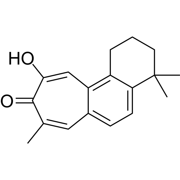
Salviolone
Based on 1 publication(s) in Google Scholar
Salviolone is a natural diterpenoid derivative that can against melanoma cells. Salviolone exhibits a pleiotropic effect against melanoma by hampering cell cycle progression, STAT3 signaling, and malignant phenotype of A375 melanoma cells.
Nur für Forschungszwecke. Wir verkaufen nicht an Patienten.
- Reinheit : 99.32%
- CAS. Nr.: 119400-86-1
- Formel: C18H20O2
- Molecular Weight:268.35
-
Speicherung:
-20°C, protect from light
* In solvent : -80°C, 6 months; -20°C, 1 month (protect from light)

To place orders, for customer services and technical support, please contact: MedChemExpress USA
Tel: 609-228-6898 E-mail: [email protected] [email protected]


-
 Biologische Aktivität
-
Chemical Information
- Protokoll
- Reinheit & Dokumentation
- Verweise
-
 Help & FAQs


Publications Citing Use of MedChemExpress (MCE) Salviolone
More

Biologische Aktivität
Beschreibung
In Vitro
Salviolone (5-60 μM; 72 hours) reduces cell viability in the A375 and MeWo melanoma cell lines with EC50 values of 17 µM and 22 µM, respectively. Salviolone does not affect the growth of normal melanocytes[1].
Salviolone (20 μM; 48-72 hours) strongly reduces pRb, pCdk2, and cyclin A2, Tyr705-STAT3 phosphorylation expression levels in A375 cells. Salviolone also strongly increases the P21 and P53 protein expression level. Salviolone induces sustained activation of the phosphorylation of ERK1/2 and Akt[1].
Salviolone (10-20 μM) inhibits MMP2 gelatinase activity in the A375 melanoma cell line[1].
MedChemExpress (MCE) has not independently confirmed the accuracy of these methods. They are for reference only.
-
Cell Line:A375, MeWo melanoma cells, and NHEM cells
-
Concentration:5 μM, 10 μM, 20 μM, 30 μM, 40 μM, 50 μM, 60 μM
-
Incubation Time:72 hours
-
Result:Impaired the viability of melanoma cells without affecting the growth of normal melanocytes.
-
Cell Line:A375 cells
-
Concentration:20 μM
-
Incubation Time:48 hours, 72 hours
-
Result:Reduced the expression of the active forms of Cdk2 (pCdk2) and cyclin A2, and the phosphorylation of Rb.


Chemical Information
-
CAS. Nr. 119400-86-1
-
Appearance Solid
-
Molecular Weight 268.35
-
Formel C18H20O2
-
Color Light yellow to yellow
-
SMILES
O=C1C(C)=CC2=CC=C3C(C)(C)CCCC3=C2C=C1O
-
Structure Classification
-
Initial Source
-
Versand
Room temperature in continental US; may vary elsewhere.
-
Speicherung
-20°C, protect from light
* In solvent : -80°C, 6 months; -20°C, 1 month (protect from light)


Publications (1)
-
Journal Impact Factor
-
Most Recent
-
Adv Sci (Weinh)
Multi-Targeting Carnosic Acid Kills Drug-Resistant Helicobacter pylori With Narrow-Spectrum Activity. [Abstract]2026 Jun 11:e76080. PMID: 42272345


Reinheit & Dokumentation
-
Data Sheet (272 KB)
-
SDS (252 KB)
- English - EN (252 KB)
- Français - FR (252 KB)
- Deutsch - DE (252 KB)
- Norwegian - NO (252 KB)
- Español - ES (252 KB)
- Swedish - SV (252 KB)
- Italian - IT (252 KB)
- Korean - KR (252 KB)
- Portuguese - PT (252 KB)
-
Handling Instructions (2659 KB)
Verweise
[1]. Valentina Zanrè, et al. Salviolone from Salvia miltiorrhiza Roots Impairs Cell Cycle Progression, Colony Formation, and Metalloproteinase-2 Activity in A375 Melanoma Cells: Involvement of P21(Cip1/Waf1) Expression and STAT3 Phosphorylation. Int J Mol Sci. 2022 Jan 20;23(3):1121. [Content Brief]

Calculators
Konzentration (Stammlösung) × Volumen (Stammlösung) = Konzentration (Ziellösung) × Volumen (Ziellösung)


Salviolone Related Classifications
HY-122506 Related Classifications
 Powered by Bioz