



Briobacept

Based on 1 Customer Validation
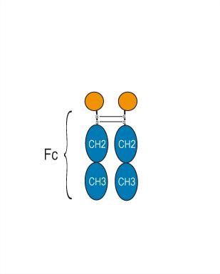
Briobacept (BR 3FC) is a recombinant glucoprotein, consists of 2 molecules from the BLyS receptor (BR3)and a Fc domain of human IgG1. Briobacept selectively targets to BLyS (BAFF), induces B cells apoptosis. Briobacept can be used in studies of rheumatoid arthritis (RA).
Nos produits utilisent uniquement pour la recherche. Nous ne vendons pas aux patients.
- Pureté : 97.66%
- CAS No.: 869881-54-9
- Masse moléculaire:65.75 kDa
-
Stockage:
Please store the product under the recommended conditions in the Certificate of Analysis.

To place orders, for customer services and technical support, please contact: MedChemExpress USA
Tel: 609-228-6898 E-mail: [email protected] [email protected]


-
 Activité biologique
-
Chemical Information
- Protocole
- Pureté et documentation
- Références
-
 Help & FAQs




Activité biologique
Description
IC50 & Target
BLyS (BAFF)[1]
In Vitro
In Vivo
Briobacept (100 μg/mouse; i.p.; 3 times per week for 5 weeks) inhibits p52 generation and development of spontaneous lupus-like disease in NZB/WF1 mice[4].
MedChemExpress (MCE) has not independently confirmed the accuracy of these methods. They are for reference only.
Conjugated
Unconjugated
Reconsititution
The product can be reconstituted/diluted with sterile PBS or saline.
Format
-
[TNFRSF13C (tumor necrosis factor receptor (TNFR)superfamily member 13C, BAFFR, BR3, B cell activating factor receptor, CD268)]2-IGHG1 Fc (Fragment constant)
Application
ELISA, FACS, Functional assay


Chemical Information
-
CAS No. 869881-54-9
-
Appearance Liquid
-
Masse moléculaire 65.75 kDa
-
Color Colorless to light yellow
-
SMILES
[Briobacept]
-
Synonyms
BR 3FC
-
Livraison
Shipping with dry ice.
-
Formulation
Please refer to the lot-specific COA for specific buffer information.
-
Stockage
Please store the product under the recommended conditions in the Certificate of Analysis.


Pureté et documentation
-
Fiche technique (258 KB)
-
SDS (251 KB)
- English - EN (251 KB)
- Français - FR (251 KB)
- Deutsch - DE (251 KB)
- Norwegian - NO (251 KB)
- Español - ES (251 KB)
- Swedish - SV (251 KB)
- Italian - IT (251 KB)
- Korean - KR (251 KB)
- Portuguese - PT (251 KB)
-
Inhibitory Antibodies User Guide (603 KB)
Références
[1]. Sifuentes Giraldo WA, et al. New targets in systemic lupus (part 2/2). Reumatol Clin. 2012 Sep-Oct;8(5):263-9. [Content Brief]
[3]. La Cava A. Targeting B cells with biologics in systemic lupus erythematosus. Expert Opin Biol Ther. 2010 Nov;10(11):1555-61. [Content Brief]
[4]. Kayagaki N, et al. BAFF/BLyS receptor 3 binds the B cell survival factor BAFF ligand through a discrete surface loop and promotes processing of NF-kappaB2. Immunity. 2002 Oct;17(4):515-24. [Content Brief]

Calculators
Concentration (start) × Volume (start) = Concentration (final) × Volume (final)


Briobacept Related Classifications
HY-P99487 Related Classifications
 Powered by Bioz