



4A7C-301
Based on 1 publication(s) in Google Scholar
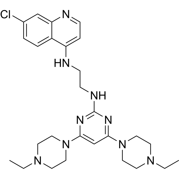
4A7C-301 is a blood-brain barrier-permeable Nurr1 agonist, with IC50 values of 48.22 nM and 107.71 nM for binding to Nurr1-LBD in the [3H]-CQ competition assay and TR-FRET assay, respectively. The EC50 of 4A7C-301 for activating Nurr1 transcriptional activity is 6.53 μM, and its EC50 in N27-A dopaminergic cells is approximately 0.2 μM. 4A7C-301 binds directly to Nurr1-LBD, enhances the transcriptional function of Nurr1, and restores the reduction of Nurr1 protein induced by MPP+ or αSyn. 4A7C-301 alleviates oxidative stress and mitochondrial dysfunction, protects midbrain dopaminergic neurons, inhibits microglial activation, and restores impaired autophagic flux. 4A7C-301 can be used in studies related to neuroprotection and Parkinson's disease.
Nur für Forschungszwecke. Wir verkaufen nicht an Patienten.
- Reinheit : 99.86%
- CAS. Nr.: 3137918-83-0
- Formel: C27H38ClN9
- Molecular Weight:524.10
-
Speicherung:Powder -20°C, 3 years ; In solvent -80°C, 6 months , -20°C, 1 month

To place orders, for customer services and technical support, please contact: MedChemExpress USA
Tel: 609-228-6898 E-mail: [email protected] [email protected]


-
 Biologische Aktivität
-
Chemical Information
-
Lösungsmittel & Löslichkeit
- Protokoll
- Reinheit & Dokumentation
- Verweise
-
 Help & FAQs


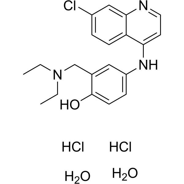
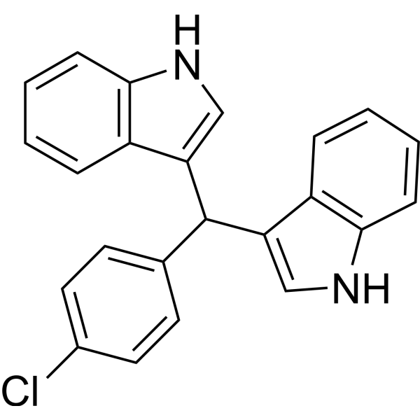
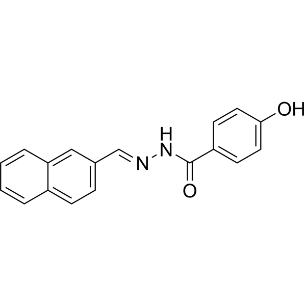
Publications Citing Use of MedChemExpress (MCE) 4A7C-301
MoreAlle Nuclear Hormone Receptor 4A/NR4A Isoform-spezifische Produkte anzeigen
More

Biologische Aktivität
Beschreibung
IC50 & Target
[1]|
Nurr1 6.53 μM (EC50) |
Nurr1-LBD 48.22 nM (IC50, [3H]-CQ competition assay) |
In Vitro
4A7C-301 activates Nurr1 transcriptional activity with an EC50 of 6.53 μM and a maximum induction fold of 18.12-fold, and exhibits an EC50 of approximately 0.2 μM in N27-A dopaminergic cells[1].
4A7C-301 (5-1000 nM; 30 min pre-treatment) exerts dose-dependent protection against MPP+ (0.5 μM)- or LPS (HY-D1056) (15 ng/mL)-induced injury in TH+ dopaminergic neurons in primary rat VM neuron-glia co-cultures, with the maximal neuroprotective effect observed at 500 nM after 7 days of treatment[1].
MedChemExpress (MCE) has not independently confirmed the accuracy of these methods. They are for reference only.
-
Cell Line:N27-A cells with Nurr1 OE or KD
-
Concentration:4A7C-301: 1 μM; MPP+: 1 mM
-
Incubation Time:Overnight
-
Result:Increased cell viability in a Nurr1-dependent manner.
In Vivo
MedChemExpress (MCE) has not independently confirmed the accuracy of these methods. They are for reference only.
-
Animal Model:C57BL/6 J (male, 8-10 weeks old, 25-30 g, subchronic MPTP-induced Parkinson’s disease model)[1]
-
Dosage:5 mg/kg/day
-
Administration:i.p.; daily; 16 days
-
Result:Rescued MPTP-induced motor deficits in rotarod, pole, and cylinder tests to a degree comparable to higher doses of CQ and L-DOPA.
Restored olfactory dysfunction impaired by MPTP lesion.
Detected no abnormal involuntary movements (AIMs, dyskinesia-like behaviors).
Significantly retained tyrosine hydroxylase-positive (TH+) fibers in the striatum, TH+ dopamine neurons, and NeuN+ neurons in the substantia nigra compared to MPTP-only group.
Significantly reduced Iba-1+ activated microglia in both the striatum and substantia nigra.
Restored dopamine levels in both the striatum and substantia nigra.
-
Animal Model:C57BL/6 J (male, 8-10 weeks old, 25-30 g, AAV2-mediated wild-type α-synuclein and mutant α-synuclein overexpression Parkinson’s disease model)[1]
-
Dosage:5 mg/kg/day
-
Administration:i.p.; daily; 5 weeks
-
Result:Improved motor deficits in cylinder, rotarod, and pole tests with greater potency than CQ in both αSynWT- and αSynA53T-induced mice.
Rescued olfactory dysfunction in both αSynWT- and αSynA53T-induced mice.
Significantly prevented the loss of TH+ and NeuN+ neurons in the substantia nigra and TH+ fibers in the striatum in both αSynWT- and αSynA53T-induced mice.
Significantly reduced phosphorylated αSyn at serine 129 (αSynS129) in both αSynWT- and αSynA53T-induced mice.
Restored dopamine levels in both the striatum and substantia nigra in both models.


Chemical Information
-
CAS. Nr. 3137918-83-0
-
Appearance Solid
-
Molecular Weight 524.10
-
Formel C27H38ClN9
-
Color White to light yellow
-
SMILES
ClC1=CC=C2C(NCCNC3=NC(N4CCN(CC)CC4)=CC(N5CCN(CC)CC5)=N3)=CC=NC2=C1
-
Versand
Room temperature in continental US; may vary elsewhere.
-
Speicherung
Powder -20°C 3 years In solvent -80°C 6 months -20°C 1 month


Publications (1)
-
Journal Impact Factor
-
Most Recent
-
J Med Chem
Comparative Profiling and Chemogenomics Application of Chemical Tools for NR4A Nuclear Receptors. [Abstract]2025 Oct 9;68(19):19955-19970. PMID: 40968635


Lösungsmittel & Löslichkeit
In Vitro:
DMSO : 140 mg/mL (267.12 mM; Need ultrasonic; Hygroscopic DMSO has a significant impact on the solubility of product, please use newly opened DMSO)
Please refer to the solubility information to select the appropriate solvent. Once prepared, please aliquot and store the solution to prevent product inactivation from repeated freeze-thaw cycles.
Storage method and period of stock solution: -80°C, 6 months; -20°C, 1 month. When stored at -80°C, please use it within 6 months. When stored at -20°C, please use it within 1 month.
Please refer to the solubility information to select the appropriate solvent. Once prepared, please aliquot and store the solution to prevent product inactivation from repeated freeze-thaw cycles.
Storage method and period of stock solution: -80°C, 6 months; -20°C, 1 month. When stored at -80°C, please use it within 6 months. When stored at -20°C, please use it within 1 month.
Konzentration (Stammlösung) × Volumen (Stammlösung) = Konzentration (Ziellösung) × Volumen (Ziellösung)


Reinheit & Dokumentation
-
Data Sheet (293 KB)
-
SDS (251 KB)
- English - EN (251 KB)
- Français - FR (251 KB)
- Deutsch - DE (251 KB)
- Norwegian - NO (251 KB)
- Español - ES (251 KB)
- Swedish - SV (251 KB)
- Italian - IT (251 KB)
- Korean - KR (251 KB)
- Portuguese - PT (251 KB)
-
Handling Instructions (2659 KB)
Verweise


Complete Stock Solution Preparation Table
Please refer to the solubility information to select the appropriate solvent. Once prepared, please aliquot and store the solution to prevent product inactivation from repeated freeze-thaw cycles.
Storage method and period of stock solution: -80°C, 6 months; -20°C, 1 month. When stored at -80°C, please use it within 6 months. When stored at -20°C, please use it within 1 month.
| Optional Solvent | Concentration Solvent Mass | 1 mg | 5 mg | 10 mg | 25 mg |
|---|---|---|---|---|---|
| DMSO | 1 mM | 1.9080 mL | 9.5402 mL | 19.0803 mL | 47.7008 mL |
| 5 mM | 0.3816 mL | 1.9080 mL | 3.8161 mL | 9.5402 mL | |
| 10 mM | 0.1908 mL | 0.9540 mL | 1.9080 mL | 4.7701 mL | |
| 15 mM | 0.1272 mL | 0.6360 mL | 1.2720 mL | 3.1801 mL | |
| 20 mM | 0.0954 mL | 0.4770 mL | 0.9540 mL | 2.3850 mL | |
| 25 mM | 0.0763 mL | 0.3816 mL | 0.7632 mL | 1.9080 mL | |
| 30 mM | 0.0636 mL | 0.3180 mL | 0.6360 mL | 1.5900 mL | |
| 40 mM | 0.0477 mL | 0.2385 mL | 0.4770 mL | 1.1925 mL | |
| 50 mM | 0.0382 mL | 0.1908 mL | 0.3816 mL | 0.9540 mL | |
| 60 mM | 0.0318 mL | 0.1590 mL | 0.3180 mL | 0.7950 mL | |
| 80 mM | 0.0239 mL | 0.1193 mL | 0.2385 mL | 0.5963 mL | |
| 100 mM | 0.0191 mL | 0.0954 mL | 0.1908 mL | 0.4770 mL |


4A7C-301 Related Classifications
HY-155659 Related Classifications
 Powered by Bioz