



Dynorphin A (1-8)

Based on 1 Customer Validation
Dynorphin A (1-8) is a ligand with preference for the κ-opioid receptor, and its IC50 value against human placental κ-opioid receptor is 330 nM. Dynorphin A (1-8) mediates neuroprotective effects by inhibiting oxidative stress, suppressing apoptosis, and competitively displacing the binding of κ partial agonists. Dynorphin A (1-8) is the dominant opioid peptide in human placental villi and tissue extracts; it undergoes rapid hydrolysis and can enter the central nervous system via intranasal administration. Dynorphin A (1-8) can be used in studies related to ischemic stroke.
Nur für Forschungszwecke. Wir verkaufen nicht an Patienten.
- Reinheit : 99.69%
- CAS. Nr.: 75790-53-3
- Formel: C46H72N14O10
- Molecular Weight:981.15
-
Speicherung:
Stored under nitrogen, away from moisture.
Powder -80°C, 2 years , -20°C, 1 year* In solvent : -80°C, 6 months; -20°C, 1 month (stored under nitrogen, away from moisture)

To place orders, for customer services and technical support, please contact: MedChemExpress USA
Tel: 609-228-6898 E-mail: [email protected] [email protected]


-
 Biologische Aktivität
-
Chemical Information
-
Lösungsmittel & Löslichkeit
- Protokoll
- Reinheit & Dokumentation
- Verweise
-
 Help & FAQs


-
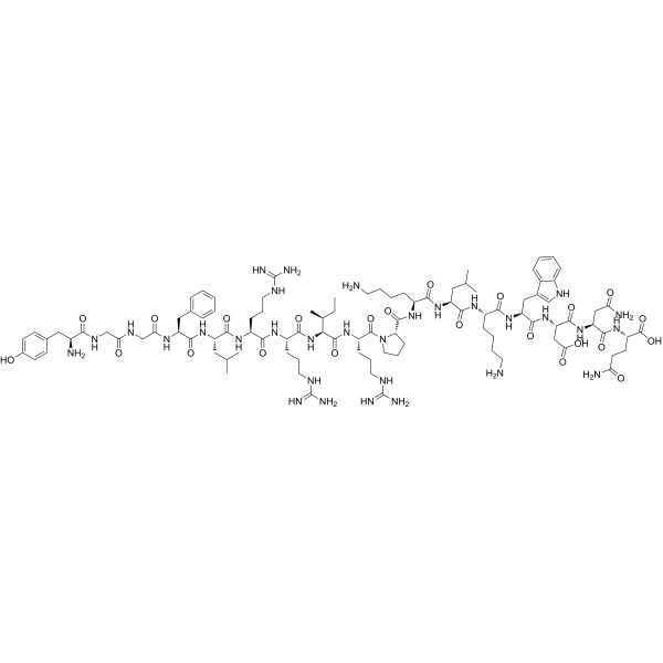
HY-P1333
Dynorphin AKOR Agonist -
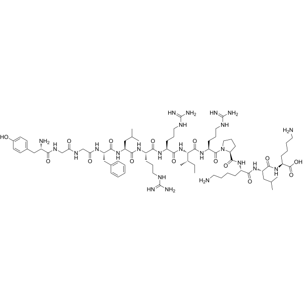
HY-P0088
Porcine dynorphin A(1-13)Opioid Receptor Agonist -
![[Leu5]-Enkephalin](https://file.medchemexpress.com/product_pic/hy-p0288.gif)
HY-P0288
[Leu5]-EnkephalinEndogenous Metabolite -

HY-P1502
β-Endorphin, humanOpioid Receptor Agonist -
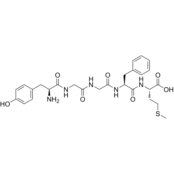
HY-P0073
Tyr-Gly-Gly-Phe-Met-OHOpioid Receptor Activator -

HY-P1594
Dynorphin A (1-10)Opioid Receptor Agonist -

HY-P1337
Dynorphin B (1-13)Kappa Opioid Receptor Agonist -
![[Leu5]-Enkephalin, amide](https://file.medchemexpress.com/product_pic/hy-p1470.gif)
HY-P1470
[Leu5]-Enkephalin, amideOpioid Receptor Agonist -

HY-P0179
β-Casomorphin, bovineOpioid Peptide -
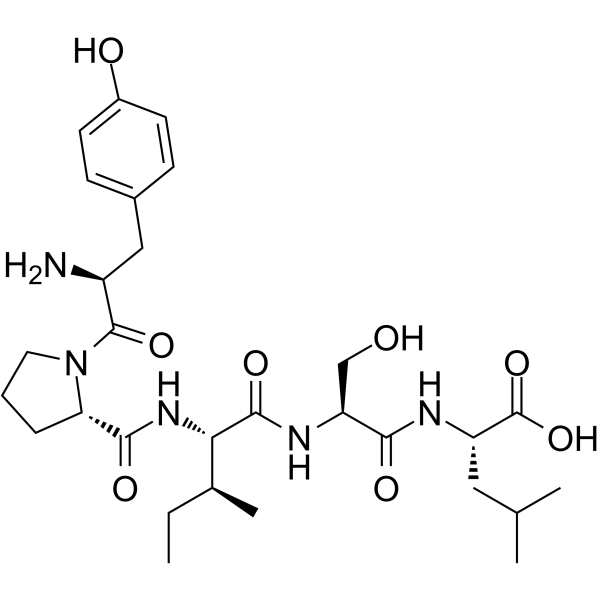
HY-P1596
Gluten Exorphin Cμ/δ Opioid Inhibitor -

HY-P1331
BAM-22POpioid Receptor Agonist -
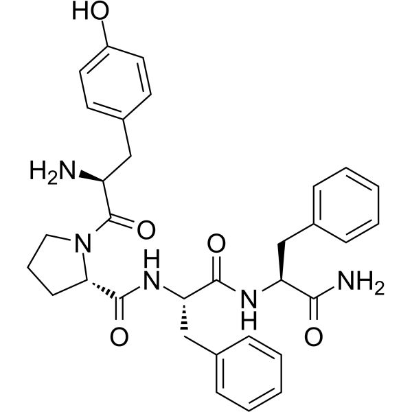
HY-P0186
Endomorphin 2μ-Opioid Receptor Agonist
Alle Opioid Receptor Isoform-spezifische Produkte anzeigen
MoreAlle Endogenous Metabolite Isoform-spezifische Produkte anzeigen
More

Biologische Aktivität
Beschreibung
IC50 & Target
|
κ Opioid Receptor/KOR |
Human Endogenous Metabolite |
In Vitro
Dynorphin A (1-8) inhibits 3H-EKC binding to the purified 63 kDa kappa opioid receptor from human placental villus tissue with an IC50 of 330 nM[2].
Dynorphin A (1-8) inhibits 3H-Bremazocine binding to the purified 63 kDa kappa opioid receptor from human placental villus tissue with an IC50 of 610 nM and a Ki of 303 nM[2].
MedChemExpress (MCE) has not independently confirmed the accuracy of these methods. They are for reference only. Further protocols information, click here.
In Vivo
MedChemExpress (MCE) has not independently confirmed the accuracy of these methods. They are for reference only.
-
Animal Model:Sprague-Dawley (SD) (male, 240-270 g, middle cerebral artery occlusion modeling)[1]
-
Dosage:100 nmol (intranasal, neuroprotective); 100 nmol (intravenous, no effect)
-
Administration:intranasal; single dose at start of surgery; intravenous; single dose at start of surgery
-
Result:Significantly reduced neurological deficit scores.
Significantly reduced cerebral infarction volume.
Significantly reduced brain water content.
Increased neuron number, improved cell arrangement, and reduced neuronal degeneration, necrosis, and pyknosis.
Significantly reduced the number of TUNEL-positive apoptotic neurons.
Significantly increased Bcl-2 expression and decreased Bax and Caspase-3 expression in the ischemic cortex.
Significantly increased serum SOD activity and decreased serum MDA concentration.
Showed no statistically significant effects on neurological deficit scores, cerebral infarction volume, brain water content, TUNEL-positive neuron count, or apoptosis-related protein expression compared to the MCAO model group.


Chemical Information
-
CAS. Nr. 75790-53-3
-
Appearance Solid
-
Molecular Weight 981.15
-
Formel C46H72N14O10
-
Color White to off-white
-
Sequence
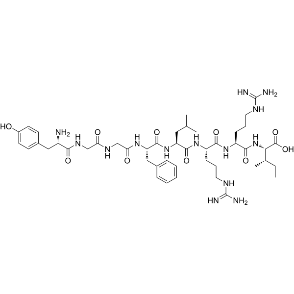
Tyr-Gly-Gly-Phe-Leu-Arg-Arg-Ile
-
Sequence Shortening
YGGFLRRI
-
Structure Classification
-
Initial Source
-
Versand
Room temperature in continental US; may vary elsewhere.
-
Speicherung
Stored under nitrogen, away from moisture
Powder -80°C 2 years -20°C 1 year * In solvent : -80°C, 6 months; -20°C, 1 month (stored under nitrogen, away from moisture)


Lösungsmittel & Löslichkeit
In Vitro:
H2O : 100 mg/mL (101.92 mM; Need ultrasonic)
Please refer to the solubility information to select the appropriate solvent. Once prepared, please aliquot and store the solution to prevent product inactivation from repeated freeze-thaw cycles.
Storage method and period of stock solution: -80°C, 6 months; -20°C, 1 month (stored under nitrogen, away from moisture). When stored at -80°C, please use it within 6 months. When stored at -20°C, please use it within 1 month.
* Note: If you choose water as the stock solution, please dilute it to the working solution, then filter and sterilize it with a 0.22 μm filter before use.
Please refer to the solubility information to select the appropriate solvent. Once prepared, please aliquot and store the solution to prevent product inactivation from repeated freeze-thaw cycles.
Storage method and period of stock solution: -80°C, 6 months; -20°C, 1 month (stored under nitrogen, away from moisture). When stored at -80°C, please use it within 6 months. When stored at -20°C, please use it within 1 month.
* Note: If you choose water as the stock solution, please dilute it to the working solution, then filter and sterilize it with a 0.22 μm filter before use.
Konzentration (Stammlösung) × Volumen (Stammlösung) = Konzentration (Ziellösung) × Volumen (Ziellösung)

Protokoll
-
Apoptosis
Apoptosis, also called programmed cell death, is generally characterized by distinct morphological characteristics.
-
TUNEL staining for apoptotic DNA fragmentation
TUNEL staining detects DNA strand breaks by using terminal deoxynucleotidyl transferase to add labeled nucleotides to exposed 3′-OH DNA termini, generating either microscopic staining in fixed cells or tissue sections, or fluorescence/cytometric signal in cell suspensions. TUNEL positivity reflects DNA fragmentation but should not be interpreted alone as definitive apoptosis, because TUNEL can also label necrotic, autolytic, mechanically damaged, or DNA-repair-associated DNA breaks.
-
Cell Cytotoxicity Assay
Cytotoxicity assays are usually based on the assessment of cell membrane damage, which can also be indirectly detected by measuring cell viability. Detection methods include MTT assay, CKK-8 assay, LDH assay and ATP assay, etc.
-
Annexin V plus membrane-impermeant dye apoptosis staining
Annexin V-based apoptosis assays rely on the detection of phosphatidylserine (PS) externalization from the inner leaflet of the plasma membrane to the outer leaflet, an early biochemical hallmark of apoptosis. Fluorescently labeled Annexin V binds PS in a calcium-dependent manner, enabling identification of early apoptotic cells by flow cytometry or fluorescence microscopy. When combined with a membrane-impermeant DNA-binding dye (e. g. , propidium iodide), this approach allows discrimination between viable (Annexin V−/dye−), early apoptotic (Annexin V+/dye−), and late apoptotic or necrotic (Annexin V+/dye+) cell populations by assessing membrane integrity and PS exposure.
-
ROS/oxidative-stress fluorescent staining
ROS/oxidative-stress fluorescent staining uses cell-permeant fluorogenic probes that become fluorescent after oxidation inside cells or tissues; commonly used examples include DCFH-DA/DCFDA for broad cellular oxidant detection, DHE for superoxide-related signal detection, MitoSOX for mitochondrial superoxide-related signal detection, and CellROX probes for oxidative-stress-associated fluorescence readouts. The assay detects probe oxidation rather than a single ROS species unless the probe and analysis method have been chemically validated for that species. DCFH-DA enters cells, is deacetylated by intracellular esterases to DCFH, and produces fluorescent DCF after oxidation, so the readout is used as an operational measure of total cellular oxidative stress rather than a species-specific ROS measurement. DHE and MitoSOX can report superoxide-related oxidation, but red fluorescence alone can include non-specific ethidium-like oxidation products; HPLC or optimized spectral approaches are
-
Apoptosis Solutions
Apoptosis is a regulated, generally non-lytic cell-death pathway that removes unwanted, damaged, infected, or abnormal cells through coordinated morphological changes, caspase activation, DNA fragmentation, and membrane remodeling. The intrinsic apoptosis pathway is controlled mainly by mitochondrial outer membrane permeabilization, BCL-2 family proteins, cytochrome c release, apoptosome formation, caspase-9 activation, and downstream executioner caspase-3/7 activation. The extrinsic apoptosis pathway is initiated by death receptors such as Fas, TNFR, and TRAIL receptors, which recruit adaptor proteins and activate caspase-8 before engaging executioner caspases or mitochondrial amplification through BID cleavage. Apoptosis is linked to many phenotypes, including cancer cell killing, tissue homeostasis, immune regulation, neurodegeneration, infection response, and treatment-induced cytotoxicity; unresolved questions include how apoptosis interacts with necroptosis, pyroptosis, ferroptos


Reinheit & Dokumentation
-
Data Sheet (274 KB)
-
SDS (254 KB)
- English - EN (254 KB)
- Français - FR (254 KB)
- Deutsch - DE (254 KB)
- Norwegian - NO (254 KB)
- Español - ES (254 KB)
- Swedish - SV (254 KB)
- Italian - IT (254 KB)
- Korean - KR (254 KB)
- Portuguese - PT (254 KB)
-
Handling Instructions (2659 KB)
Verweise
[1]. Chen M, et al. Dynorphin A (1-8) inhibits oxidative stress and apoptosis in MCAO rats, affording neuroprotection through NMDA receptor and κ-opioid receptor channels. Neuropeptides. 2021;89:102182. [Content Brief]
[2]. Ahmed MS, et al. Opioid binding properties of the purified kappa receptor from human placenta. Life Sci. 1989;44(13):861-871. [Content Brief]
[3]. Agbas A, et al. Dynorphin A(1-8) in human placenta: amino acid sequence determined by tandem mass spectrometry. Peptides. 1995;16(4):623-627. [Content Brief]


Complete Stock Solution Preparation Table
Please refer to the solubility information to select the appropriate solvent. Once prepared, please aliquot and store the solution to prevent product inactivation from repeated freeze-thaw cycles.
Storage method and period of stock solution: -80°C, 6 months; -20°C, 1 month (stored under nitrogen, away from moisture). When stored at -80°C, please use it within 6 months. When stored at -20°C, please use it within 1 month.
| Optional Solvent | Concentration Solvent Mass | 1 mg | 5 mg | 10 mg | 25 mg |
|---|---|---|---|---|---|
| H2O | 1 mM | 1.0192 mL | 5.0961 mL | 10.1921 mL | 25.4803 mL |
| 5 mM | 0.2038 mL | 1.0192 mL | 2.0384 mL | 5.0961 mL | |
| 10 mM | 0.1019 mL | 0.5096 mL | 1.0192 mL | 2.5480 mL | |
| 15 mM | 0.0679 mL | 0.3397 mL | 0.6795 mL | 1.6987 mL | |
| 20 mM | 0.0510 mL | 0.2548 mL | 0.5096 mL | 1.2740 mL | |
| 25 mM | 0.0408 mL | 0.2038 mL | 0.4077 mL | 1.0192 mL | |
| 30 mM | 0.0340 mL | 0.1699 mL | 0.3397 mL | 0.8493 mL | |
| 40 mM | 0.0255 mL | 0.1274 mL | 0.2548 mL | 0.6370 mL | |
| 50 mM | 0.0204 mL | 0.1019 mL | 0.2038 mL | 0.5096 mL | |
| 60 mM | 0.0170 mL | 0.0849 mL | 0.1699 mL | 0.4247 mL | |
| 80 mM | 0.0127 mL | 0.0637 mL | 0.1274 mL | 0.3185 mL | |
| 100 mM | 0.0102 mL | 0.0510 mL | 0.1019 mL | 0.2548 mL |
* Note: If you choose water as the stock solution, please dilute it to the working solution, then filter and sterilize it with a 0.22 μm filter before use.


Dynorphin A (1-8) Related Classifications
HY-P2159 Related Classifications
 Powered by Bioz