



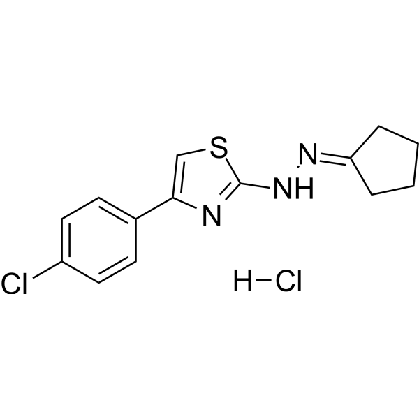
CPTH2 hydrochloride
Based on 3 publication(s) in Google Scholar
CPTH2 hydrochloride is a potent histone acetyltransferase (HAT) inhibitor. CPTH2 hydrochloride selectively inhibits the acetylation of histone H3 by Gcn5. CPTH2 hydrochloride induces apoptosis and decreases the invasiveness of a clear cell renal carcinoma (ccRCC) cell line through the inhibition of acetyltransferase p300 (KAT3B).
For research use only. We do not sell to patients.
- CAS No.: 2108899-91-6
- Formula: C14H15Cl2N3S
- Molecular Weight:328.26
-
Storage:
Please store the product under the recommended conditions in the Certificate of Analysis.

To place orders, for customer services and technical support, please contact: MedChemExpress USA
Tel: 609-228-6898 E-mail: [email protected] [email protected]


-
 Biological Activity
-
Chemical Information
- Protocols
- Purity & Documentation
- References
-
 Help & FAQs


-
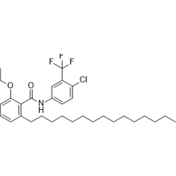
HY-124960
CTPBHistone Acetyltransferase Activator -
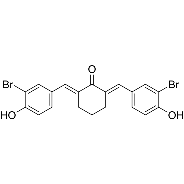
HY-100734
Histone Acetyltransferase Inhibitor IIp300 Histone Acetyltransferase Inhibitor -
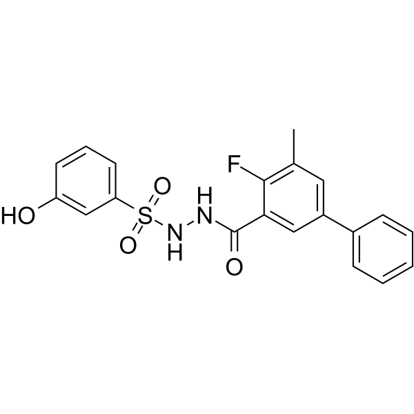
HY-134901
WM-3835HBO1 Inhibitor -
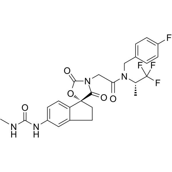
HY-107455
A-485p300/CBP Inhibitor -
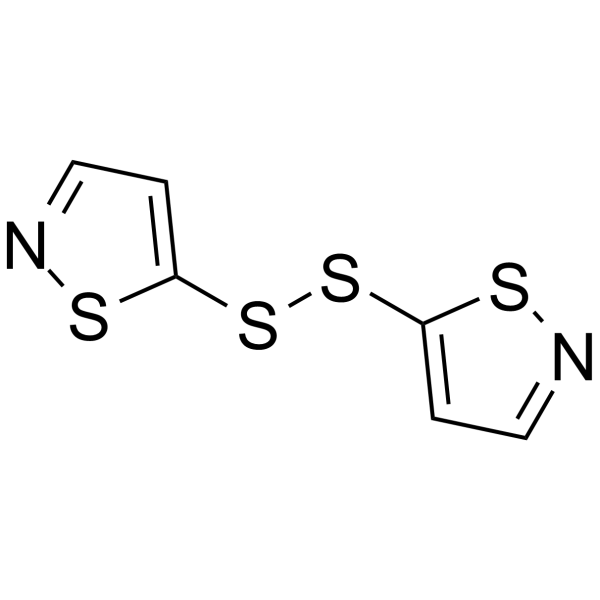
HY-110127
NU9056Tip60 Inhibitor -
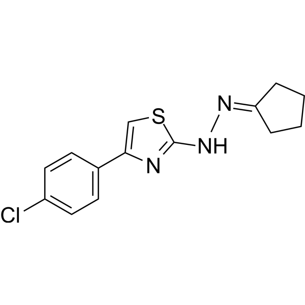
HY-W013274
CPTH2Histone Acetyltransferase Inhibitor -
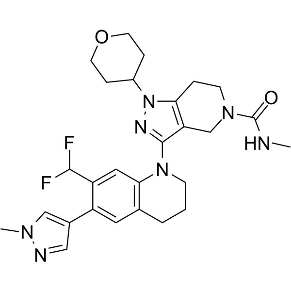
HY-108696
GNE-781CBP Inhibitor -
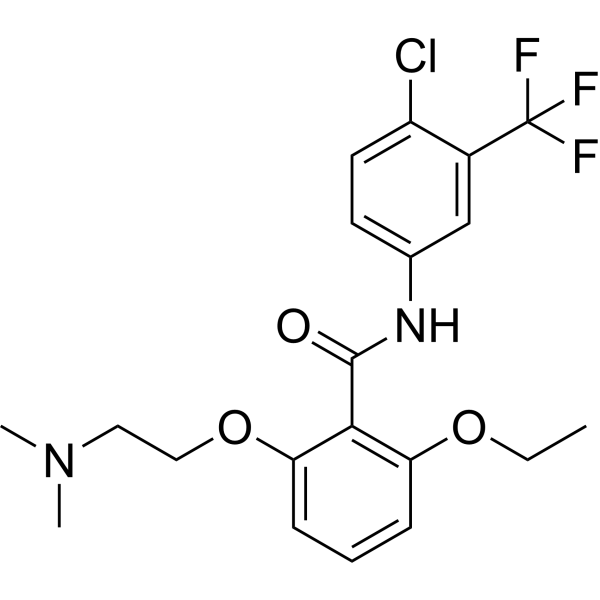
HY-16531
YF-2Histone Acetyltransferase -
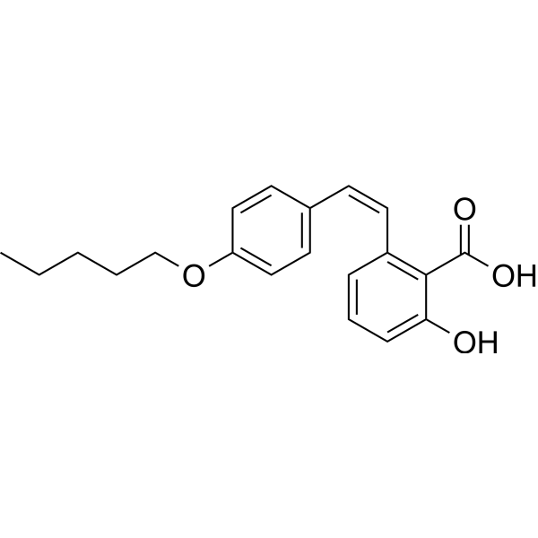
HY-117617
CAY10669PCAF Inhibitor -
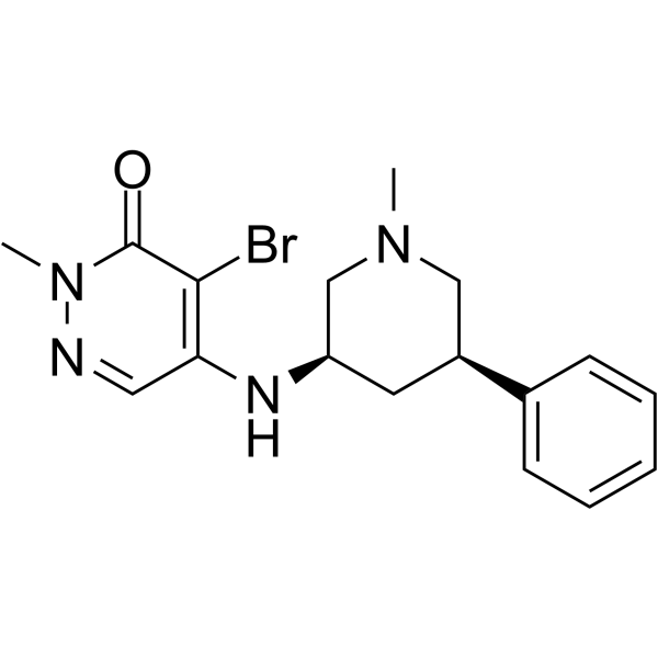
HY-101027
GSK 4027PCAF Probe -

HY-107455R
A-485 (Standard)Reference Standard -
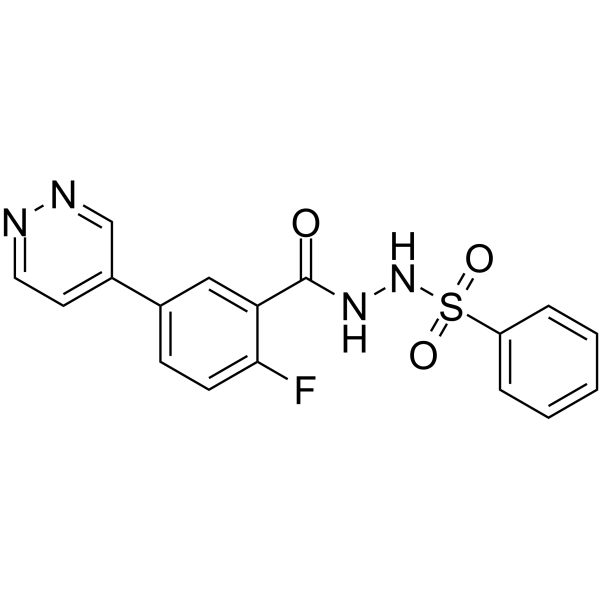
HY-102059
MOZ-IN-2MOZ Inhibitor
Publications Citing Use of MedChemExpress (MCE) CPTH2 hydrochloride
More

Biological Activity
Description
|
GCN5 |
In Vitro
CPTH2 (100 μM; 12, 24, 48 hours) hydrochloride causes a decrease in cell proliferation after as early as 12 h with a further significant reduction after 48 h stimulation[1].
CPTH2 (100 μM; 12 or 48 hours) hydrochloride causes a comparable drop of the activity in both cell lines[1].
CPTH2 (100 μM; 48 hours) hydrochloride produces a drastic increase in apoptotic/dead cell population after 48 h[1].
CPTH2 (100 μM; 12, 24, 48 hours) hydrochloride shows a reduced acetylation of both global AcH3 histone and H3AcK18[1].
CPTH2 (100 μM; 24, 48 hours) hydrochloride is capable to counteract invasion and migration of ccRCC-786-O cells in culture[1].
CPTH2 (0.2, 0.5, 1 mM) hydrochloride inhibits the growth of a GCN5 deleted strain and a single catalytic mutant E173H[2].
CPTH2 (0.6, 0.8 mM; for 24 hours) hydrochloride inhibits histone H3 acetylation in yeast cell cultures[2].
CPTH2 hydrochloride inhibits the Gcn5p dependent functional network[2].
MedChemExpress (MCE) has not independently confirmed the accuracy of these methods. They are for reference only.
-
Cell Line:Papillary thyroid (K1) and clear cell Renal Cell Carcinoma (ccRCC-786-O) cell lines
-
Concentration:100 μM
-
Incubation Time:12, 24, 48 hours
-
Result:Caused a decrease in cell proliferation after as early as 12 h with a further significant reduction after 48 h stimulation.
-
Cell Line:K1 and ccRCC-786-O cell lines
-
Concentration:100 μM
-
Incubation Time:24 hours (K1 cell) and 48 hours (ccRCC-786-O cell)
-
Result:Caused a comparable drop of the activity in both cell lines.
-
Cell Line:ccRCC-786-O cells
-
Concentration:100 μM
-
Incubation Time:48 hours
-
Result:Produced a drastic increase in apoptotic/dead cell population after 48 h.
-
Cell Line:ccRCC-786-O cells
-
Concentration:100 μM
-
Incubation Time:12, 24, 48 hours
-
Result:Showed a reduced acetylation of both global AcH3 histone and H3AcK18.


Chemical Information
-
CAS No. 2108899-91-6
-
Molecular Weight 328.26
-
Formula C14H15Cl2N3S
-
SMILES
ClC1=CC=C(C2=CSC(N/N=C3CCCC/3)=N2)C=C1.[H]Cl
-
Shipping
Room temperature in continental US; may vary elsewhere.
-
Storage
Please store the product under the recommended conditions in the Certificate of Analysis.


Publications (3)
-
Journal Impact Factor
-
Most Recent
-
Immunity
Palmitic acid reprograms neutrophils to compromise vascular integrity and promote breast cancer lung metastasis. [Abstract]2026 Jul 14;59(7):1964-1981.e9. PMID: 42034064 -
Autophagy
MAP1S limits autoimmune uveitis by suppressing Th17 differentiation through dual Control of the EGR2-LCN2 axis and autophagic flux. [Abstract]2026 Jul 23:1-20. PMID: 42454709 -


Purity & Documentation
References
[1]. Cocco E, et al. KAT3B-p300 and H3AcK18/H3AcK14 levels are prognostic markers for kidney ccRCC tumoraggressiveness and target of KAT inhibitor CPTH2. Clin Epigenetics. 2018 Apr 4;10:44. [Content Brief]
[2]. Chimenti F, et al. A novel histone acetyltransferase inhibitor modulating Gcn5 network: cyclopentylidene-[4-(4'-chlorophenyl)thiazol-2-yl)hydrazone. J Med Chem. 2009 Jan 22;52(2):530-6. [Content Brief]

Calculators
Concentration (start) × Volume (start) = Concentration (final) × Volume (final)


CPTH2 hydrochloride Related Classifications
HY-W013274A Related Classifications
 Powered by Bioz