



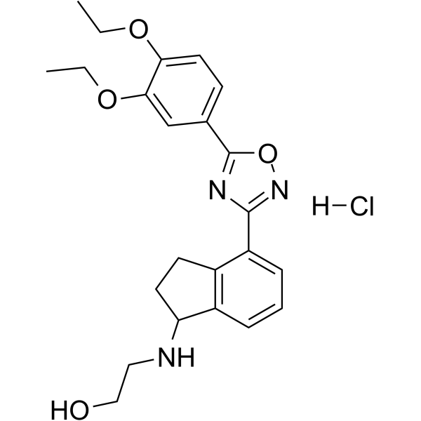
CYM5442 hydrochloride
Based on 5 publication(s) in Google Scholar
CYM5442 hydrochloride is a potent, highly-selective and orally active sphingosine 1-phosphate (S1P1) receptor agonist with an EC50 of 1.35 nM. CYM5442 hydrochloride is inactive against S1P2, S1P3, S1P4, and S1P5. CYM5442 hydrochloride activates S1P1-dependent p42/p44-MAPK phosphorylation. CYM5442 exerts retinal neuroprotection. CYM5442 hydrochloride can easily penetrate the central nervous system (CNS).
Nos produits utilisent uniquement pour la recherche. Nous ne vendons pas aux patients.
- Pureté : 98.58%
- CAS No.: 1783987-80-3
- Formule: C23H28ClN3O4
- Masse moléculaire:445.94
-
Stockage:Powder -20°C, 3 years , 4°C, 2 years ; In solvent -80°C, 6 months , -20°C, 1 month

To place orders, for customer services and technical support, please contact: MedChemExpress USA
Tel: 609-228-6898 E-mail: [email protected] [email protected]


-
 Activité biologique
-
Chemical Information
- Protocole
- Pureté et documentation
- Références
-
 Help & FAQs


-
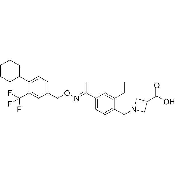
HY-12355
SiponimodS1P1/S1P5 Agonist -
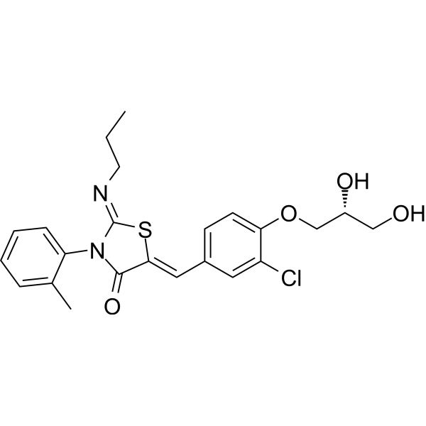
HY-10569
PonesimodS1P1 Agonist -
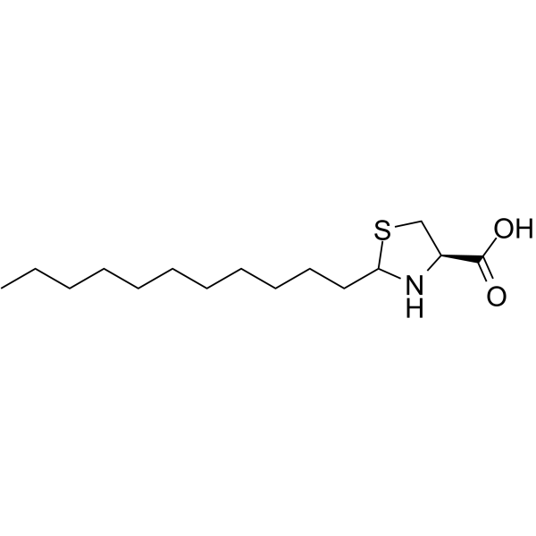
HY-119401
CAY10444S1P3 Antagonist -
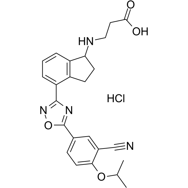
HY-101939A
RP-001 hydrochlorideS1P1 Agonist -
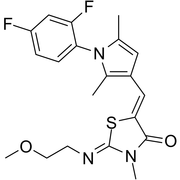
HY-108495
CYM50308S1P4 Receptor Agonist -
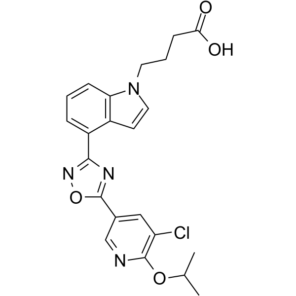
HY-19511
GSK2018682LPL Receptor Agonist -
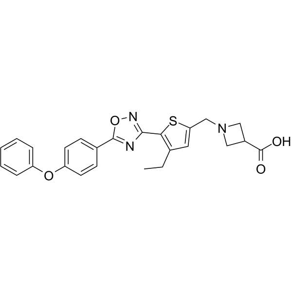
HY-108493
CS-2100S1P1 Agonist -
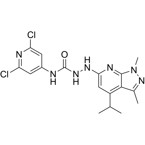
HY-100675
JTE-013S1P2 Antagonist -
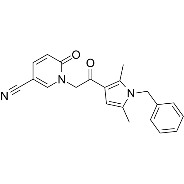
HY-100953
CYM-5520S1PR2 Agonist -
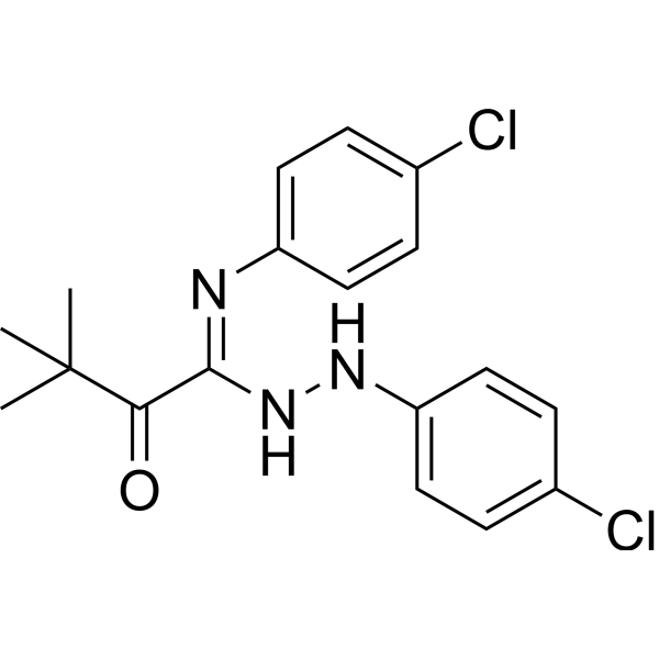
HY-19736
TY-52156LPL Receptor Antagonist -
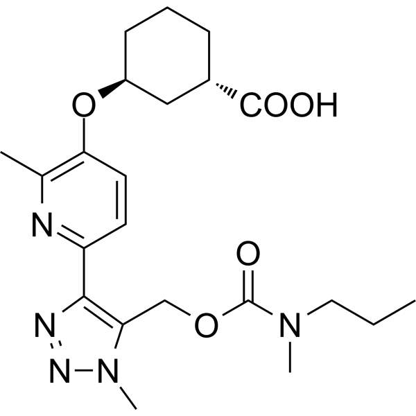
HY-139853
BMS-986278LPA1 Antagonist -

HY-12355R
Siponimod (Standard)Standard of Siponimod
Publications Citing Use of MedChemExpress (MCE) CYM5442 hydrochloride
More

Activité biologique
Description
IC50 & Target
EC50: 1.35 nM (Sphingosine 1-phosphate (S1P1) receptor)[1]
In Vitro
CYM5442 (0.5 µM; 0-60 minutes; HEK293 cells) treatment stimulates S1P1 phosphorylation in a time-dependent manner in P32-orthophosphate labeled cells[1].
CYM5442 activates S1P1-dependent p42/p44-MAPK phosphorylation in CHO-K1 cells transfected with S1P1 with an EC50 of 46 nM. The R120 for alanine (R120A) mutant is still able to maintain p42/p44-MAPK activity when incubated with CYM5442 (EC50 of 67 nM). Activation of p42/p44-MAPK by CYM-5442 in E121A S1P1 cells is concentration dependent, with a mean EC50 value of 134 nM[1].
MedChemExpress (MCE) has not independently confirmed the accuracy of these methods. They are for reference only.
-
Cell Line:HEK293 cells stably expressing S1P1 fused to GFP on the carboxy-terminus
-
Concentration:0.5 µM
-
Incubation Time:0 minutes, 2 minutes, 5 minutes, 10 minutes, 30 minutes, 60 minutes
-
Result:Stimulated S1P1 phosphorylation in a time-dependent manner.
In Vivo
MedChemExpress (MCE) has not independently confirmed the accuracy of these methods. They are for reference only.
-
Animal Model:Adult male albino Wistar rats (8-10 weeks old; 180-230 g) infected ocular endothelin-1 (ET-1)[2]
-
Dosage:1 mg/kg
-
Administration:Intraperitoneal injection; daily; for 5 days
-
Result:Visual evoked potentials (VEP) showed preserved visual function. Showed significantly higher retinal ganglion cells (RGCs) numbers.


Chemical Information
-
CAS No. 1783987-80-3
-
Appearance Solid
-
Masse moléculaire 445.94
-
Formule C23H28ClN3O4
-
Color White to off-white
-
SMILES
CCOC1=CC=C(C2=NC(C3=CC=CC4=C3CCC4NCCO)=NO2)C=C1OCC.[H]Cl
-
Livraison
Room temperature in continental US; may vary elsewhere.
-
Stockage
Powder -20°C 3 years 4°C 2 years In solvent -80°C 6 months -20°C 1 month


Publications (5)
-
Journal Impact Factor
-
Most Recent
-
PLoS Biol
Structural insights into subtype-specific agonist recognition by sphingosine-1-phosphate receptors. [Abstract]2026 Apr 10;24(4):e3003381. PMID: 41961881 -
Int Immunopharmacol
CD83 mediates the inhibitory effect of the S1PR1 agonist CYM5442 on LPS-induced M1 polarization of macrophages through the ERK-STAT-1 signaling pathway. [Abstract]2024 Oct 31;143(Pt 3):113526. PMID: 39486189 -
Bioorg Chem
Identification of FTY720 and COH29 as novel topoisomerase I catalytic inhibitors by experimental and computational studies. [Abstract]2024 Jun:147:107412. PMID: 38696845 -
Hepatobiliary Pancreat Dis Int
Tripartite motif-containing 67 promotes progression of hepatocellular carcinoma by regulating MAPK signaling pathway. [Abstract]2026 Apr 27. PMID: 42106217 -


Pureté et documentation
-
Fiche technique (276 KB)
-
SDS (252 KB)
- English - EN (252 KB)
- Français - FR (252 KB)
- Deutsch - DE (252 KB)
- Norwegian - NO (252 KB)
- Español - ES (252 KB)
- Swedish - SV (252 KB)
- Italian - IT (252 KB)
- Korean - KR (252 KB)
- Portuguese - PT (252 KB)
-
Instruction de manipulation (2659 KB)
Références
[1]. Gonzalez-Cabrera PJ, et al. Full pharmacological efficacy of a novel S1P1 agonist that does not require S1P-like headgroup interactions. Mol Pharmacol. 2008 Nov;74(5):1308-18. [Content Brief]
[2]. Blanco R, et al. The S1P1 receptor-selective agonist CYM-5442 protects retinal ganglion cells in endothelin-1 induced retinal ganglion cell loss. Exp Eye Res. 2017 Nov;164:37-45. [Content Brief]

Calculators
Concentration (start) × Volume (start) = Concentration (final) × Volume (final)


CYM5442 hydrochloride Related Classifications
HY-10968A Related Classifications
 Powered by Bioz