



Chicoric acid
Based on 4 publication(s) in Google Scholar
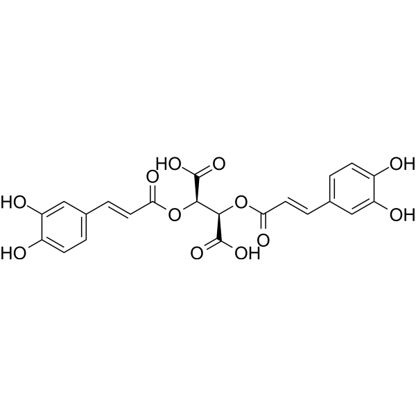
Chicoric acid (Cichoric acid), an orally active dicaffeyltartaric acid, induces reactive oxygen species (ROS) generation. Chicoric acid inhibits cell viability and induces mitochondria-dependent apoptosis in 3T3-L1 preadipocytes through ROS-mediated PI3K/Akt and MAPK signaling pathways. Chicoric acid increases glucose uptake, improves insulin resistance, and attenuates glucosamine-induced inflammation. Chicoric acid has antidiabetic properties and antioxidant, anti-inflammatory effects.
For research use only. We do not sell to patients.
- Purity : 99.86%
- CAS No.: 6537-80-0
- Formula: C22H18O12
- Molecular Weight:474.37
-
Storage:Powder -20°C, 3 years , 4°C, 2 years ; In solvent -80°C, 6 months , -20°C, 1 month

To place orders, for customer services and technical support, please contact: MedChemExpress USA
Tel: 609-228-6898 E-mail: [email protected] [email protected]


-
 Biological Activity
-
Chemical Information
-
Solvent & Solubility
- Protocols
- Purity & Documentation
- References
-
 Help & FAQs


-
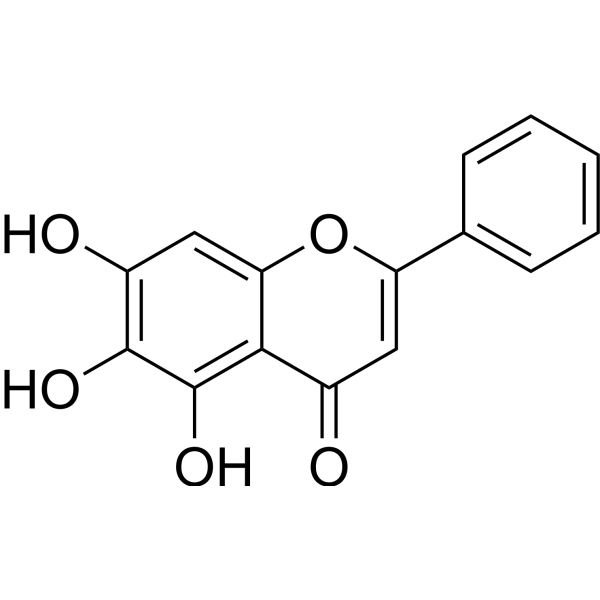
HY-N0196
BaicaleinXanthine Oxidase Inhibitor -
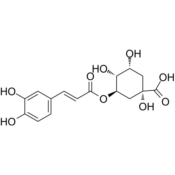
HY-N0055
Chlorogenic acidAntioxidant -
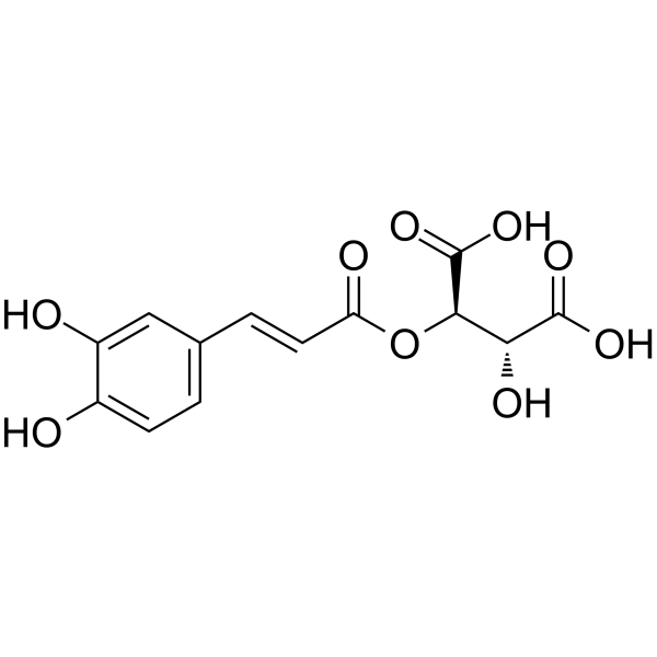
HY-N0321
Caftaric acidAnti-apoptotic Agent -
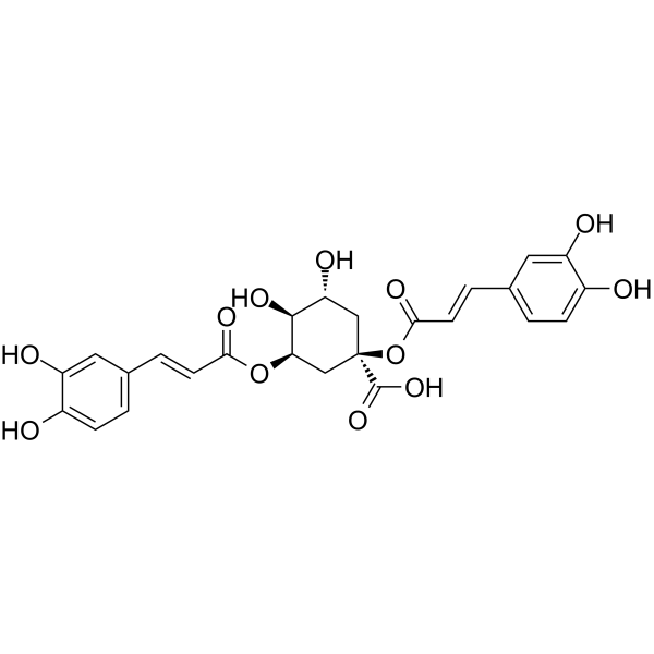
HY-N0359
CynarinAntioxidant -
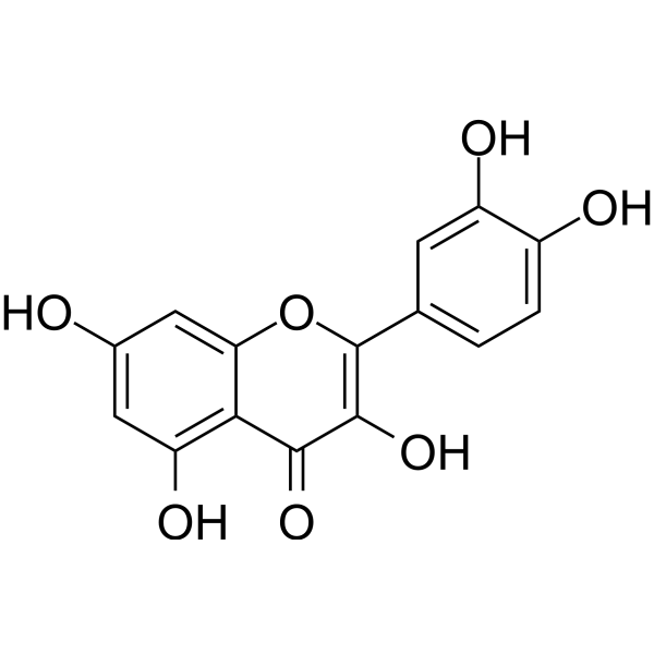
HY-18085
QuercetinFlavonoid Antioxidant -
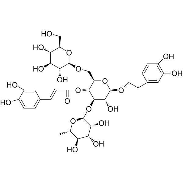
HY-N0020
EchinacosideWnt/β-catenin Signaling Inhibitor -

HY-10219
RapamycinmTOR Inhibitor -
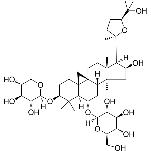
HY-N0431
Astragaloside IVMMP Inhibitor -
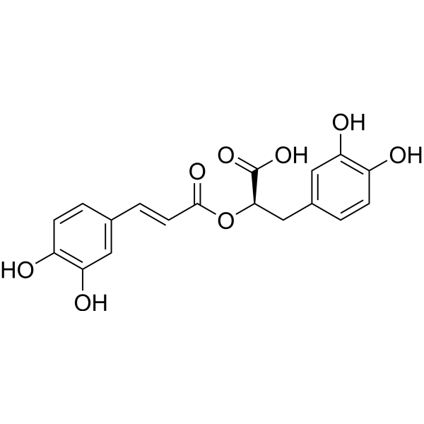
HY-N0529
Rosmarinic acidAnti-Inflammatory Agent -
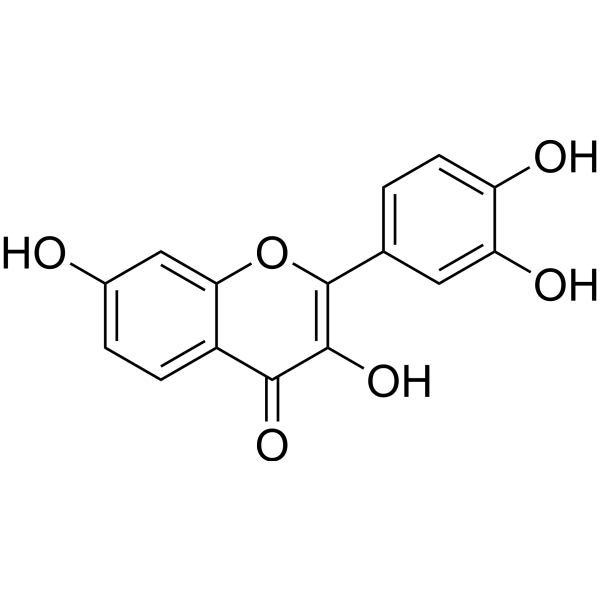
HY-N0182
FisetinTNF Receptor Inhibitor -
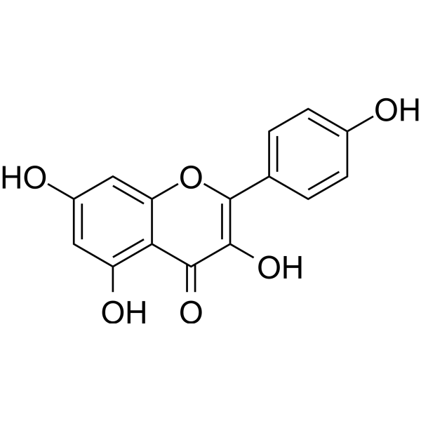
HY-14590
KaempferolEstrogen Receptor α Inhibitor -

HY-N0457R
Chicoric acid (Standard)ROS Inducer
Publications Citing Use of MedChemExpress (MCE) Chicoric acid
More Customer Validation & Images

-
Bio/Physico-chemical Assay

-
Bio/Physico-chemical Assay

-
Bio/Physico-chemical Assay


Biological Activity
Description
Cellular Effect
|
Cell Line

|
Type | Value | Description | References |
|---|---|---|---|---|
| CEM-SS | IC50 |
20.1 μM
Compound: 3 (-)-L-chichoric acid
|
Compound was evaluated for the cytoprotection of CEM-SS cells by XTT cytoprotection assay through the NCI AIDS Screen
Compound was evaluated for the cytoprotection of CEM-SS cells by XTT cytoprotection assay through the NCI AIDS Screen
|
[PMID: 10212126] |
| MT2 | EC50 |
0.81 μM
Compound: L-CA
|
Antiviral activity against HIV LAI infected in human MT2 cells assessed as protection against virus-induced cytopathic effect
Antiviral activity against HIV LAI infected in human MT2 cells assessed as protection against virus-induced cytopathic effect
|
[PMID: 20977258] |
| MT4 | CC50 |
115 μM
Compound: L-CA
|
Cytotoxicity against human MT4 cells by MTT assay
Cytotoxicity against human MT4 cells by MTT assay
|
[PMID: 18243421] |
| MT4 | CC50 |
115 μM
Compound: L-chicoric acid
|
Antiviral activity against HIV1 3B infected in human MT4 cells assessed as inhibition of virus-induced cytopathic effect by MTT assay
Antiviral activity against HIV1 3B infected in human MT4 cells assessed as inhibition of virus-induced cytopathic effect by MTT assay
|
[PMID: 20541944] |
| MT4 | EC50 |
12.7 μM
Compound: L-CA
|
Antiviral activity against HIV1 3B infected in human MT4 cells assessed as inhibition of virus-induced cytopathic effect by MTT assay
Antiviral activity against HIV1 3B infected in human MT4 cells assessed as inhibition of virus-induced cytopathic effect by MTT assay
|
[PMID: 18243421] |
| MT4 | EC50 |
12.7 μM
Compound: L-chicoric acid
|
Cytotoxicity gainst human MT4 cells after 3 days by MTT assay
Cytotoxicity gainst human MT4 cells after 3 days by MTT assay
|
[PMID: 20541944] |
| MT4 | EC50 |
21.2 μM
Compound: 3 (-)-L-chichoric acid
|
Concentration of compound required to protect 50% of cells against retroviral cytopathic effects in HIV-2 (ROD)
Concentration of compound required to protect 50% of cells against retroviral cytopathic effects in HIV-2 (ROD)
|
[PMID: 10212126] |
| MT4 | EC50 |
5.3 μM
Compound: 3 (-)-L-chichoric acid
|
Concentration of compound required to protect 50% of MT-4 cells against retroviral cytopathic effects by HIV-1 (IIIa)
Concentration of compound required to protect 50% of MT-4 cells against retroviral cytopathic effects by HIV-1 (IIIa)
|
[PMID: 10212126] |
| MT4 | IC50 |
45 μM
Compound: 3 (-)-L-chichoric acid
|
Concentration of compound required to reduce MT-4 cell viability by 50%
Concentration of compound required to reduce MT-4 cell viability by 50%
|
[PMID: 10212126] |
In Vitro
Chicoric acid (Cichoric acid; 10-200 μM; for 24, 48, and 72 h) causes a dose- and time-dependent decrease in cell viability[1].
Chicoric acid (100 μM; 48 h) induces apoptosis through caspase-3-dependent pathway[1].
Chicoric acid (100 μM; 48 h) decreases the protein level of p-Akt[1].
Chicoric acid (25, 50, 100 μM; for 24 hours) dramatically improves glucose uptake in a dose-dependent manner, and Chicoric acid further enhances insulin-induced (100 nM; 30 min) glucose uptake by 57.7% in HepG2 cells[2].
Chicoric acid (100 μM; for 24 hours) restores glucosamine-induced impairment of GLUT2 translocation through activating PI3K/Akt pathway in HepG2 cells[2].
Chicoric acid (100 μM) has no effects on HepG2 cell viability[2].
MedChemExpress (MCE) has not independently confirmed the accuracy of these methods. They are for reference only. Further protocols information, click here.
-
Cell Line:3T3-L1 preadipocytes
-
Concentration:10-200 μM
-
Incubation Time:24, 48, and 72 hours
-
Result:Had no effect on the viability of 3T3-L1 preadipocytes with 10-50 μM for 24 h, but significantly decreased cell viability with 100 μM and 200 μM.
-
Cell Line:3T3-L1 preadipocytes
-
Concentration:100 μM
-
Incubation Time:48 hours
-
Result:Demonstrated typical characteristics of apoptosis such as cell shrinkage, chromatin condensation, and the increased permeability of cell membranes after DAPI and AO/EB staining.
-
Cell Line:3T3-L1 preadipocytes
-
Concentration:100 μM
-
Incubation Time:48 hours
-
Result:Decreased the protein level of p-Akt in a dose- and time-dependent manner.
The protein level of total Akt was not affected
In Vivo
MedChemExpress (MCE) has not independently confirmed the accuracy of these methods. They are for reference only.
-
Animal Model:C57BL/6J mice with STZ (50 mg/kg; ip; for consecutive 5 days)[3]
-
Dosage:60 mg/kg
-
Administration:Drinking water; daily; for 4 weeks
-
Result:Inhibited pancreas apoptosis and adjusted islet function in diabetic mice, leading to an increase in insulin generation and secretion.
Regulated mitochondrial biogenesis, glycogen synthesis, and inhibited inflammation via activating antioxidant responses.
Showed a remarkable increase in body weight starting at week 7.


Chemical Information
-
CAS No. 6537-80-0
-
Appearance Solid
-
Molecular Weight 474.37
-
Formula C22H18O12
-
Color White to yellow
-
SMILES
O=C(O)[C@H](OC(/C=C/C1=CC=C(O)C(O)=C1)=O)[C@@H](OC(/C=C/C2=CC=C(O)C(O)=C2)=O)C(O)=O
-
Synonyms
Cichoric acid; Dicaffeoyltartaric acid
-
Structure Classification
-
Initial Source
-
Shipping
Room temperature in continental US; may vary elsewhere.
-
Storage
Powder -20°C 3 years 4°C 2 years In solvent -80°C 6 months -20°C 1 month


Publications (4)
-
Journal Impact Factor
-
Most Recent
-
Phytomedicine
Cichoric acid-loaded hydroxyapatite nanorods remodel the immune microenvironment to enhance bone regeneration. [Abstract]2026 Jan:150:157701. PMID: 41421279 -
Antioxidants (Basel)
Bioactive Extracts and Constituents from Taraxacum mongolicum: Antioxidant, Anti-Inflammatory, Enzyme-Inhibitory, and Molecular Docking Studies. [Abstract]2026 May 29;15(6):688. PMID: 42351994
Chicoric acid purchased from MedChemExpress. Usage Cited in: Antioxidants (Basel). 2026 May 29;15(6):688. [Abstract]
Antioxidant activities of T. mongolicum isolated components.
Chicoric acid purchased from MedChemExpress. Usage Cited in: Antioxidants (Basel). 2026 May 29;15(6):688. [Abstract]
Enzyme suppression activities of Taraxacum mongolicum isolated components.
Chicoric acid purchased from MedChemExpress. Usage Cited in: Antioxidants (Basel). 2026 May 29;15(6):688. [Abstract]
NO inhibitory activities of Taraxacum mongolicum isolated components.
-
Foods
Design of a Plant-Based Smoothie: Exploiting Ingredient Complementarity for a Diversified (Poly)phenolic Profile Quantified by Targeted LC-MS/MS Analysis. [Abstract]2026 Apr 9;15(8):1293. PMID: 42073181 -


Solvent & Solubility
In Vitro:
DMSO : 100 mg/mL (210.81 mM; Need ultrasonic; Hygroscopic DMSO has a significant impact on the solubility of product, please use newly opened DMSO)
H2O : 100 mg/mL (210.81 mM; Need ultrasonic)
Please refer to the solubility information to select the appropriate solvent. Once prepared, please aliquot and store the solution to prevent product inactivation from repeated freeze-thaw cycles.
Storage method and period of stock solution: -80°C, 6 months; -20°C, 1 month. When stored at -80°C, please use it within 6 months. When stored at -20°C, please use it within 1 month.
* Note: If you choose water as the stock solution, please dilute it to the working solution, then filter and sterilize it with a 0.22 μm filter before use.
Please refer to the solubility information to select the appropriate solvent. Once prepared, please aliquot and store the solution to prevent product inactivation from repeated freeze-thaw cycles.
Storage method and period of stock solution: -80°C, 6 months; -20°C, 1 month. When stored at -80°C, please use it within 6 months. When stored at -20°C, please use it within 1 month.
* Note: If you choose water as the stock solution, please dilute it to the working solution, then filter and sterilize it with a 0.22 μm filter before use.
Concentration (start) × Volume (start) = Concentration (final) × Volume (final)

Protocols
-
Mammalian live/dead viability and cytotoxicity staining
Live/dead viability and cytotoxicity staining assays are based on the simultaneous detection of intracellular esterase activity in metabolically active (viable) cells and membrane integrity loss in non-viable cells. In commonly used dual-staining approaches, membrane-permeant fluorogenic substrates are converted by intracellular esterases into fluorescent products in live cells, while impermeant DNA-binding dyes selectively enter cells with compromised plasma membranes and label nucleic acids in dead or dying cells, enabling discrimination between viable and non-viable populations by fluorescence microscopy or flow cytometry.
-
Annexin V plus membrane-impermeant dye apoptosis staining
Annexin V-based apoptosis assays rely on the detection of phosphatidylserine (PS) externalization from the inner leaflet of the plasma membrane to the outer leaflet, an early biochemical hallmark of apoptosis. Fluorescently labeled Annexin V binds PS in a calcium-dependent manner, enabling identification of early apoptotic cells by flow cytometry or fluorescence microscopy. When combined with a membrane-impermeant DNA-binding dye (e. g. , propidium iodide), this approach allows discrimination between viable (Annexin V−/dye−), early apoptotic (Annexin V+/dye−), and late apoptotic or necrotic (Annexin V+/dye+) cell populations by assessing membrane integrity and PS exposure.
-
ROS/oxidative-stress fluorescent staining
ROS/oxidative-stress fluorescent staining uses cell-permeant fluorogenic probes that become fluorescent after oxidation inside cells or tissues; commonly used examples include DCFH-DA/DCFDA for broad cellular oxidant detection, DHE for superoxide-related signal detection, MitoSOX for mitochondrial superoxide-related signal detection, and CellROX probes for oxidative-stress-associated fluorescence readouts. The assay detects probe oxidation rather than a single ROS species unless the probe and analysis method have been chemically validated for that species. DCFH-DA enters cells, is deacetylated by intracellular esterases to DCFH, and produces fluorescent DCF after oxidation, so the readout is used as an operational measure of total cellular oxidative stress rather than a species-specific ROS measurement. DHE and MitoSOX can report superoxide-related oxidation, but red fluorescence alone can include non-specific ethidium-like oxidation products; HPLC or optimized spectral approaches are
-
CCK-8/WST-8 Cell Proliferation Assay
The CCK-8/WST-8 assay is based on the reduction of the water-soluble tetrazolium salt WST-8 to a water-soluble formazan product by cellular dehydrogenases in metabolically active cells, where the generated formazan amount is proportional to the number of living cells and is quantified by measuring absorbance in the visible range, providing a colorimetric readout for cell viability and proliferation assessment. This class of tetrazolium-based assays improves upon earlier MTT-based systems by producing a water-soluble formazan, eliminating the need for organic solubilization steps and enabling direct spectrophotometric measurement in culture medium.
-
Research Protocol for Inflammation-related Diseases
The NLRP3 inflammasome is a cytosolic innate immune signaling platform that integrates priming signals and danger-signal activation to promote caspase-1 activation, maturation of IL-1β and IL-18, and gasdermin D-mediated pyroptotic cell death. The core experimental logic is to determine whether inflammatory disease phenotypes are driven by increased NLRP3 expression, ASC-containing inflammasome assembly, caspase-1 cleavage, GSDMD cleavage, and extracellular release of IL-1β/IL-18 rather than by nonspecific cell injury alone. The pathway is strongly linked to inflammation-related disease phenotypes because monosodium urate crystals activate NALP3/NLRP3 inflammasome signaling in gout-like crystal inflammation, cholesterol crystals activate NLRP3 inflammasomes in atherogenesis models, and DSS-induced intestinal inflammation has been reported to involve NLRP3 inflammasome activity. However, experimental colitis studies also show context-dependent protective effects of NLRP3 inflammasome co
-
Research Protocol for Metabolic Diseases
AMP-activated protein kinase, AMPK, is a conserved cellular energy sensor that responds to reduced cellular energy status and coordinates metabolism by increasing ATP-generating catabolic pathways while suppressing ATP-consuming anabolic processes. In metabolic disease research, the AMPK pathway is experimentally relevant because it regulates hepatic lipid synthesis, fatty acid oxidation, glucose production, skeletal-muscle glucose disposal, mTORC1-linked biosynthesis, autophagy, mitochondrial homeostasis, and whole-body energy balance. The central pathway logic is that energy stress, metformin, exercise-like stimulation, or direct AMPK activators increase AMPKα Thr172 phosphorylation and downstream substrate phosphorylation, including ACC and RAPTOR. Phosphorylation of ACC suppresses lipogenesis and supports fatty acid oxidation, whereas phosphorylation of RAPTOR suppresses mTORC1 signaling and links cellular energy status to growth and protein synthesis control. The pathway is linked
-
Cell Viability Determination by MTT Colorimetric Assay
The following protocol uses the MTT colorimetric assay as a classic literature-established method for assessing cell viability/metabolic activity in cultured mammalian cells. MTT[3-(4,5-dimethylthiazol-2-yl)-2,5-diphenyltetrazolium bromide] is reduced by metabolically active cells to a colored formazan product; the amount of formazan is quantified spectrophotometrically and provides an indirect measure of metabolically active viable cells. Importantly, MTT reduction reflects cellular oxidoreductase/metabolic activity rather than an absolute direct count of living cells, so changes in cellular metabolism can alter the signal independently of cell number.
-
Apoptosis
Apoptosis, also called programmed cell death, is generally characterized by distinct morphological characteristics.
-
TUNEL staining for apoptotic DNA fragmentation
TUNEL staining detects DNA strand breaks by using terminal deoxynucleotidyl transferase to add labeled nucleotides to exposed 3′-OH DNA termini, generating either microscopic staining in fixed cells or tissue sections, or fluorescence/cytometric signal in cell suspensions. TUNEL positivity reflects DNA fragmentation but should not be interpreted alone as definitive apoptosis, because TUNEL can also label necrotic, autolytic, mechanically damaged, or DNA-repair-associated DNA breaks.
-
Cell Cytotoxicity Assay
Cytotoxicity assays are usually based on the assessment of cell membrane damage, which can also be indirectly detected by measuring cell viability. Detection methods include MTT assay, CKK-8 assay, LDH assay and ATP assay, etc.
-
Research Protocol for Endocrine Diseases
Endocrine diseases often arise from disrupted hormone production, hormone signaling, or target-tissue responsiveness; for diabetes-focused endocrine disease models, insulin signaling regulates glucose uptake, hepatic glucose output, lipid metabolism, and β-cell compensation. Type 2 diabetes develops through interacting defects in insulin resistance, β-cell dysfunction, adipose inflammation, hepatic glucose overproduction, altered incretin signaling, and ectopic lipid metabolism. A major unresolved question is whether endocrine dysfunction is driven primarily by target-tissue insulin resistance, intrinsic β-cell failure, immune/inflammatory stress, or combined multi-organ failure that differs by disease stage.
-
Apoptosis Solutions
Apoptosis is a regulated, generally non-lytic cell-death pathway that removes unwanted, damaged, infected, or abnormal cells through coordinated morphological changes, caspase activation, DNA fragmentation, and membrane remodeling. The intrinsic apoptosis pathway is controlled mainly by mitochondrial outer membrane permeabilization, BCL-2 family proteins, cytochrome c release, apoptosome formation, caspase-9 activation, and downstream executioner caspase-3/7 activation. The extrinsic apoptosis pathway is initiated by death receptors such as Fas, TNFR, and TRAIL receptors, which recruit adaptor proteins and activate caspase-8 before engaging executioner caspases or mitochondrial amplification through BID cleavage. Apoptosis is linked to many phenotypes, including cancer cell killing, tissue homeostasis, immune regulation, neurodegeneration, infection response, and treatment-induced cytotoxicity; unresolved questions include how apoptosis interacts with necroptosis, pyroptosis, ferroptos
-
MTT Cell Proliferation Assay
The MTT assay is a colorimetric endpoint assay for estimating viable cell number, cell growth, cytotoxicity, or cell activation in cultured mammalian cells. Living cells reduce the yellow tetrazolium salt MTT into purple/blue formazan, while dead cells do not generate the same signal; the resulting color can be quantified with a multiwell spectrophotometer. MTT reduction is commonly interpreted as a readout of metabolic activity that often correlates with viable cell number, but it should not be treated as a direct cell-counting method unless the assay is optimized for the cell type and experimental condition. Studies show that MTT reduction can involve mitochondrial and non-mitochondrial reducing systems, and formazan may accumulate in intracellular lipid droplets rather than simply marking mitochondria.


Purity & Documentation
-
Data Sheet (277 KB)
-
SDS (396 KB)
- English - EN (396 KB)
- Français - FR (396 KB)
- Deutsch - DE (396 KB)
- Norwegian - NO (396 KB)
- Español - ES (396 KB)
- Swedish - SV (396 KB)
- Italian - IT (396 KB)
- Korean - KR (396 KB)
- Portuguese - PT (396 KB)
-
Handling Instructions (2659 KB)
References
[1]. Haifang Xiao, et al. Chicoric acid induces apoptosis in 3T3-L1 preadipocytes through ROS-mediated PI3K/Akt and MAPK signaling pathways. J Agric Food Chem. 2013 Feb 20;61(7):1509-20. [Content Brief]
[2]. Di Zhu, et al. Cichoric Acid Reverses Insulin Resistance and Suppresses Inflammatory Responses in the Glucosamine-Induced HepG2 Cells. J Agric Food Chem. 2015 Dec 30;63(51):10903-13. [Content Brief]
[3]. Di Zhu, et al. Cichoric acid improved hyperglycaemia and restored muscle injury via activating antioxidant response in MLD-STZ-induced diabetic mice. Food Chem Toxicol. 2017 Sep;107(Pt A):138-149. [Content Brief]


Complete Stock Solution Preparation Table
Please refer to the solubility information to select the appropriate solvent. Once prepared, please aliquot and store the solution to prevent product inactivation from repeated freeze-thaw cycles.
Storage method and period of stock solution: -80°C, 6 months; -20°C, 1 month. When stored at -80°C, please use it within 6 months. When stored at -20°C, please use it within 1 month.
| Optional Solvent | Concentration Solvent Mass | 1 mg | 5 mg | 10 mg | 25 mg |
|---|---|---|---|---|---|
| DMSO / H2O | 1 mM | 2.1081 mL | 10.5403 mL | 21.0806 mL | 52.7015 mL |
| 5 mM | 0.4216 mL | 2.1081 mL | 4.2161 mL | 10.5403 mL | |
| 10 mM | 0.2108 mL | 1.0540 mL | 2.1081 mL | 5.2701 mL | |
| 15 mM | 0.1405 mL | 0.7027 mL | 1.4054 mL | 3.5134 mL | |
| 20 mM | 0.1054 mL | 0.5270 mL | 1.0540 mL | 2.6351 mL | |
| 25 mM | 0.0843 mL | 0.4216 mL | 0.8432 mL | 2.1081 mL | |
| 30 mM | 0.0703 mL | 0.3513 mL | 0.7027 mL | 1.7567 mL | |
| 40 mM | 0.0527 mL | 0.2635 mL | 0.5270 mL | 1.3175 mL | |
| 50 mM | 0.0422 mL | 0.2108 mL | 0.4216 mL | 1.0540 mL | |
| 60 mM | 0.0351 mL | 0.1757 mL | 0.3513 mL | 0.8784 mL | |
| 80 mM | 0.0264 mL | 0.1318 mL | 0.2635 mL | 0.6588 mL | |
| 100 mM | 0.0211 mL | 0.1054 mL | 0.2108 mL | 0.5270 mL |
* Note: If you choose water as the stock solution, please dilute it to the working solution, then filter and sterilize it with a 0.22 μm filter before use.


Chicoric acid Related Classifications
HY-N0457 Related Classifications
 Powered by Bioz