



EB-3D
Based on 2 publication(s) in Google Scholar
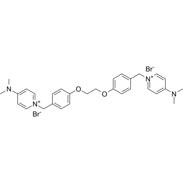
EB-3D is a potent and selective choline kinase α (ChoKα) inhibitor, with an IC50 of 1 μM for ChoKα1. EB-3D exerts effects on ChoKα expression, AMPK activation, apoptosis, endoplasmic reticulum stress and lipid metabolism. EB-3D exhibits a potent antiproliferative activity in a panel of T-leukemia cell lines. Anti-cancer activity.
For research use only. We do not sell to patients.
- Purity : 99.64%
- CAS No.: 1839150-63-8
- Formula: C30H36Br2N4O2
- Molecular Weight:644.44
-
Storage:
4°C, sealed storage, away from moisture
* In solvent : -80°C, 6 months; -20°C, 1 month (sealed storage, away from moisture)

To place orders, for customer services and technical support, please contact: MedChemExpress USA
Tel: 609-228-6898 E-mail: [email protected] [email protected]


-
 Biological Activity
-
Chemical Information
-
Solvent & Solubility
- Protocols
- Purity & Documentation
- References
-
 Help & FAQs


-
HY-15142
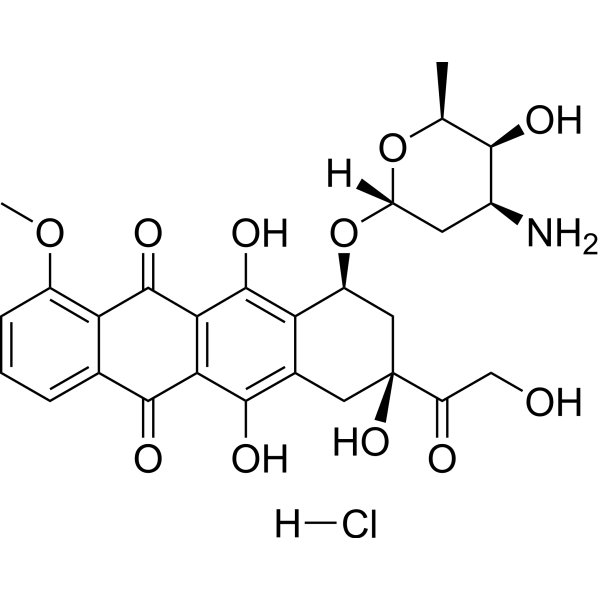
Doxorubicin hydrochlorideTopoisomerase Inhibitor -
HY-B0627
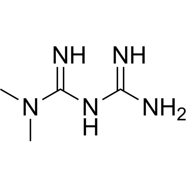
MetforminAntihyperglycemic Agent -
HY-111363
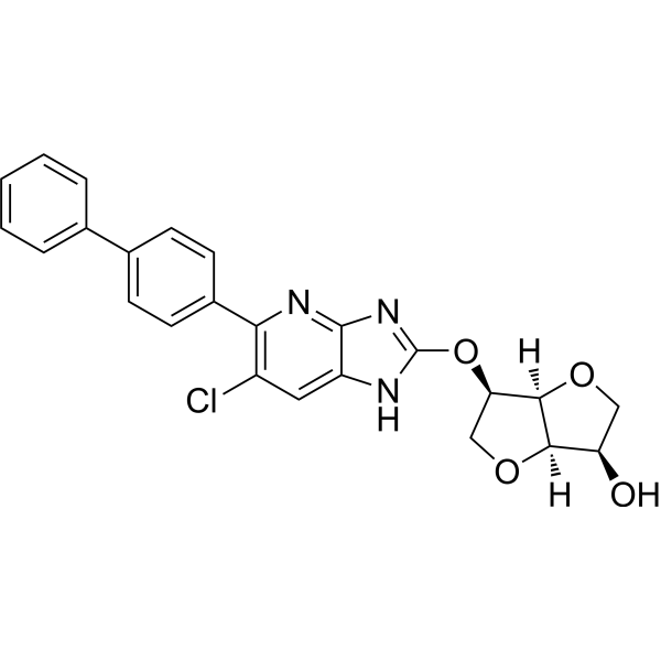
MK8722AMPK Activator -
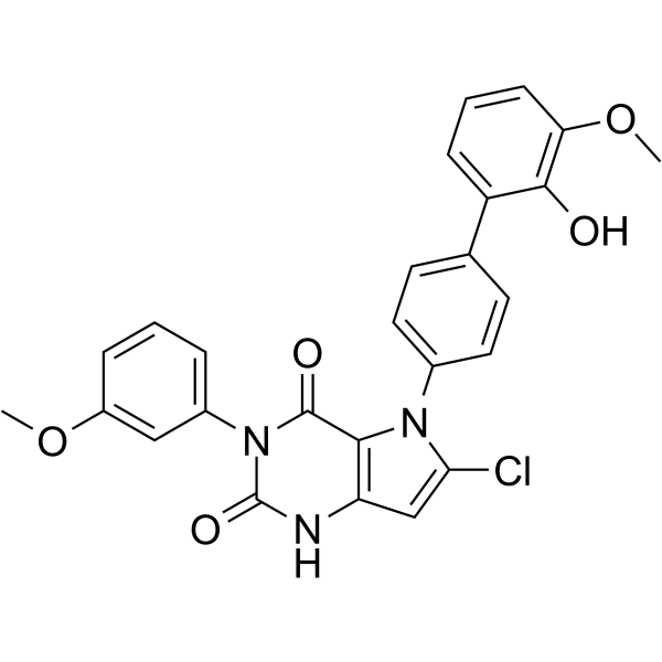
HY-100548
GSK621AMPK Activator -
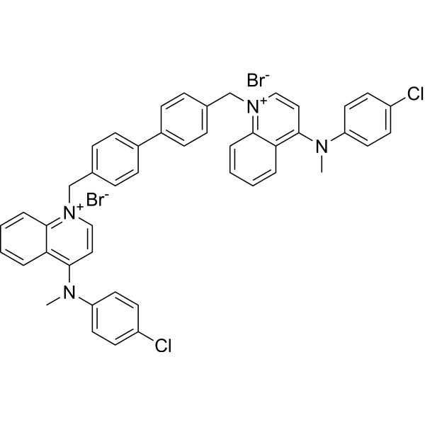
HY-101144
RSM-932AChoKα Inhibitor -
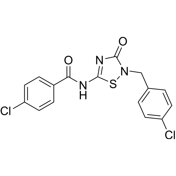
HY-112233
O-304AMPK Activator -
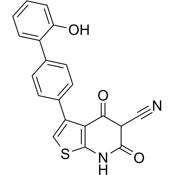
HY-50662
A-769662AMPK Activator -
HY-12357
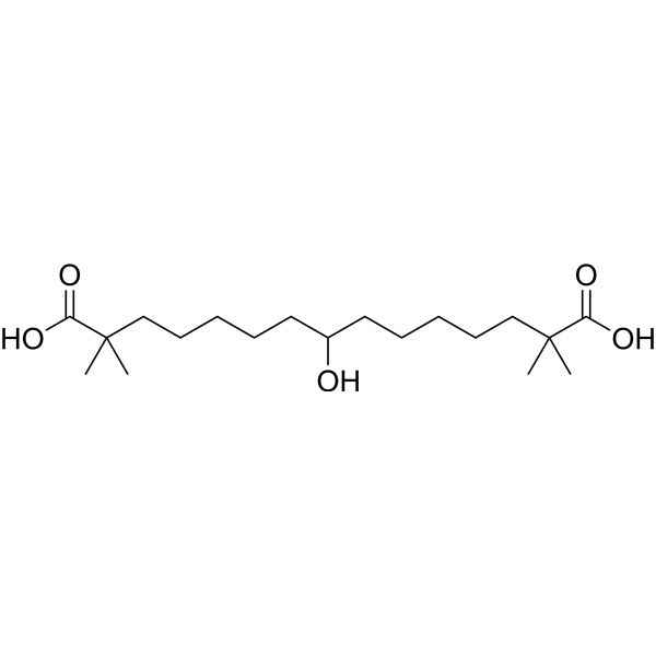
Bempedoic acidACL Inhibitor/AMPK Activator -
HY-12377
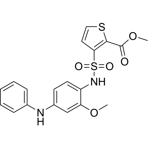
GSK0660PPAR Antagonist -
HY-N6631
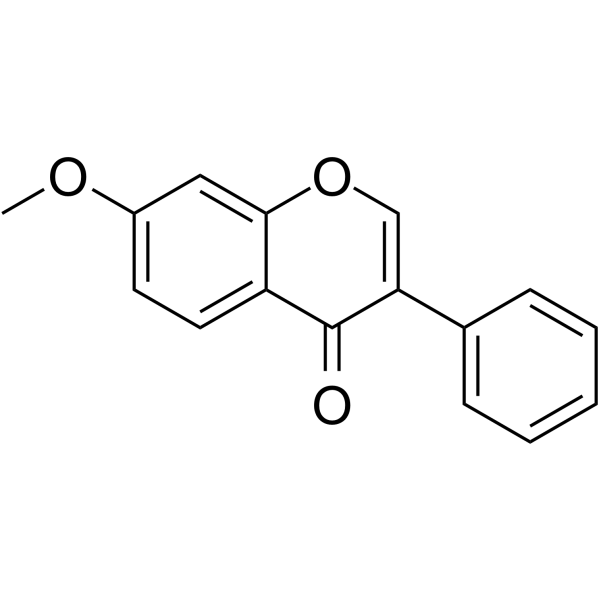
7-MethoxyisoflavoneAMPK Activator -
HY-N1419
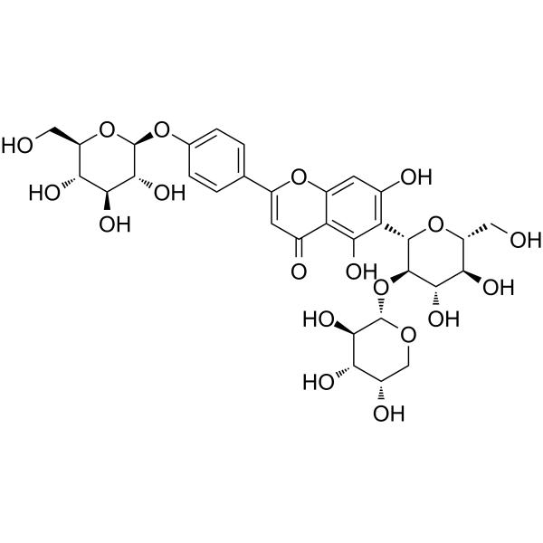
VaccarinAKT/ERK/AMPK Agonists -
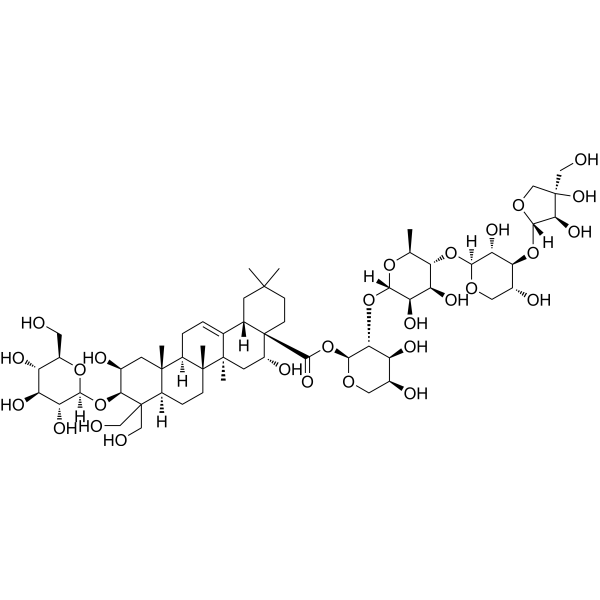
HY-N1411
Platycodin DAMPK Activator
Publications Citing Use of MedChemExpress (MCE) EB-3D
MoreAll AMPK Isoforms
More

Biological Activity
Description
IC50 & Target
IC50: 1 μM; Kd: 0.7 μM[3]
In Vitro
EB-3D displays (0-100 μM; 72 hours) excellent antiproliferative activity against a wide cohort of T-leukemic cell lines, with GI GI50s 13 values in the nanomolar range[1].
EB-3D (1.25-5μM; 24 hours) induced apoptosis in leukemia cell lines[1].
EB-3D (0.5-1 μM; 24 hours) induces a G0/G1 arrest that lead to apoptosis[1].
EB-3D (0.3 μM; 48 hours) shows a first spike of activation of AMPKα after 30 minutes and a later increase in the phosphorylation of T172[1].
EB-3D (1-40 μM; 48 hours) inhibits cell growth in HepG2 cells with a GI50 of 14.55 μM[2].
EB-3D induces deregulation of the AMPK-mTOR pathway and apoptosis in leukemia T-cells[1].
MedChemExpress (MCE) has not independently confirmed the accuracy of these methods. They are for reference only.
-
Cell Line:JURKAT, CCRF-CEM, HSB-2, MOLT-16, DNA-41, LOUCY, PEER, ALL-SIL cells
-
Concentration:0.001, 0.01, 0.1, 1, 10, 100 μM
-
Incubation Time:72 hours
-
Result:Inhibited JURKAT, CCRF-CEM, HSB-2, MOLT-16, DNA-41, LOUCY, PEER, and ALL-SIL cells growth with GI50s of 136.2, 478.8, 17.7, 0.9, 60.6, 200, 265, and 132 nM, respectively.
-
Cell Line:Jurkat, CCRF-CEM and HSB-2 cells
-
Concentration:1.25, 2.5, 5 μM
-
Incubation Time:24 hours
-
Result:Induced apoptosis in leukemia cell lines.
-
Cell Line:Jurkat, CCRF-CEM and HSB-2 cells
-
Concentration:0.5, 1 μM
-
Incubation Time:24 hours
-
Result:Induces cell cycle arrest in G0/G1 phase.
-
Cell Line:Jurkat cells
-
Concentration:0.3 μM
-
Incubation Time:48 hours
-
Result:Showed a first spike of activation of AMPKα after 30 minutes of treatment and a later increase in the phosphorylation of T172. The increase in S79 phosphorylation of its main target ACC (acetyl-coenzyme A (CoA) carboxylase), followed the same pattern. This rapid activation of AMPK, in turn induced a consequent reduction in mTOR phosphorylation that is visible already at 30’ and that becomes amplified at longer time probably due to the interruption of feedback loops that are characteristic of mTOR connecting pathways.
In Vivo
EB-3D (2.5 mg/kg; every other day for 4 weeks) shows a reduction of the number of spontaneous lung macro- and micrometastasis[4].
MedChemExpress (MCE) has not independently confirmed the accuracy of these methods. They are for reference only.
-
Animal Model:E0771-C57BL/6 mice[4]
-
Dosage:I.p.; every other day for 4 weeks
-
Administration:2.5 mg/kg
-
Result:A reduction of the number of spontaneous lung macro- and micrometastasis.


Chemical Information
-
CAS No. 1839150-63-8
-
Appearance Solid
-
Molecular Weight 644.44
-
Formula C30H36Br2N4O2
-
Color White to off-white
-
SMILES
CN(C1=CC=[N+](CC2=CC=C(OCCOC3=CC=C(C[N+]4=CC=C(N(C)C)C=C4)C=C3)C=C2)C=C1)C.[Br-].[Br-]
-
Shipping
Room temperature in continental US; may vary elsewhere.
-
Storage
4°C, sealed storage, away from moisture
* In solvent : -80°C, 6 months; -20°C, 1 month (sealed storage, away from moisture)


Publications (2)
-
Journal Impact Factor
-
Most Recent


Solvent & Solubility
In Vitro:
DMSO : 50 mg/mL (77.59 mM; Need ultrasonic; Hygroscopic DMSO has a significant impact on the solubility of product, please use newly opened DMSO)
H2O : ≥ 9.09 mg/mL (14.11 mM)
* "≥" means soluble, but saturation unknown.
Please refer to the solubility information to select the appropriate solvent. Once prepared, please aliquot and store the solution to prevent product inactivation from repeated freeze-thaw cycles.
Storage method and period of stock solution: -80°C, 6 months; -20°C, 1 month (sealed storage, away from moisture). When stored at -80°C, please use it within 6 months. When stored at -20°C, please use it within 1 month.
* Note: If you choose water as the stock solution, please dilute it to the working solution, then filter and sterilize it with a 0.22 μm filter before use.
Please refer to the solubility information to select the appropriate solvent. Once prepared, please aliquot and store the solution to prevent product inactivation from repeated freeze-thaw cycles.
Storage method and period of stock solution: -80°C, 6 months; -20°C, 1 month (sealed storage, away from moisture). When stored at -80°C, please use it within 6 months. When stored at -20°C, please use it within 1 month.
* Note: If you choose water as the stock solution, please dilute it to the working solution, then filter and sterilize it with a 0.22 μm filter before use.
Concentration (start) × Volume (start) = Concentration (final) × Volume (final)


Purity & Documentation
-
Data Sheet (276 KB)
-
SDS (393 KB)
- English - EN (393 KB)
- Français - FR (393 KB)
- Deutsch - DE (393 KB)
- Norwegian - NO (393 KB)
- Español - ES (393 KB)
- Swedish - SV (393 KB)
- Italian - IT (393 KB)
- Korean - KR (393 KB)
- Portuguese - PT (393 KB)
-
Handling Instructions (2659 KB)
References
[1]. Mariotto E, et al. EB-3D a novel choline kinase inhibitor induces deregulation of the AMPK-mTOR pathway and apoptosis in leukemia T-cells. Biochem Pharmacol. 2018 Sep;155:213-223. [Content Brief]
[2]. Sola-Leyva A, et al. Choline kinase inhibitors EB-3D and EB-3P interferes with lipid homeostasis in HepG2 cells. Sci Rep. 2019 Mar 25;9(1):5109. [Content Brief]
[3]. Schiaffino-Ortega S, et al. Design, synthesis, crystallization and biological evaluation of new symmetrical biscationic compounds as selective inhibitors of human Choline Kinase α1 (ChoKα1). [Content Brief]
[4]. Mariotto E, et al. Choline Kinase Alpha Inhibition by EB-3D Triggers Cellular Senescence, Reduces Tumor Growth and Metastatic Dissemination in Breast Cancer. Cancers (Basel). 2018;10(10):391. Published 2018 Oct 22. [Content Brief]


Complete Stock Solution Preparation Table
Please refer to the solubility information to select the appropriate solvent. Once prepared, please aliquot and store the solution to prevent product inactivation from repeated freeze-thaw cycles.
Storage method and period of stock solution: -80°C, 6 months; -20°C, 1 month (sealed storage, away from moisture). When stored at -80°C, please use it within 6 months. When stored at -20°C, please use it within 1 month.
| Optional Solvent | Concentration Solvent Mass | 1 mg | 5 mg | 10 mg | 25 mg |
|---|---|---|---|---|---|
| H2O / DMSO | 1 mM | 1.5517 mL | 7.7587 mL | 15.5173 mL | 38.7934 mL |
| 5 mM | 0.3103 mL | 1.5517 mL | 3.1035 mL | 7.7587 mL | |
| 10 mM | 0.1552 mL | 0.7759 mL | 1.5517 mL | 3.8793 mL | |
| DMSO | 15 mM | 0.1034 mL | 0.5172 mL | 1.0345 mL | 2.5862 mL |
| 20 mM | 0.0776 mL | 0.3879 mL | 0.7759 mL | 1.9397 mL | |
| 25 mM | 0.0621 mL | 0.3103 mL | 0.6207 mL | 1.5517 mL | |
| 30 mM | 0.0517 mL | 0.2586 mL | 0.5172 mL | 1.2931 mL | |
| 40 mM | 0.0388 mL | 0.1940 mL | 0.3879 mL | 0.9698 mL | |
| 50 mM | 0.0310 mL | 0.1552 mL | 0.3103 mL | 0.7759 mL | |
| 60 mM | 0.0259 mL | 0.1293 mL | 0.2586 mL | 0.6466 mL |
* Note: If you choose water as the stock solution, please dilute it to the working solution, then filter and sterilize it with a 0.22 μm filter before use.


EB-3D Related Classifications
HY-115463 Related Classifications
 Powered by Bioz