



TVB-3166
Based on 12 publication(s) in Google Scholar
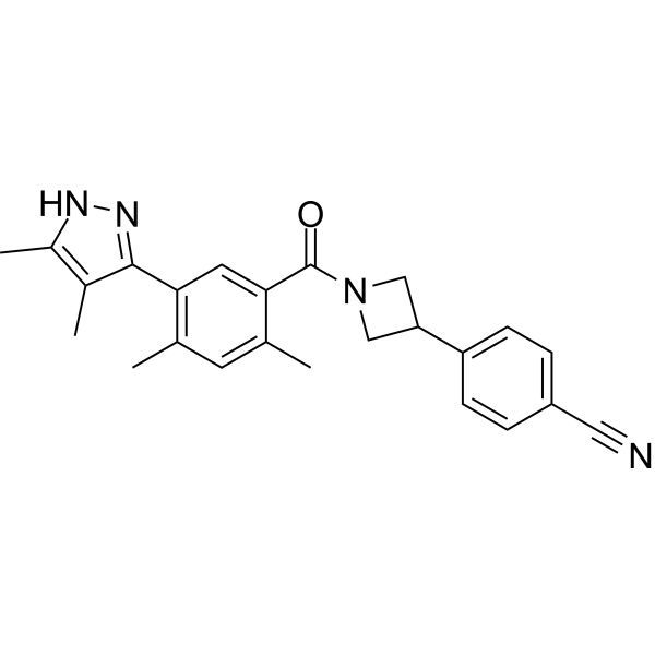
TVB-3166 is an orally-available, reversible, and selective fatty acid synthase (FASN) inhibitor with IC50s of 42 nM and 81 nM for biochemical FASN and cellular palmitate synthesis, respectively. TVB-3166 induces apoptosis, and inhibits in-vivo xenograft tumor growth.
연구목적의 판매만을 진행합니다. 환자를 대상으로 한 판매는 하지 않습니다.
- Purity : 99.69%
- CAS No.: 1533438-83-3
- 화학식: C24H24N4O
- 분자량:384.47
-
보관:Powder -20°C, 3 years , 4°C, 2 years ; In solvent -80°C, 2 years , -20°C, 1 year

To place orders, for customer services and technical support, please contact: MedChemExpress USA
Tel: 609-228-6898 E-mail: [email protected] [email protected]


-
 Biological Activity
-
Chemical Information
-
용액&용해도
- Protocol
- 순도&문서
- References
-
 Help & FAQs


-
HY-112829
DenifanstatFASN Inhibitor -
HY-B0218
OrlistatFASN Inhibitor -
HY-120062
TVB-3664FASN Inhibitor -
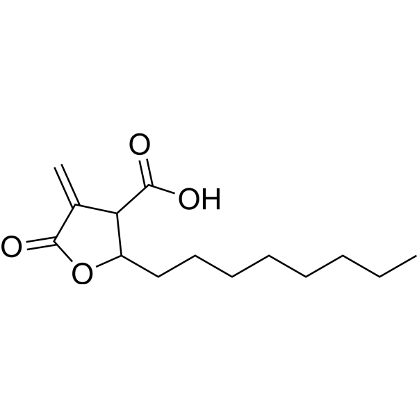
HY-12364
C75Fatty Acid Synthase Inhibitor -
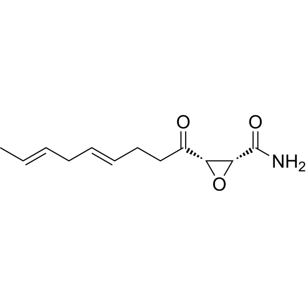
HY-A0210
CeruleninFASN Inhibitor -
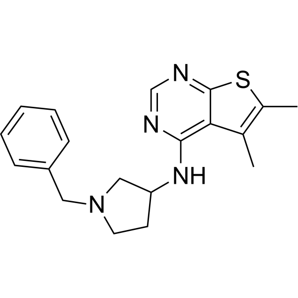
HY-121250
FasnallFASN Inhibitor -
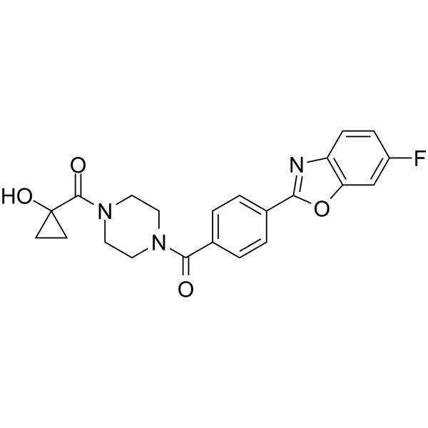
HY-111551
FT113FASN Inhibitor -
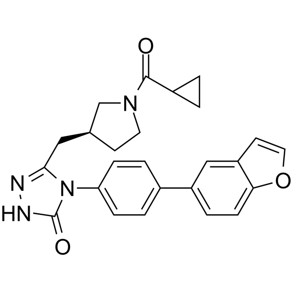
HY-12325
GSK2194069FASN Inhibitor -
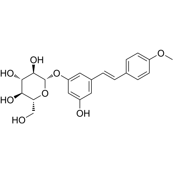
HY-N2486
DesoxyrhaponticinFASN Inhibitor -
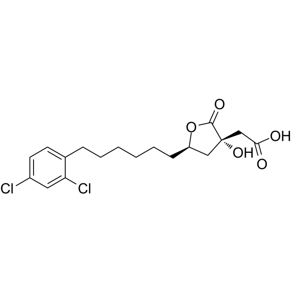
HY-16450
SB 204990ATP Citrate Lyase Inhibitor -
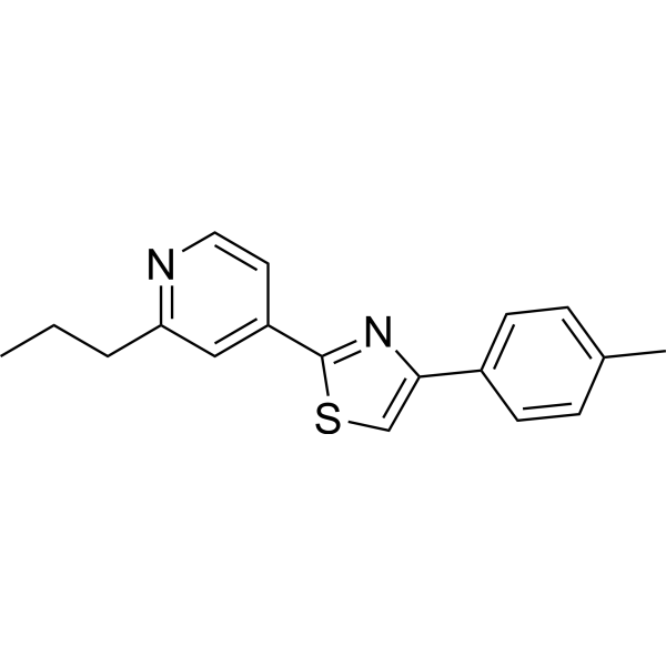
HY-14452
FatostatinSREBP Activation Inhibitor -
HY-B0218R
Orlistat (Standard)
Publications Citing Use of MedChemExpress (MCE) TVB-3166
More- Nat Metab. 2021 Nov;3(11):1466-1475. [Abstract]
- Nat Commun. 2025 Feb 19;16(1):1762. [Abstract]
- J Immunother Cancer. 2025 Apr 12;13(4):e010924. [Abstract]
- J Transl Med. 2024 Jan 13;22(1):55. [Abstract]
- Br J Cancer. 2023 Mar;128(7):1344-1359. [Abstract]
- Neurosci Bull. 2025 Jun 28. [Abstract]
- EMBO Rep. 2023 Dec 6;24(12):e49561. [Abstract]
- ACS Omega. 2024 Feb 28;9(10):11870-11882. [Abstract]
- J Cell Sci. 2025 May 15;138(10):jcs263688. [Abstract]
- Biomed Pharmacother. 2025 Nov:192:118656. [Abstract]
- University of Rijeka. 2023.
- McGill University. 2021 Aug, 30347448.
 Customer Validation & Images

-
WB
-
Cell Proliferation/Viability Assay
-
In Vivo Efficacy Study
-
2D/3D Cell Culture and Differentiation
-
ELISA


Biological Activity
제품 설명
IC50 & Target
IC50: 42 nM (FASN) and 81 nM (cellular palmitate synthesis)[1]
In Vitro
ISX-9 promotes neurogenesis in vivo, enhancing the proliferation and differentiation of hippocampal subgranular zone (SGZ) neuroblasts, and the dendritic arborization of adult-generated dentate gyrus neurons. At 2.5-20 μM, ISX-9 has been shown to dose-dependently trigger neurogenesis and block gliogenesis in adult rat hippocampal stem cells through a calcium-activated signaling pathway dependent on myocyte-enhancer factor 2-dependent gene expression[1].
Molecular exploration of ISX-9-induced regulation of neurogenesis (via FACS and microarray of SGZ stem and progenitor cells) suggested the involvement of the myocyte-enhancer family of proteins (Mef2)[1].
MedChemExpress (MCE) has not independently confirmed the accuracy of these methods. They are for reference only.
-
Cell Line:CALU-6 tumor cells
-
Concentration:0.001, 0.01, 0.1, 1, 10 μM
-
Incubation Time:24 hours
-
Result:Caused cell death in CALU-6 non-small-cell lung tumor cells with a cellular IC50 value of 0.10 μM.
-
Cell Line:90 different tumor cell lines (such as CALU-6 NSCLC cell line, NCI-H1975 NSCLC cell line)
-
Concentration:0.02 or 0.20 μM
-
Incubation Time:7 days
-
Result:Dose-dependent induction of cell death was observed in all tumor cell lines.
-
Cell Line:COLO-205 and A549 cells
-
Concentration:0.2 μM
-
Incubation Time:48 hours
-
Result:Inhibited β-catenin pathway signal transduction and transcriptional activity.
In Vivo
TVB-3166 (Oral gavage; 30-100 mg/kg/day) has the concentration is approximately 3-fold higher in plasma than tumor. The 100 and 30 mg/kg groups had plasma and tumor concentrations of 7 and 2.9 μM, respectively[1].
MedChemExpress (MCE) has not independently confirmed the accuracy of these methods. They are for reference only.
-
Animal Model:Female BALB-c-nude mice[1]
-
Dosage:30, 60, or 100 mg/kg
-
Administration:Oral gavage; once daily
-
Result:Inhibited xenograft tumor growth.
-
Animal Model:Female BALB-c-nude mice[1]
-
Dosage:30, 60, or 100 mg/kg (Pharmacokinetic Study)
-
Administration:Oral gavage; once daily
-
Result:The concentration was approximately 3-fold higher in plasma than tumor. The 100 and 30 mg/kg groups had plasma and tumor concentrations of 7 and 2.9 μM, respectively.
- Detection of Apoptosis
- TUNEL staining for apoptotic DNA fragmentation
- Annexin V plus membrane-impermeant dye apoptosis staining
- Patient-Derived Xenograft (PDX)
- Apoptosis Solutions
- Patient-Derived Orthotopic Xenograft (PDOX)
- Splenic/Portal-Vein Liver Metastasis Xenograft
- Subcutaneous Cell-Line-Derived Xenograft
- Orthotopic Cell-Line Xenograft
- Intraperitoneal/Peritoneal Dissemination Xenograft


Chemical Information
-
CAS No. 1533438-83-3
-
Appearance Solid
-
분자량 384.47
-
화학식 C24H24N4O
-
Color White to off-white
-
SMILES
N#CC1=CC=C(C2CN(C(C3=CC(C4=NNC(C)=C4C)=C(C)C=C3C)=O)C2)C=C1
-
선적
Room temperature in continental US; may vary elsewhere.
-
보관
Powder -20°C 3 years 4°C 2 years In solvent -80°C 2 years -20°C 1 year


Publications (12)
-
Journal Impact Factor
-
Most Recent
-
Nat Metab
2021 Nov;3(11):1466-1475. PMID: 34580494 -
Nat Commun
Lipogenic enzyme FASN promotes mutant p53 accumulation and gain-of-function through palmitoylation. [Abstract]2025 Feb 19;16(1):1762. PMID: 39971971
TVB-3166 purchased from MedChemExpress. Usage Cited in: Nat Commun. 2025 Feb 19;16(1):1762. [Abstract]
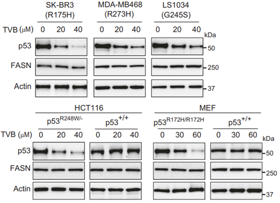
Inhibition of FASN by TVB-3166 reduced the protein levels of mutp53, but not wtp53, in different cells. Cells were treated with the small-molecule FASN inhibitor TVB-3166 (TVB) at the indicated concentrations for 24 h and subjected to Western blot analysis.
TVB-3166 purchased from MedChemExpress. Usage Cited in: Nat Commun. 2025 Feb 19;16(1):1762. [Abstract]
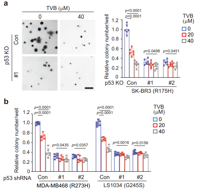
The FASN inhibitor TVB-3166 (TVB, 72 h) displayed a more pronounced inhibitory effect on AIG in cancer cells carrying mutp53 compared with cells with mutp53 KO (a) or knockdown (b). Left in a: Representative images. Scale bar: 200 μm.
TVB-3166 purchased from MedChemExpress. Usage Cited in: Nat Commun. 2025 Feb 19;16(1):1762. [Abstract]
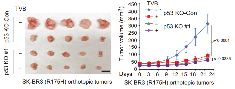
TVB-3166 (TVB) displayed a more pronounced inhibitory effect on the growth of orthotopic tumors formed by SK-BR3 (R175H) cells than tumors formed by SK-BR3 with mutp53 KO. Mice were treated with TVB-3166 (60 mg/kg; i.g.) once every two days for 3 weeks when tumor sizes reached ~30 mm3.
TVB-3166 purchased from MedChemExpress. Usage Cited in: Nat Commun. 2025 Feb 19;16(1):1762. [Abstract]
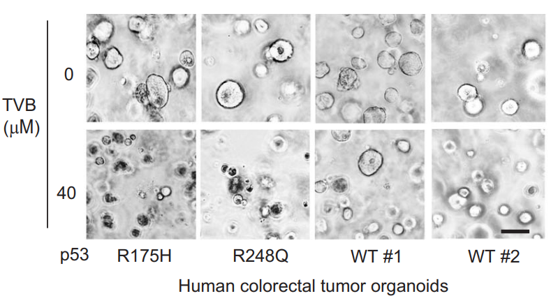
TVB-3166 (40 μM) treatment displayed a more pronounced inhibitory effect on human colorectal tumor organoids expressing mutp53 compared with organoids expressing wtp53. Left panels: Representative images of different organoids expressing mutp53 or wtp53, which were treated with or without TVB-3166 for 48 h. Scale bar: 50 μm. Right panel: The relative viability of mutp53 and wtp53 organoids.
-
J Immunother Cancer
Targeting B7-H3 inhibition-induced activation of fatty acid synthesis boosts anti-B7-H3 immunotherapy in triple-negative breast cancer. [Abstract]2025 Apr 12;13(4):e010924. PMID: 40221152 -
J Transl Med
Metabolic reprogramming based on RNA sequencing of gemcitabine-resistant cells reveals the FASN gene as a therapeutic for bladder cancer. [Abstract]2024 Jan 13;22(1):55. PMID: 38218866 -
Br J Cancer
Transcriptome analysis of newly established carboplatin-resistant ovarian cancer cell model reveals genes shared by drug resistance and drug-induced EMT. [Abstract]2023 Mar;128(7):1344-1359. PMID: 36717670 -
Neurosci Bull
2025 Jun 28. PMID: 40580390 -
EMBO Rep
2023 Dec 6;24(12):e49561. PMID: 37943703 -
ACS Omega
Identification of New Modulators and Inhibitors of Palmitoyl-Protein Thioesterase 1 for CLN1 Batten Disease and Cancer. [Abstract]2024 Feb 28;9(10):11870-11882. PMID: 38496939 -
J Cell Sci
Tumor acidosis supports cancer cell lipid uptake via a rapid transporter-independent mechanism. [Abstract]2025 May 15;138(10):jcs263688. PMID: 40190115 -
Biomed Pharmacother
Modeling of glucocorticoid resistance in multiple myeloma reveals mechanisms and markers of glucocorticoid resistance. [Abstract]2025 Nov:192:118656. PMID: 41138658
TVB-3166 purchased from MedChemExpress. Usage Cited in: Biomed Pharmacother. 2025 Nov:192:118656. [Abstract]
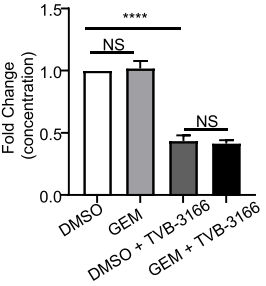
Enzyme-linked immunosorbent assay (ELISA) was used to determine the FASN content of xenograft tumors in each group. The ELISA results showed that TVB-3166 (60 mg/kg; i.g.; 5 weeks) reduced the FASN gene of the tumor.
TVB-3166 purchased from MedChemExpress. Usage Cited in: Biomed Pharmacother. 2025 Nov:192:118656. [Abstract]
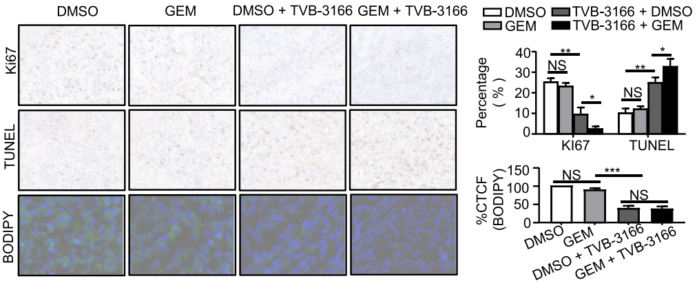
Statistical analysis was performed on the rate of KI67- and TUNEL-positive cells in each group of xenograft tumors by immunohistochemical staining (IHC). The corrected total cell fluorescence (CTCF) of BODIPY staining was used to quantitatively detect the lipid content of xenograft tumors in each group. The results showed that TVB-3166 (60 mg/kg; i.g.; 5 weeks) treatment can reduce lipid accumulation, inhibit cell proliferation and increase the apoptosis rate.
-
-


용액&용해도
In Vitro:
DMSO : 62.5 mg/mL (162.56 mM; Need ultrasonic; Hygroscopic DMSO has a significant impact on the solubility of product, please use newly opened DMSO)
Please refer to the solubility information to select the appropriate solvent. Once prepared, please aliquot and store the solution to prevent product inactivation from repeated freeze-thaw cycles.
Storage method and period of stock solution: -80°C, 2 years; -20°C, 1 year. When stored at -80°C, please use it within 2 years. When stored at -20°C, please use it within 1 year.
Please refer to the solubility information to select the appropriate solvent. Once prepared, please aliquot and store the solution to prevent product inactivation from repeated freeze-thaw cycles.
Storage method and period of stock solution: -80°C, 2 years; -20°C, 1 year. When stored at -80°C, please use it within 2 years. When stored at -20°C, please use it within 1 year.
Concentration (start) × Volume (start) = Concentration (final) × Volume (final)
In Vivo:
Select the appropriate dissolution method based on your experimental animal and administration route.
- For the following dissolution methods, please ensure to first prepare a clear stock solution using an In Vitro approach and then sequentially add co-solvents:
- To ensure reliable experimental results, the clarified stock solution can be appropriately stored based on storage conditions. As for the working solution for In Vivo experiments, it is recommended to prepare freshly and use it on the same day.
- The percentages shown for the solvents indicate their volumetric ratio in the final prepared solution. If precipitation or phase separation occurs during preparation, heat and/or sonication can be used to aid dissolution.
Add each solvent one by one: 10% DMSO 40% PEG300 5% Tween-80 45% Saline
Solubility: ≥ 2.08 mg/mL (5.41 mM); Clear solution
This protocol yields a clear solution of ≥ 2.08 mg/mL (saturation unknown).
Taking 1 mL working solution as an example, add 100 μL DMSO stock solution (20.8 mg/mL) to 400 μL PEG300, and mix evenly; then add 50 μL Tween-80 and mix evenly; then add 450 μL Saline to adjust the volume to 1 mL.
Preparation of Saline: Dissolve 0.9 g sodium chloride in ddH₂O and dilute to 100 mL to obtain a clear Saline solution.
Add each solvent one by one: 10% DMSO 90% (20% SBE-β-CD in Saline)
Solubility: 2.08 mg/mL (5.41 mM); Suspended solution; Need ultrasonic
This protocol yields a suspended solution of 2.08 mg/mL. Suspended solution can be used for oral and intraperitoneal injection.
Taking 1 mL working solution as an example, add 100 μL DMSO stock solution (20.8 mg/mL) to 900 μL 20% SBE-β-CD in Saline, and mix evenly.
Preparation of 20% SBE-β-CD in Saline (4°C, storage for one week): 2 g SBE-β-CD powder is dissolved in 10 mL Saline, completely dissolve until clear.
In Vivo Dissolution Calculator
Please enter the basic information of animal experiments:
-
-
-
-
Recommended: Prepare an additional quantity of animals to account for potential losses during experiments.
Please enter your animal formula composition:
-
%DMSO +
Recommended: Keep the proportion of DMSO in working solution below 2% if your animal is weak.
-
%+
-
+%Tween-80 + +
-
%Saline +
The co-solvents required include: DMSO, . All of co-solvents are available by MedChemExpress (MCE). , Tween 80. All of co-solvents are available by MedChemExpress (MCE).
Working solution concentration: 0.22 mg/mL
Method for preparing stock solution: mg drug dissolved in μL DMSO. Stock solution concentration: mg/mL.
1. Take μL DMSO stock solution;
2. Add μL .
μL , mix evenly;
3. Then add μL Tween 80, mix evenly;
4. Then add μL
Please ensure that the stock solution in the first step is dissolved to a clear state, and add co-solvents in sequence. You can use ultrasonic heating (ultrasonic cleaner, recommended frequency 20-40 kHz), vortexing, etc. to assist dissolution.


순도&문서
-
Data Sheet (278 KB)
-
SDS (393 KB)
- English - EN (393 KB)
- Français - FR (393 KB)
- Deutsch - DE (393 KB)
- Norwegian - NO (393 KB)
- Español - ES (393 KB)
- Swedish - SV (393 KB)
- Italian - IT (393 KB)
- Korean - KR (393 KB)
- Portuguese - PT (393 KB)
-
Handling Instructions (2659 KB)
References


Complete Stock Solution Preparation Table
Please refer to the solubility information to select the appropriate solvent. Once prepared, please aliquot and store the solution to prevent product inactivation from repeated freeze-thaw cycles.
Storage method and period of stock solution: -80°C, 2 years; -20°C, 1 year. When stored at -80°C, please use it within 2 years. When stored at -20°C, please use it within 1 year.
| Optional Solvent | Concentration Solvent Mass | 1 mg | 5 mg | 10 mg | 25 mg |
|---|---|---|---|---|---|
| DMSO | 1 mM | 2.6010 mL | 13.0049 mL | 26.0098 mL | 65.0246 mL |
| 5 mM | 0.5202 mL | 2.6010 mL | 5.2020 mL | 13.0049 mL | |
| 10 mM | 0.2601 mL | 1.3005 mL | 2.6010 mL | 6.5025 mL | |
| 15 mM | 0.1734 mL | 0.8670 mL | 1.7340 mL | 4.3350 mL | |
| 20 mM | 0.1300 mL | 0.6502 mL | 1.3005 mL | 3.2512 mL | |
| 25 mM | 0.1040 mL | 0.5202 mL | 1.0404 mL | 2.6010 mL | |
| 30 mM | 0.0867 mL | 0.4335 mL | 0.8670 mL | 2.1675 mL | |
| 40 mM | 0.0650 mL | 0.3251 mL | 0.6502 mL | 1.6256 mL | |
| 50 mM | 0.0520 mL | 0.2601 mL | 0.5202 mL | 1.3005 mL | |
| 60 mM | 0.0433 mL | 0.2167 mL | 0.4335 mL | 1.0837 mL | |
| 80 mM | 0.0325 mL | 0.1626 mL | 0.3251 mL | 0.8128 mL | |
| 100 mM | 0.0260 mL | 0.1300 mL | 0.2601 mL | 0.6502 mL |


TVB-3166 Related Classifications
HY-120394 Related Classifications
 Powered by Bioz