



ML-184

Based on 1 Customer Validation
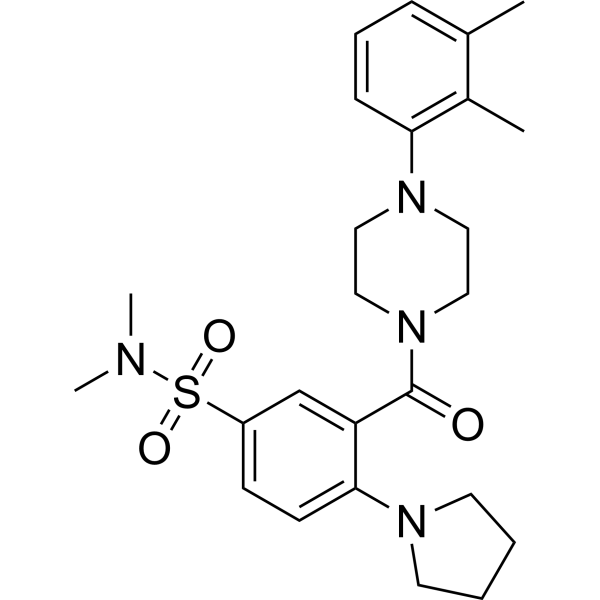
ML-184 (CID244033) is a selective GPR55 agonist with an EC50 of 250 nM, more than 100-fold selectivity for GPR55 over GPR35, CB1, and CB2. ML-184 induces ERK1/2 phosphorylation and PKCβII translocation to the plasma membrane via activation of GPR55. ML-184 (CID2440433) increases the proliferation of neural stem cells and promotes neuronal differentiation in vitro.
For research use only. We do not sell to patients.
- Purity : 99.57%
- CAS No.: 794572-10-4
- Formula: C25H34N4O3S
- Molecular Weight:470.63
-
Storage:Powder -20°C, 3 years , 4°C, 2 years ; In solvent -80°C, 6 months , -20°C, 1 month

To place orders, for customer services and technical support, please contact: MedChemExpress USA
Tel: 609-228-6898 E-mail: [email protected] [email protected]


-
 Biological Activity
-
Chemical Information
-
Solvent & Solubility
- Protocols
- Purity & Documentation
- References
-
 Help & FAQs


-
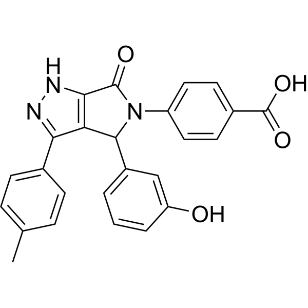
HY-16697
CID 16020046GPR55 Antagonist -
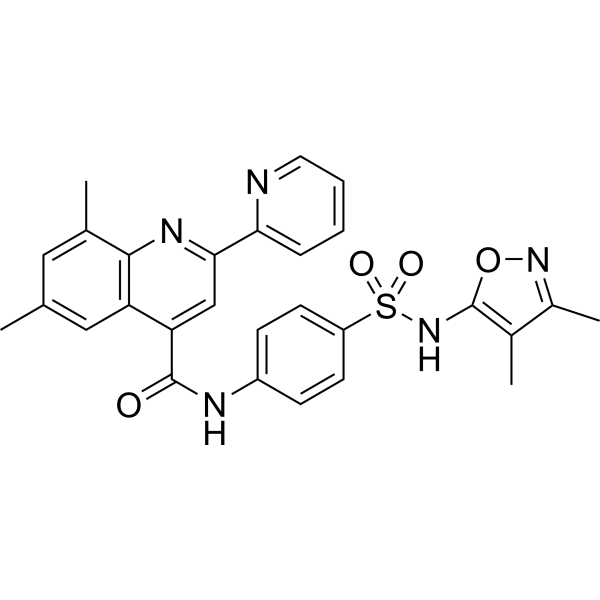
HY-110125
ML-193GPR55 Antagonist -
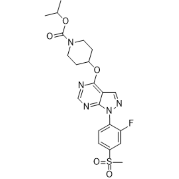
HY-15565
APD668GPR119 Agonist -
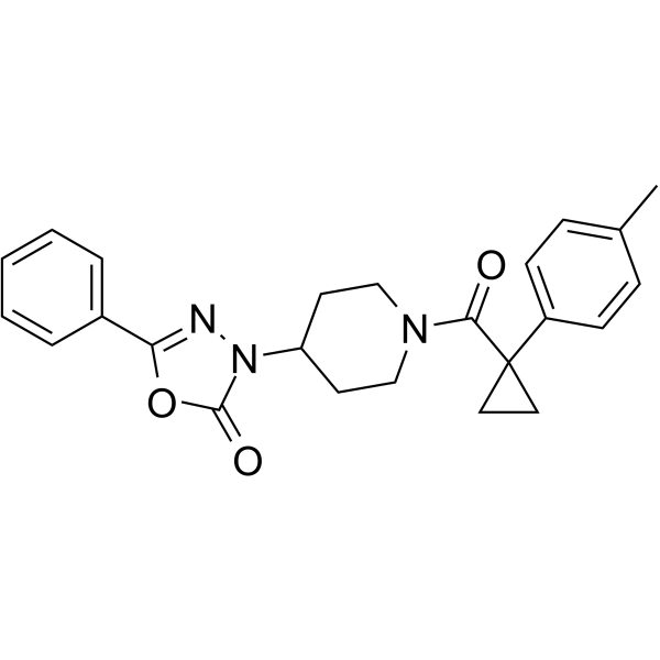
HY-111083
ML191GPR55 Antagonist -
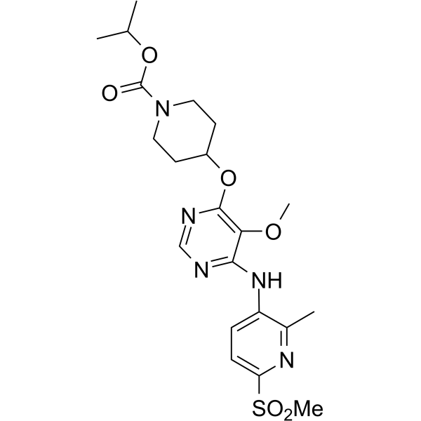
HY-15566
APD597GPR119 Agonist -

HY-16697R
CID 16020046 (Standard)GPR55 Antagonist -
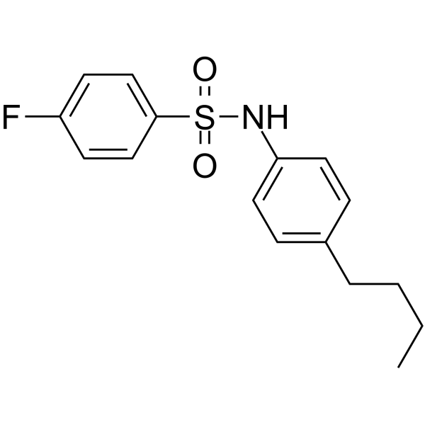
HY-101906
DC260126GPR40 antagonist -
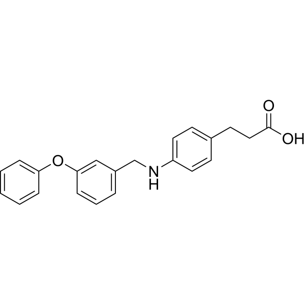
HY-15589
GW9508GPR40 Agonist -
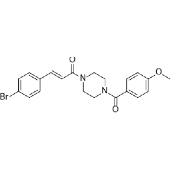
HY-12336
NIBR189EBI2 Antagonist -
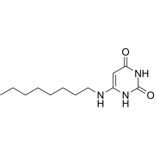
HY-12764
6-OAUGPR84 Agonist -
HY-19838
JNJ-63533054GPR139 Agonist -
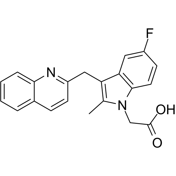
HY-15342
TimapiprantProstaglandin Receptor Antagonist


Biological Activity
Description
In Vitro
ML-184 (1 μM; 48 h) increases the proportion of cells in the S phase and promotes cell proliferation in human neural stem cells (hNSCs), an effect that could be blocked by pretreatment with 5 μM ML193[2].
ML-184 (1 μM; 10 d) significantly increases the mRNA and protein expressions of neuronal markers βIII-tubulin and microtubule-associated protein 2 (MAP2) in hNSCs, without significant effect on the astrocyte marker S100β[2].
MedChemExpress (MCE) has not independently confirmed the accuracy of these methods. They are for reference only. Further protocols information, click here.
-
Cell Line:Human neural stem cells (hNSCs)
-
Concentration:1 μM
-
Incubation Time:48 h
-
Result:Increased the percentage of BrdU-positive cells in S phase and promoted cell proliferation.
Attenuated by co-treatment with the GPR55 antagonist ML193 (5 µM).
Flow cytometric analysis showed a significant shift of cells from G0/G1 to S phase, indicating enhanced proliferation.
-
Cell Line:Human neural stem cells (hNSCs)
-
Concentration:1 μM
-
Incubation Time:10 d
-
Result:Induced neuronal differentiation, as evidenced by increased immunofluorescence staining for βIII-tubulin and upregulated mRNA expression of βIII-tubulin and MAP2.
-
Cell Line:Human neural stem cells (hNSCs)
-
Concentration:1 μM
-
Incubation Time:10 d
-
Result:Resulted a 1.5- to 2-fold increase in neuronal marker genes compared to vehicle control, with no significant changes in glial marker S100β.
In Vivo
ML-184 can increase β-cell area and β-cell mass and reduce α-cell area and mass in high-fat-fed (HFF) mice. ML-184 or combined with Sitagliptin (HY-13749) can significantly increase circulating GIP levels and reduce food intake in mice; it can also increase jejunal GPR55 and JNK1 gene expression levels[3].
MedChemExpress (MCE) has not independently confirmed the accuracy of these methods. They are for reference only.


Chemical Information
-
CAS No. 794572-10-4
-
Appearance Solid
-
Molecular Weight 470.63
-
Formula C25H34N4O3S
-
Color White to off-white
-
SMILES
CC1=CC=CC(N2CCN(CC2)C(C3=CC(S(N(C)C)(=O)=O)=CC=C3N4CCCC4)=O)=C1C
-
Synonyms
CID2440433
-
Shipping
Room temperature in continental US; may vary elsewhere.
-
Storage
Powder -20°C 3 years 4°C 2 years In solvent -80°C 6 months -20°C 1 month


Solvent & Solubility
In Vitro:
DMSO : 12.5 mg/mL (26.56 mM; ultrasonic and warming and heat to 60°C; Hygroscopic DMSO has a significant impact on the solubility of product, please use newly opened DMSO)
Please refer to the solubility information to select the appropriate solvent. Once prepared, please aliquot and store the solution to prevent product inactivation from repeated freeze-thaw cycles.
Storage method and period of stock solution: -80°C, 6 months; -20°C, 1 month. When stored at -80°C, please use it within 6 months. When stored at -20°C, please use it within 1 month.
Please refer to the solubility information to select the appropriate solvent. Once prepared, please aliquot and store the solution to prevent product inactivation from repeated freeze-thaw cycles.
Storage method and period of stock solution: -80°C, 6 months; -20°C, 1 month. When stored at -80°C, please use it within 6 months. When stored at -20°C, please use it within 1 month.
Concentration (start) × Volume (start) = Concentration (final) × Volume (final)

Protocols
-
Kinase activity and phosphorylation assays
Kinase activity assays measure the ability of kinases to transfer phosphate groups from ATP to specific substrates, while phosphorylation assays detect the presence and levels of phosphorylated proteins. Common methods include radiolabeled ATP incorporation (e. g. ,), ADP release detection via bioluminescence (e. g. ,[3]), enzyme-linked immunosorbent assays (ELISA) for phospho-specific epitopes (e. g. ,[6]), and microtiter-based formats for high-throughput screening (e. g. ,[8]). The ADP-Glo assay quantifies kinase activity by measuring ADP produced during phosphorylation using a luciferase-based system. Radiometric assays involve autoradiography or scintillation counting after incorporation of 32P-labeled ATP into substrate proteins. ELISA-based approaches rely on phospho-specific antibodies to detect activated kinases in cell lysates or purified samples.
-
PC12 NGF-Induced Neuronal Differentiation Culture
PC12 cells are a rat adrenal pheochromocytoma clonal line that responds to NGF by stopping proliferation and extending branching neurite-like processes; after longer NGF exposure, cells develop long processes and neuronal-like ultrastructural and functional features. NGF-induced differentiation is read out mainly by neurite outgrowth, reduced proliferation, microtubule assembly, and neuronal differentiation-associated proteins such as MAPs, tau, GAP-43, and synapsin-1.
-
Western Blot
Western blotting (WB) is a commonly used experimental method in molecular biology, biochemistry, and immunogenetics for identifying and quantifying target proteins. It combines gel electrophoresis with immunoassay, enabling researchers to analyze protein expression, post-translational modifications, and molecular weight.
-
iPSC/hPSC-Derived Neuron Differentiation Culture
iPSC/hPSC-derived neuron differentiation culture directs pluripotent cells toward neuroectoderm and then neuronal lineages by suppressing developmental signals that maintain non-neural fates; the classic monolayer dual-SMAD approach blocks BMP and Activin/TGF-β signaling with Noggin or dorsomorphin/LDN193189 plus SB431542, producing PAX6-positive neural progenitors that can be further matured into neurons. The readout is generated by morphology, neural progenitor markers, neuronal markers, subtype markers, and functional assays: PAX6/SOX1/NESTIN indicate neural progenitor induction, βIII-tubulin/TUJ1 and MAP2 indicate neuronal differentiation, cortical programs can be assessed by FOXG1, TBR1, CTIP2, SATB2, and synaptic maturation can be assessed by synaptic proteins, calcium activity, multielectrode arrays, or patch-clamp electrophysiology.
-
SH-SY5Y Neuronal Differentiation Culture
SH-SY5Y neuronal differentiation culture uses sequential exposure to retinoic acid and neurotrophic factors to reduce proliferative neuroblastoma-like behavior and induce neuron-like morphology, including neurite extension, neuronal marker expression, and, in RA/BDNF protocols, greater synaptic-marker expression than undifferentiated culture. Retinoic acid is commonly used as the initiating differentiation cue, while BDNF in serum-reduced or serum-free medium supports later maturation and neurotrophic-factor-dependent neuron-like survival.
-
Human pluripotent stem cell neural induction and neuron differentiation
Human pluripotent stem cell neural induction can be achieved by blocking BMP and TGFβ/Activin/Nodal SMAD signaling, which suppresses non-neural differentiation and promotes early neuroectodermal identity; the expected readout is loss of pluripotency markers such as OCT4 and induction of neural markers such as PAX6, followed by neural progenitor and neuron marker acquisition during differentiation. This protocol uses dual-SMAD neural induction as the core induction method, followed by cortical neuron differentiation as a representative neuron differentiation model; published cortical protocols describe generation of cortical progenitors, temporally ordered cortical projection neurons, action-potential firing, synaptogenesis, and neural network formation over an approximately 80-day process.
-
SH-SY5Y neuronal-like differentiation
SH-SY5Y neuronal-like differentiation uses defined culture conditions to shift proliferative human neuroblastoma cells toward a neuron-like state, mainly assessed by reduced proliferation, neurite extension, neuronal-marker expression, and, in some protocols, increased dependence on neurotrophic support. Retinoic acid (RA) is commonly used for the first differentiation phase, and sequential RA followed by brain-derived neurotrophic factor (BDNF) in serum-free medium is a well-characterized approach for generating neuron-like SH-SY5Y cultures with extensive neurite outgrowth. The primary readouts are morphology-based neurite outgrowth and marker-based confirmation using proteins such as βIII-tubulin, MAP2, GAP43, synaptophysin, NeuN, NSE, TH, or related neuronal/synaptic markers, depending on the study endpoint.
-
Cell differentiation
Cell differentiation refers to the process in which cells of the same origin gradually produce cell groups with different morphological structure and functional characteristics.
-
Protocol for Kinase activity and phosphorylation assays
Kinase activity assays measure transfer of phosphate from ATP to a protein or peptide substrate, generating phosphorylated substrate, ADP, or incorporated radiolabeled phosphate as the readout; phosphorylation assays measure site-specific phosphorylation in cells or tissues as a proxy for kinase-pathway activation, inhibition, or substrate regulation. Phosphorylation can be detected by phospho-specific Western blot, immunoprecipitation kinase assay, phospho-immunofluorescence, phospho-flow cytometry, luminescent ADP detection, radiolabeled ATP incorporation, or reporter-based pathway assays, and these readouts can be applied to cancer cells, primary neurons, mouse tumors, organoids, inflammatory macrophages, ferroptosis studies, and mitophagy studies when the kinase target is biologically relevant.
-
PC12 NGF-induced neuronal-like differentiation
PC12 cells are a rat adrenal pheochromocytoma-derived clonal cell line that responds to nerve growth factor by stopping proliferation and extending neurites, producing a sympathetic neuron-like phenotype used to study neuronal differentiation and neurite outgrowth. NGF acts through TrkA-dependent signaling, and neurite outgrowth is associated with ERK/Akt signaling, microtubule organization, neuronal-marker expression, and increased electrophysiological neuronal features such as sodium-channel density. The main assay readout is morphological differentiation, usually measured as the percentage of neurite-bearing cells, neurite length, neurite number, or total neurite length per cell. Additional readouts include GAP-43, tyrosine hydroxylase, βIII-tubulin, neurofilament, synapsin I, synaptophysin, ERK phosphorylation, Akt phosphorylation, and sodium-channel current density.
-
Cell Viability Determination by MTT Colorimetric Assay
The following protocol uses the MTT colorimetric assay as a classic literature-established method for assessing cell viability/metabolic activity in cultured mammalian cells. MTT[3-(4,5-dimethylthiazol-2-yl)-2,5-diphenyltetrazolium bromide] is reduced by metabolically active cells to a colored formazan product; the amount of formazan is quantified spectrophotometrically and provides an indirect measure of metabolically active viable cells. Importantly, MTT reduction reflects cellular oxidoreductase/metabolic activity rather than an absolute direct count of living cells, so changes in cellular metabolism can alter the signal independently of cell number.
-
Directly Induced Neuron Culture
Directly induced neuron culture converts somatic cells, most commonly fibroblasts, into induced neurons without passing through a pluripotent or neural progenitor stage; classic evidence shows that mouse fibroblasts can be converted by Ascl1, Brn2/Pou3f2, and Myt1l, human fibroblasts can be converted by defined neuronal transcription factors, and human fibroblasts can also be converted by miR-9/9-124 with neurogenic or subtype-specifying transcription factors. The readout is acquisition of neuronal identity and function, assessed by neuronal morphology, neuronal markers such as Tuj1/βIII-tubulin, MAP2, synapsin, and subtype markers when relevant, together with functional assays such as action-potential firing, synaptic activity, and electrophysiology.


Purity & Documentation
-
Data Sheet (279 KB)
-
SDS (393 KB)
- English - EN (393 KB)
- Français - FR (393 KB)
- Deutsch - DE (393 KB)
- Norwegian - NO (393 KB)
- Español - ES (393 KB)
- Swedish - SV (393 KB)
- Italian - IT (393 KB)
- Korean - KR (393 KB)
- Portuguese - PT (393 KB)
-
Handling Instructions (2659 KB)
References
[1]. Kotsikorou E, et al. Identification of the GPR55 agonist binding site using a novel set of high-potency GPR55 selective ligands. Biochemistry. 2011 Jun 28;50(25):5633-47. [Content Brief]
[2]. Hill JD, et al. Activation of GPR55 increases neural stem cell proliferation and promotes early adult hippocampal neurogenesis. Br J Pharmacol. 2018 Aug;175(16):3407-3421. [Content Brief]


Complete Stock Solution Preparation Table
Please refer to the solubility information to select the appropriate solvent. Once prepared, please aliquot and store the solution to prevent product inactivation from repeated freeze-thaw cycles.
Storage method and period of stock solution: -80°C, 6 months; -20°C, 1 month. When stored at -80°C, please use it within 6 months. When stored at -20°C, please use it within 1 month.
| Optional Solvent | Concentration Solvent Mass | 1 mg | 5 mg | 10 mg | 25 mg |
|---|---|---|---|---|---|
| DMSO | 1 mM | 2.1248 mL | 10.6241 mL | 21.2481 mL | 53.1203 mL |
| 5 mM | 0.4250 mL | 2.1248 mL | 4.2496 mL | 10.6241 mL | |
| 10 mM | 0.2125 mL | 1.0624 mL | 2.1248 mL | 5.3120 mL | |
| 15 mM | 0.1417 mL | 0.7083 mL | 1.4165 mL | 3.5414 mL | |
| 20 mM | 0.1062 mL | 0.5312 mL | 1.0624 mL | 2.6560 mL | |
| 25 mM | 0.0850 mL | 0.4250 mL | 0.8499 mL | 2.1248 mL |


ML-184 Related Classifications
HY-116461 Related Classifications
 Powered by Bioz