



Dipeptide 2

Based on 1 Customer Validation
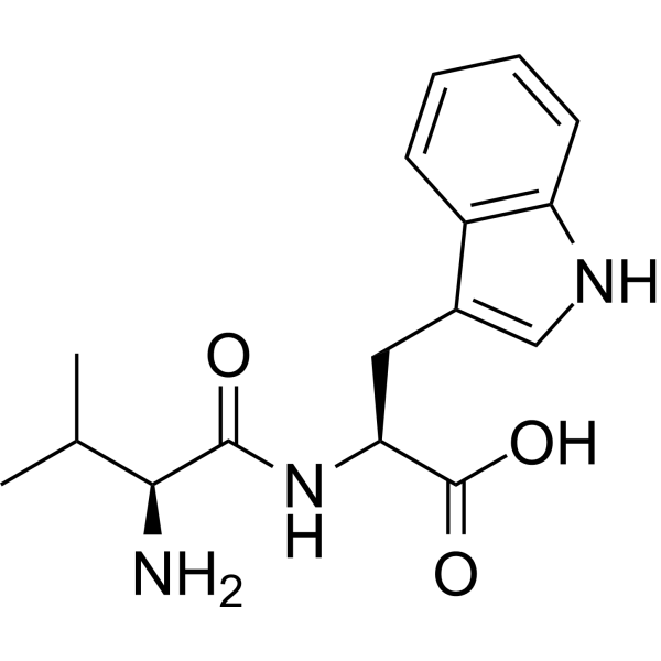
Dipeptide 2 (N-Valyltryptophan) is an orally active, competitive angiotensin-converting enzyme (ACE) inhibitory peptide with an IC50 of 10.50 μM. Dipeptide 2 reduces intracellular Calcium ions. Dipeptide 2 significantly increases the content of NO, inhibits the production of ET-1, and induces the phosphorylation of eNOS. Dipeptide 2 decreases the systolic and diastolic blood pressure of spontaneously hypertensive rats.
For research use only. We do not sell to patients.
- Purity : 98.12%
- CAS No.: 24587-37-9
- Formula: C16H21N3O3
- Molecular Weight:303.36
-
Storage:Powder -20°C, 3 years , 4°C, 2 years ; In solvent -80°C, 6 months , -20°C, 1 month

To place orders, for customer services and technical support, please contact: MedChemExpress USA
Tel: 609-228-6898 E-mail: [email protected] [email protected]


-
 Biological Activity
-
Chemical Information
-
Solvent & Solubility
- Protocols
- Purity & Documentation
- References
-
 Help & FAQs


-
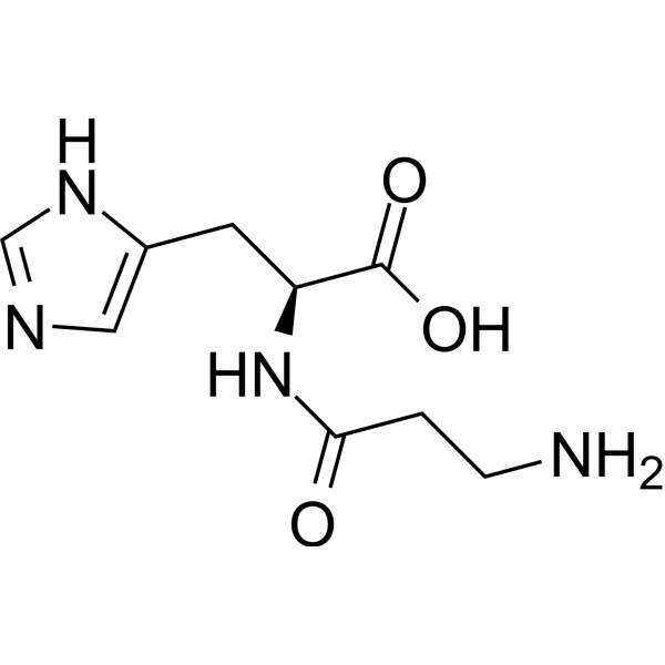
HY-W013494
L-CarnosineFree Radical Scavenger -

HY-W013494R
L-Carnosine (Standard) -
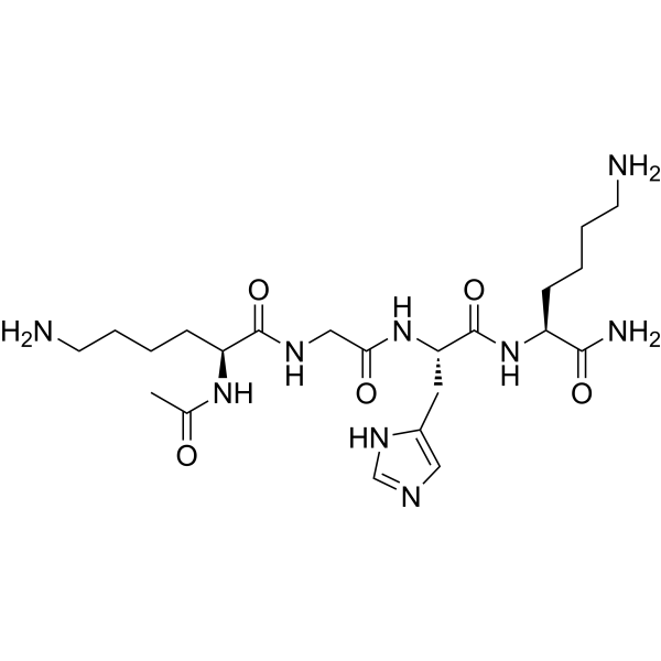
HY-W129161
Acetyl tetrapeptide-3ECM Protein Stimulant -
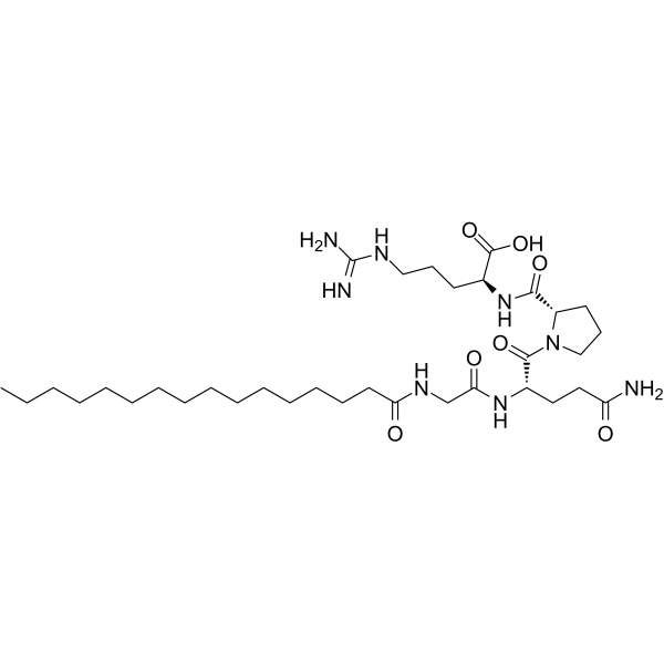
HY-P0064
Palmitoyl Tetrapeptide-3IL-6 Inhibitor -
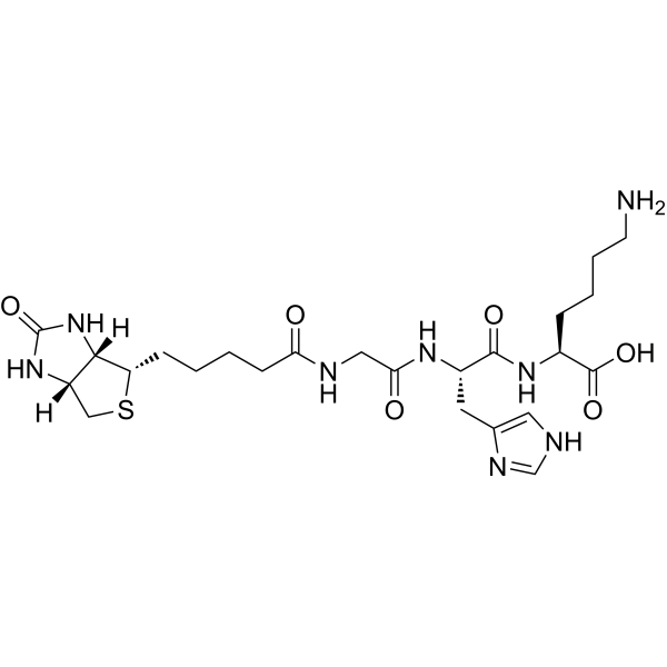
HY-125616
Biotinoyl tripeptide-1Peptide -

HY-118060R
Dipeptide 2 (Standard)Anti-aging Peptide -
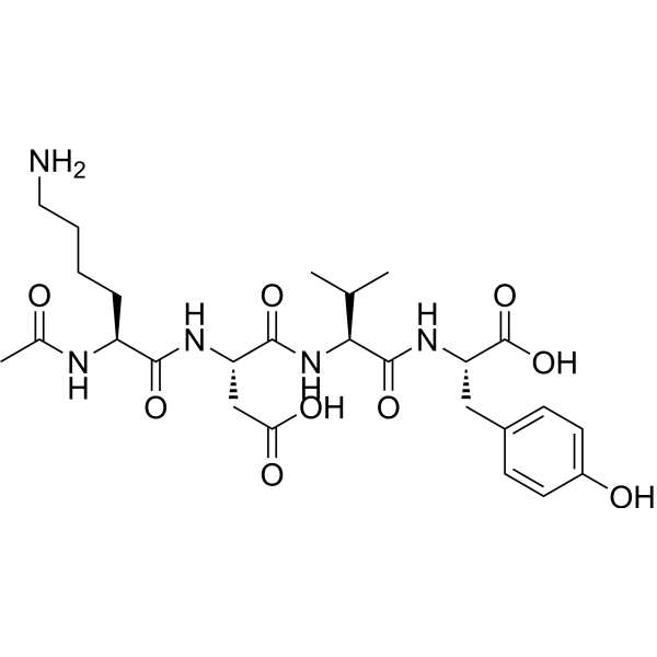
HY-P2663
Acetyl tetrapeptide-2Anti-aging Peptide -
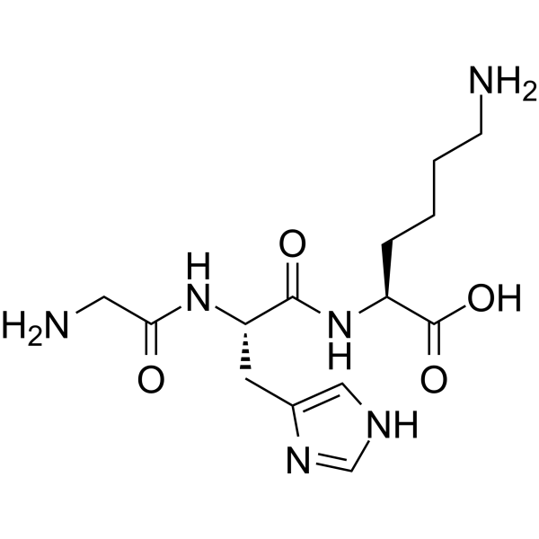
HY-P0046
Glycyl-L-histidyl-L-lysineTripeptide -
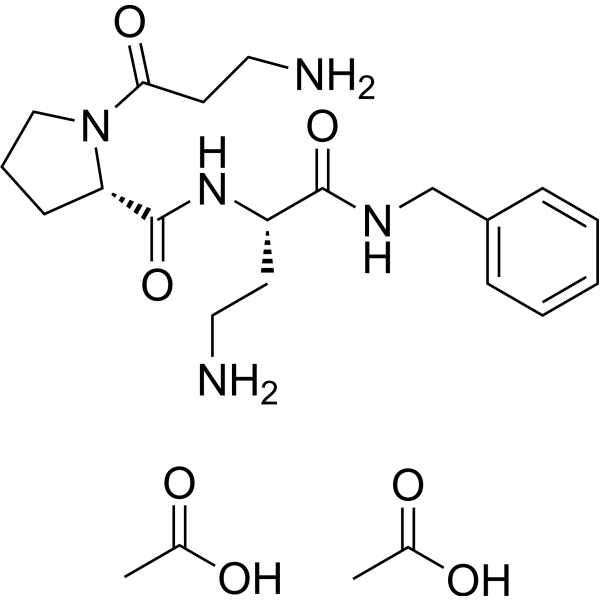
HY-P0102
Dipeptide diaminobutyroyl benzylamide diacetatenAChR Antagonist -
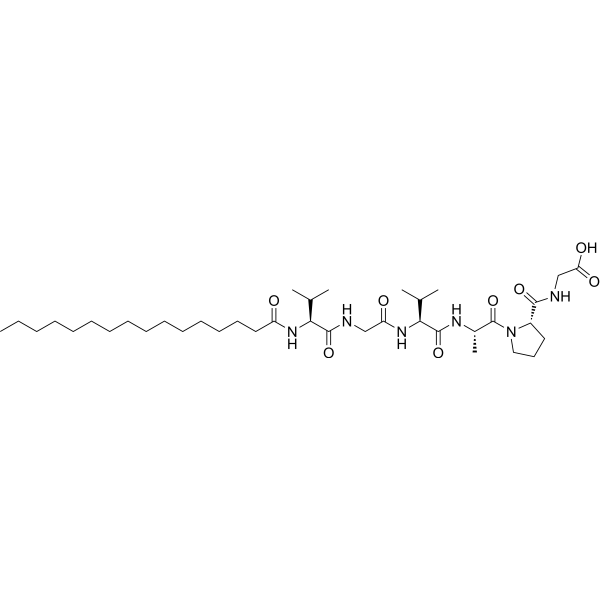
HY-66015
Palmitoyl hexapeptide-12Anti-aging Peptide -
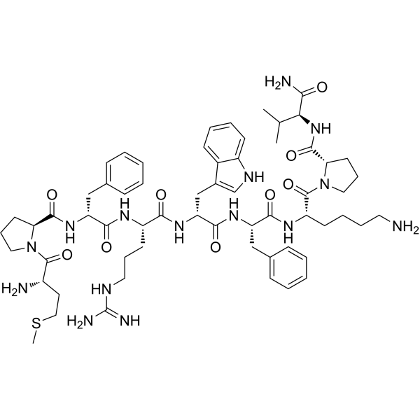
HY-P0097
Nonapeptide-1Melanocortin Receptor Antagonist -
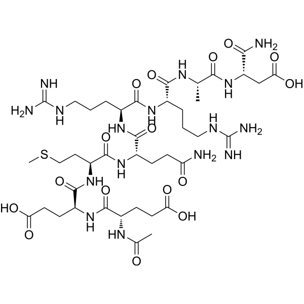
HY-P0065
Acetyl octapeptide-1SNAP-25 Inhibitor
All Calcium Channel Isoforms
More

Biological Activity
Description
IC50 & Target
[1]|
eNOS |
Cellular Effect
|
Cell Line

|
Type | Value | Description | References |
|---|---|---|---|---|
| MDCK | EC50 |
0.04 mM
Compound: 79
|
Activation of human PEPT1 expressed in MDCK cells
Activation of human PEPT1 expressed in MDCK cells
|
[PMID: 16759105] |
| MDCK | IC50 |
0.1 mM
Compound: 79
|
Binding affinity to human PEPT1 assessed as inhibition of [14C]Gly-Sar uptake in MDCK cells
Binding affinity to human PEPT1 assessed as inhibition of [14C]Gly-Sar uptake in MDCK cells
|
[PMID: 16759105] |
In Vitro
Dipeptide 2 potently inhibits purified ACE with an IC50 of 10.50 ± 0.97 μM[2].
Dipeptide 2 (12.5-50 μM; 24 h) is non-toxic to HUVECs[2].
Dipeptide 2 (12.5-50 μM; 4 h) reduces intracellular calcium concentration in Ang I-induced HUVECs, promoting vascular smooth muscle relaxation[2].
Dipeptide 2 (12.5-50 μM; 4 h) promotes NO production in Ang I-induced HUVECs, with a 12.9% increase at 50 μM[2].
MedChemExpress (MCE) has not independently confirmed the accuracy of these methods. They are for reference only. Further protocols information, click here.
-
Cell Line:Ang I-induced human umbilical vein endothelial cells (HUVECs)
-
Concentration:12.5-50 μM
-
Incubation Time:4 h
-
Result:Significantly inhibited ET-1 protein expression and stimulated eNOS phosphorylation compared to the Ang I-only group.
Caused a 60.4% reduction in ET-1 and a 92.9% increase in p-eNOS protein expression in the 50 μM group.
In Vivo
MedChemExpress (MCE) has not independently confirmed the accuracy of these methods. They are for reference only.
-
Animal Model:SHRs (male, 8 weeks old, 220 ± 20 g, spontaneously hypertensive model)[2]
-
Dosage:10 mg/kg; 40 mg/kg
-
Administration:p.o.; once daily; 12 days
-
Result:Significantly suppressed diastolic blood pressure (DBP) in SHRs between days 8 and 12, with DBP reductions ranging from 3 to 27 mmHg.
Produced a maximum DBP reduction of 27 mmHg on day 12 at 40 mg/kg.
Reduced systolic blood pressure (SBP) by 32 mmHg on day 12.


Chemical Information
-
CAS No. 24587-37-9
-
Appearance Solid
-
Molecular Weight 303.36
-
Formula C16H21N3O3
-
Color White to off-white
-
Synonyms
N-Valyltryptophan; Val-Trp
-
Sequence
Val-Trp
-
Sequence Shortening
VW
-
Shipping
Room temperature in continental US; may vary elsewhere.
-
Storage
Powder -20°C 3 years 4°C 2 years In solvent -80°C 6 months -20°C 1 month


Solvent & Solubility
In Vitro:
DMSO : 12.5 mg/mL (41.21 mM; Need ultrasonic; Hygroscopic DMSO has a significant impact on the solubility of product, please use newly opened DMSO)
Please refer to the solubility information to select the appropriate solvent. Once prepared, please aliquot and store the solution to prevent product inactivation from repeated freeze-thaw cycles.
Storage method and period of stock solution: -80°C, 6 months; -20°C, 1 month. When stored at -80°C, please use it within 6 months. When stored at -20°C, please use it within 1 month.
Please refer to the solubility information to select the appropriate solvent. Once prepared, please aliquot and store the solution to prevent product inactivation from repeated freeze-thaw cycles.
Storage method and period of stock solution: -80°C, 6 months; -20°C, 1 month. When stored at -80°C, please use it within 6 months. When stored at -20°C, please use it within 1 month.
Concentration (start) × Volume (start) = Concentration (final) × Volume (final)

Protocols
-
Kinase activity and phosphorylation assays
Kinase activity assays measure the ability of kinases to transfer phosphate groups from ATP to specific substrates, while phosphorylation assays detect the presence and levels of phosphorylated proteins. Common methods include radiolabeled ATP incorporation (e. g. ,), ADP release detection via bioluminescence (e. g. ,[3]), enzyme-linked immunosorbent assays (ELISA) for phospho-specific epitopes (e. g. ,[6]), and microtiter-based formats for high-throughput screening (e. g. ,[8]). The ADP-Glo assay quantifies kinase activity by measuring ADP produced during phosphorylation using a luciferase-based system. Radiometric assays involve autoradiography or scintillation counting after incorporation of 32P-labeled ATP into substrate proteins. ELISA-based approaches rely on phospho-specific antibodies to detect activated kinases in cell lysates or purified samples.
-
Western Blot
Western blotting (WB) is a commonly used experimental method in molecular biology, biochemistry, and immunogenetics for identifying and quantifying target proteins. It combines gel electrophoresis with immunoassay, enabling researchers to analyze protein expression, post-translational modifications, and molecular weight.
-
Cell Cytotoxicity Assay
Cytotoxicity assays are usually based on the assessment of cell membrane damage, which can also be indirectly detected by measuring cell viability. Detection methods include MTT assay, CKK-8 assay, LDH assay and ATP assay, etc.
-
Calcium Spark Assay
Calcium sparks are localized, transient increases in intracellular calcium concentration ([Ca2+]i) that occur in cardiac myocytes and represent elementary events underlying excitation-contraction coupling. These events are generated by the coordinated opening of clusters of ryanodine receptors (RyRs) on the sarcoplasmic reticulum membrane, leading to a brief release of Ca2+ into the cytosol. The detection and analysis of calcium sparks provide insights into the mechanisms of calcium handling and signaling in cardiac cells. Imaging techniques using fluorescent calcium indicators such as Fluo-3 are employed to visualize these subcellular calcium transients with high spatial and temporal resolution. The protocol is based on established methodologies described in primary literature for both experimental measurement and automated analysis of calcium sparks.
-
Ca2+ Staining Technique
Ca2+ staining is an experimental technique that utilizes specific fluorescent probes (such as Fluo-4 AM, Fura-2, etc.) to qualitatively or quantitatively detect dynamic changes in intracellular Ca2+ concentrations; this is achieved by monitoring the changes in fluorescent signals generated when these probes bind to free intracellular calcium ions. The underlying principle relies primarily on the presence of chelating groups within the probe's molecular structure that possess high affinity for calcium ions.
-
Protocol for Kinase activity and phosphorylation assays
Kinase activity assays measure transfer of phosphate from ATP to a protein or peptide substrate, generating phosphorylated substrate, ADP, or incorporated radiolabeled phosphate as the readout; phosphorylation assays measure site-specific phosphorylation in cells or tissues as a proxy for kinase-pathway activation, inhibition, or substrate regulation. Phosphorylation can be detected by phospho-specific Western blot, immunoprecipitation kinase assay, phospho-immunofluorescence, phospho-flow cytometry, luminescent ADP detection, radiolabeled ATP incorporation, or reporter-based pathway assays, and these readouts can be applied to cancer cells, primary neurons, mouse tumors, organoids, inflammatory macrophages, ferroptosis studies, and mitophagy studies when the kinase target is biologically relevant.


Purity & Documentation
-
Data Sheet (279 KB)
-
SDS (393 KB)
- English - EN (393 KB)
- Français - FR (393 KB)
- Deutsch - DE (393 KB)
- Norwegian - NO (393 KB)
- Español - ES (393 KB)
- Swedish - SV (393 KB)
- Italian - IT (393 KB)
- Korean - KR (393 KB)
- Portuguese - PT (393 KB)
-
Handling Instructions (2659 KB)
References


Complete Stock Solution Preparation Table
Please refer to the solubility information to select the appropriate solvent. Once prepared, please aliquot and store the solution to prevent product inactivation from repeated freeze-thaw cycles.
Storage method and period of stock solution: -80°C, 6 months; -20°C, 1 month. When stored at -80°C, please use it within 6 months. When stored at -20°C, please use it within 1 month.
| Optional Solvent | Concentration Solvent Mass | 1 mg | 5 mg | 10 mg | 25 mg |
|---|---|---|---|---|---|
| DMSO | 1 mM | 3.2964 mL | 16.4821 mL | 32.9641 mL | 82.4103 mL |
| 5 mM | 0.6593 mL | 3.2964 mL | 6.5928 mL | 16.4821 mL | |
| 10 mM | 0.3296 mL | 1.6482 mL | 3.2964 mL | 8.2410 mL | |
| 15 mM | 0.2198 mL | 1.0988 mL | 2.1976 mL | 5.4940 mL | |
| 20 mM | 0.1648 mL | 0.8241 mL | 1.6482 mL | 4.1205 mL | |
| 25 mM | 0.1319 mL | 0.6593 mL | 1.3186 mL | 3.2964 mL | |
| 30 mM | 0.1099 mL | 0.5494 mL | 1.0988 mL | 2.7470 mL | |
| 40 mM | 0.0824 mL | 0.4121 mL | 0.8241 mL | 2.0603 mL |


Dipeptide 2 Related Classifications
HY-118060 Related Classifications
 Powered by Bioz
Keywords