



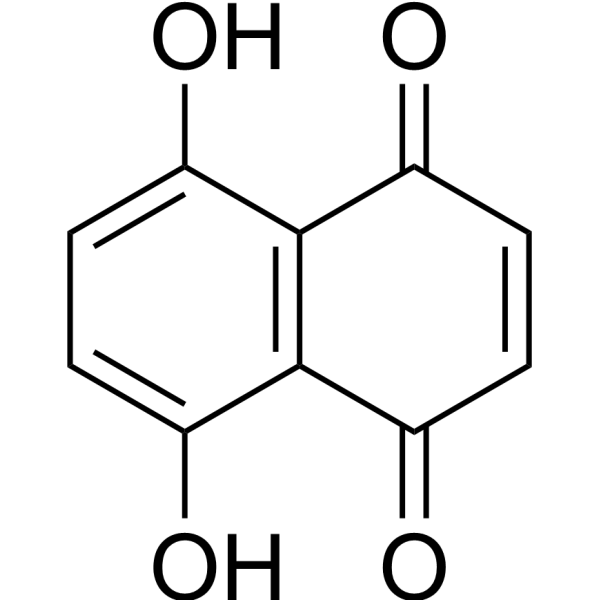
Naphthazarin
Based on 1 publication(s) in Google Scholar
Naphthazarin (DHNQ) is a microtubule depolymerizing agent. Naphthazarin can improve motor function and reduce neuroinflammation in mouse models of Parkinson's disease. Naphthazarin can induce tumor cell apoptosis, autophagy, and cell cycle arrest. Naphthazarin can also induce erythrocyte apoptosis. Naphthazarin can be used in the research of tumors and neurodegenerative diseases.
商品は「研究用試薬」です。人や動物の医療用・臨床診断用・食品用の製品ではありません。
研究用途以外に使用した場合、当社は一切の責任を負いかねます。
- 純度 : 99.59%
- CAS 番号: 475-38-7
- 分子式: C10H6O4
- 分子量:190.15
-
保管条件:Powder -20°C, 3 years , 4°C, 2 years ; In solvent -80°C, 6 months , -20°C, 1 month

To place orders, for customer services and technical support, please contact: MedChemExpress USA
Tel: 609-228-6898 E-mail: [email protected] [email protected]


-
 生物活性
生物活性
-
化学情報
-
溶剤 & 溶解度
- プロトコル
- 純度とドキュメンテーション
- 参考文献
-
 Help & FAQs


-
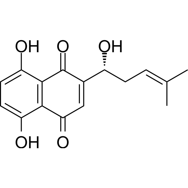
HY-N0822
ShikoninChloride Channel Inhibitor -
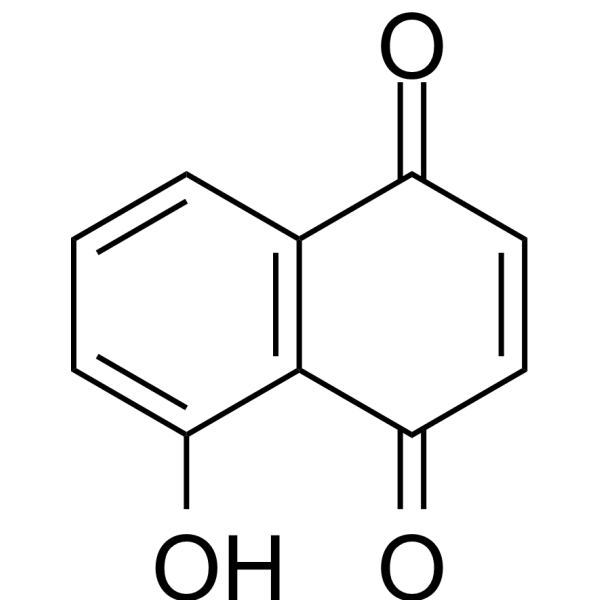
HY-N6949
JugloneAntimicrobial Drug -
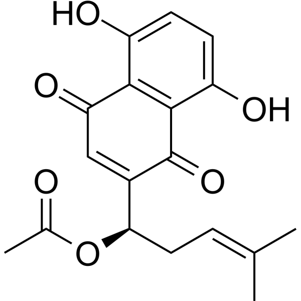
HY-N2181
AcetylshikoninAnti-inflammatory agent, AChE Inhibitor, P450 Inhibitor, Neuroprotective agents -
HY-N6012
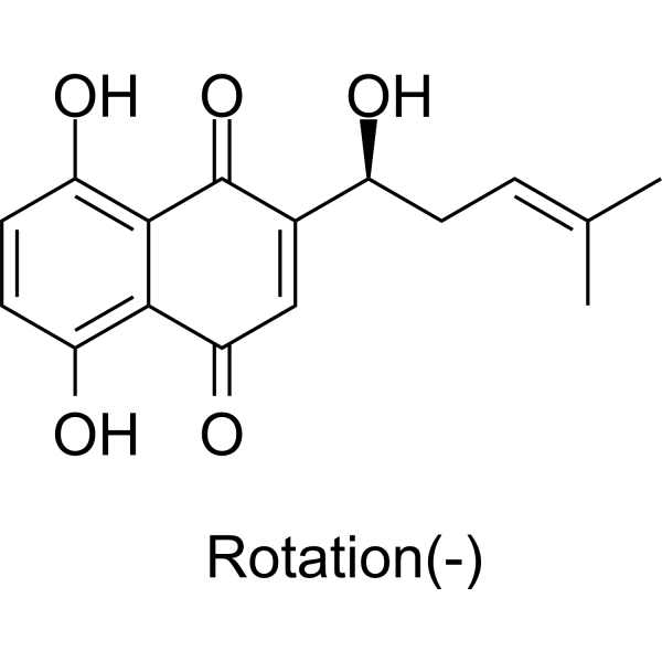
(-)-AlkanninApoptosis Inducer -
HY-N1451
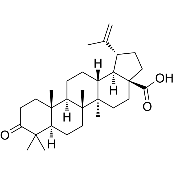
Betulonic acidParasite Inhibitor -

HY-N7526R
Naphthazarin (Standard)Apoptosis Inducer -
HY-N7610
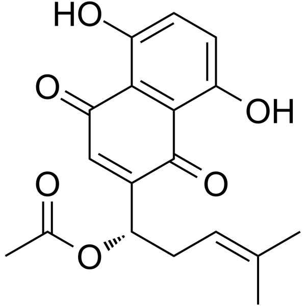
AcetylalkanninBacterial Inhibitor
MedChemExpress(MCE)の使用を引用している文献 Naphthazarin
More

生物活性
製品説明
Cellular Effect
|
Cell Line

|
Type | Value | Description | References |
|---|---|---|---|---|
| A2780 | IC50 |
0.08 μM
Compound: 15
|
Antiproliferative activity against human A2780 cells after 2 days by alamar-blue assay
Antiproliferative activity against human A2780 cells after 2 days by alamar-blue assay
|
[PMID: 19028102] |
| A-375 | IC50 |
8.85 μM
Compound: SK-6
|
Antiproliferative activity against human A-375 cells assessed as inhibition of cell proliferation measured after 3 days by MTT assay
Antiproliferative activity against human A-375 cells assessed as inhibition of cell proliferation measured after 3 days by MTT assay
|
[PMID: 39129245] |
| BT-549 | GI50 |
0.43 μM
Compound: 1
|
Antiproliferative activity against human BT-549 cells measured after 72 hrs by SRB assay
Antiproliferative activity against human BT-549 cells measured after 72 hrs by SRB assay
|
[PMID: 35687347] |
| CAL-51 | GI50 |
0.56 μM
Compound: 1
|
Antiproliferative activity against human CAL-51 cells measured after 72 hrs by SRB assay
Antiproliferative activity against human CAL-51 cells measured after 72 hrs by SRB assay
|
[PMID: 35687347] |
| HaCaT | IC50 |
0.7 μM
Compound: 7e ; Naphthazarin
|
Antiproliferative (inhibition of cell growth) activity against HaCaT cells (human keratinocyte line)
Antiproliferative (inhibition of cell growth) activity against HaCaT cells (human keratinocyte line)
|
[PMID: 9371243] |
| HCC1954 | GI50 |
0.49 μM
Compound: 1
|
Antiproliferative activity against human HCC1954 cells measured after 72 hrs by SRB assay
Antiproliferative activity against human HCC1954 cells measured after 72 hrs by SRB assay
|
[PMID: 35687347] |
| HCT-116 | GI50 |
0.16 μM
Compound: 1
|
Antiproliferative activity against human HCT-116 cells measured after 72 hrs by SRB assay
Antiproliferative activity against human HCT-116 cells measured after 72 hrs by SRB assay
|
[PMID: 35687347] |
| HCT-116 | IC50 |
1.04 μM
Compound: SK-6
|
Antiproliferative activity against human HCT-116 cells assessed as inhibition of cell proliferation measured after 3 days by MTT assay
Antiproliferative activity against human HCT-116 cells assessed as inhibition of cell proliferation measured after 3 days by MTT assay
|
[PMID: 39129245] |
| HeLa | IC50 |
9.37 μM
Compound: 15
|
Inhibition of histidine-tagged mouse MKP1 catalytic domain expressed in human Hela cells
Inhibition of histidine-tagged mouse MKP1 catalytic domain expressed in human Hela cells
|
[PMID: 19028102] |
| HepG2 | IC50 |
4.97 μM
Compound: SK-6
|
Antiproliferative activity against human HepG2 cells assessed as inhibition of cell proliferation measured after 3 days by MTT assay
Antiproliferative activity against human HepG2 cells assessed as inhibition of cell proliferation measured after 3 days by MTT assay
|
[PMID: 39129245] |
| HL-60 | IC50 |
0.8 μM
Compound: 2
|
Cytotoxicity in human HL60 cells assessed as reduction in cell viability incubated for 72 hrs by MTT assay
Cytotoxicity in human HL60 cells assessed as reduction in cell viability incubated for 72 hrs by MTT assay
|
[PMID: 28987605] |
| HL-60 | IC50 |
0.93 μM
Compound: 2
|
Cytotoxicity in human HL60 cells assessed as reduction in cell viability incubated for 48 hrs by MTT assay
Cytotoxicity in human HL60 cells assessed as reduction in cell viability incubated for 48 hrs by MTT assay
|
[PMID: 28987605] |
| K562 | GI50 |
1.7 μM
Compound: 1
|
Antiproliferative activity against human K562 cells measured after 72 hrs by SRB assay
Antiproliferative activity against human K562 cells measured after 72 hrs by SRB assay
|
[PMID: 35687347] |
| MCF7 | GI50 |
0.43 μM
Compound: 1
|
Antiproliferative activity against human MCF7 cells measured after 72 hrs by SRB assay
Antiproliferative activity against human MCF7 cells measured after 72 hrs by SRB assay
|
[PMID: 35687347] |
| MOLM-13 | GI50 |
0.28 μM
Compound: 1
|
Antiproliferative activity against human MOLM-13 cells measured after 72 hrs by SRB assay
Antiproliferative activity against human MOLM-13 cells measured after 72 hrs by SRB assay
|
[PMID: 35687347] |
| MV4-11 | GI50 |
0.19 μM
Compound: 1
|
Antiproliferative activity against human MV4-11 cells measured after 72 hrs by SRB assay
Antiproliferative activity against human MV4-11 cells measured after 72 hrs by SRB assay
|
[PMID: 35687347] |
体外実験
Naphthazarin (0-10 μM; 24 h) can stimulate apoptosis in human red blood cells by increasing oxidative stress and surface ceramide abundance[1].
Naphthazarin (0-80 μM; 24 h) can inhibit the cell viability, induce cell apoptosis, autophagy and cell cycle arrest, and suppress the PI3K/Akt pathway in A549 cells[2].
Naphthazarin (10-1000 nM; 30 h) can alleviate MPP+-induced activation in primary cultured rat astrocytes[3].
MedChemExpress (MCE) has not independently confirmed the accuracy of these methods. They are for reference only.
-
Cell Line:Primary cultured astrocyte treated MPP+
-
Concentration:10 nM, 100 nM and 1 μM
-
Incubation Time:Pretreated with 6 h, then co-incubation for 24 h
-
Result:Inhibited the levels of GFAP.
体内実験
MedChemExpress (MCE) has not independently confirmed the accuracy of these methods. They are for reference only.
-
Animal Model:Male C57BL/C mice aged 7-8 weeks old weighing approximately 20-23 g) treated MPTP (HY-W114750) to induce Parkinson's disease model[3]
-
Dosage:0.1 mg/kg and 1 mg/kg
-
Administration:Intraperitoneal injection; 5 days
-
Result:Improved the motor function of the mice at 1 mg/kg
Protected dopaminergic neurons and inhibited neuroinflammation in the mice at 0.1 mg/kg and 1 mg/kg.
Suppressed MPTP-induced glial activation at 0.1 mg/kg and 1 mg/kg.
- Detection of Apoptosis
- TUNEL staining for apoptotic DNA fragmentation
- Flow cytometric DNA-content cell-cycle staining
- Annexin V plus membrane-impermeant dye apoptosis staining
- BrdU Incorporation Assay
- Human pluripotent stem cell midbrain dopaminergic neuron differentiation
- Autophagy
- Apoptosis Solutions
- Protocol for Cell Cycle
- Lysosome and acidic-vesicle live-cell staining
- Research Protocol for Neurological Diseases
- Macroautophagy Solutions


化学情報
-
CAS 番号 475-38-7
-
性状 Solid
-
分子量 190.15
-
分子式 C10H6O4
-
Color Brown to black
-
SMILES
O=C1C=CC(C2=C1C(O)=CC=C2O)=O
-
別名
DHNQ; 5,8-Dihydroxy-1,4-naphthoquinone
-
Structure Classification
-
Initial Source
-
輸送条件
Room temperature in continental US; may vary elsewhere.
-
保管条件
Powder -20°C 3 years 4°C 2 years In solvent -80°C 6 months -20°C 1 month


Publications (1)
-
Journal Impact Factor
-
Most Recent
-
Int J Biol Macromol
Identification of Vitamin K3 and its analogues as covalent inhibitors of SARS-CoV-2 3CLpro. [Abstract]2021 Jul 31:183:182-192. PMID: 33901557


溶剤 & 溶解度
体外:
DMSO : 10 mg/mL (52.59 mM; Need ultrasonic; Hygroscopic DMSO has a significant impact on the solubility of product, please use newly opened DMSO)
Please refer to the solubility information to select the appropriate solvent. Once prepared, please aliquot and store the solution to prevent product inactivation from repeated freeze-thaw cycles.
Storage method and period of stock solution: -80°C, 6 months; -20°C, 1 month. When stored at -80°C, please use it within 6 months. When stored at -20°C, please use it within 1 month.
Please refer to the solubility information to select the appropriate solvent. Once prepared, please aliquot and store the solution to prevent product inactivation from repeated freeze-thaw cycles.
Storage method and period of stock solution: -80°C, 6 months; -20°C, 1 month. When stored at -80°C, please use it within 6 months. When stored at -20°C, please use it within 1 month.
濃度 (開始) × 体積 (開始) = 濃度 (終了) × 体積 (終了)
体内:
Select the appropriate dissolution method based on your experimental animal and administration route.
- For the following dissolution methods, please ensure to first prepare a clear stock solution using an In Vitro approach and then sequentially add co-solvents:
- To ensure reliable experimental results, the clarified stock solution can be appropriately stored based on storage conditions. As for the working solution for In Vivo experiments, it is recommended to prepare freshly and use it on the same day.
- The percentages shown for the solvents indicate their volumetric ratio in the final prepared solution. If precipitation or phase separation occurs during preparation, heat and/or sonication can be used to aid dissolution.
Add each solvent one by one: 10% DMSO 90% (20% SBE-β-CD in Saline)
Solubility: 0.71 mg/mL (3.73 mM); Suspended solution; Need ultrasonic
This protocol yields a suspended solution of 0.71 mg/mL. Suspended solution can be used for oral and intraperitoneal injection.
Taking 1 mL working solution as an example, add 100 μL DMSO stock solution (7.1 mg/mL) to 900 μL 20% SBE-β-CD in Saline, and mix evenly.
Preparation of 20% SBE-β-CD in Saline (4°C, storage for one week): 2 g SBE-β-CD powder is dissolved in 10 mL Saline, completely dissolve until clear.
In Vivo Dissolution Calculator
Please enter the basic information of animal experiments:
-
-
-
-
Recommended: Prepare an additional quantity of animals to account for potential losses during experiments.
Please enter your animal formula composition:
-
%DMSO +
Recommended: Keep the proportion of DMSO in working solution below 2% if your animal is weak.
-
%+
-
+%Tween-80 + +
-
%Saline +
The co-solvents required include: DMSO, . All of co-solvents are available by MedChemExpress (MCE). , Tween 80. All of co-solvents are available by MedChemExpress (MCE).
Working solution concentration: 0.22 mg/mL
Method for preparing stock solution: mg drug dissolved in μL DMSO. Stock solution concentration: mg/mL.
1. Take μL DMSO stock solution;
2. Add μL .
μL , mix evenly;
3. Then add μL Tween 80, mix evenly;
4. Then add μL
Please ensure that the stock solution in the first step is dissolved to a clear state, and add co-solvents in sequence. You can use ultrasonic heating (ultrasonic cleaner, recommended frequency 20-40 kHz), vortexing, etc. to assist dissolution.


純度とドキュメンテーション
-
データシート (282 KB)
-
SDS (393 KB)
- English - EN (393 KB)
- Français - FR (393 KB)
- Deutsch - DE (393 KB)
- Norwegian - NO (393 KB)
- Español - ES (393 KB)
- Swedish - SV (393 KB)
- Italian - IT (393 KB)
- Korean - KR (393 KB)
- Portuguese - PT (393 KB)
-
取扱説明書 (2659 KB)
参考文献
[1]. Omar Aljanadi, et al. Stimulation of Suicidal Erythrocyte Death by Naphthazarin. Basic Clin Pharmacol Toxicol. 2015 Dec;117(6):369-74. [Content Brief]
[2]. Acharya BR, et al. The microtubule depolymerizing agent naphthazarin induces both apoptosis and autophagy in A549 lung cancer cells. Apoptosis. 2011 Sep;16(9):924-39. doi: 10.1007/s10495-011-0613-1 [Content Brief]
[3]. Seon Young Choi, et al. Naphthazarin has a protective effect on the 1-methyl-4-phenyl-1,2,3,4-tetrahydropyridine-induced Parkinson's disease model. J Neurosci Res. 2012 Sep;90(9):1842-9. [Content Brief]


Complete Stock Solution Preparation Table
Please refer to the solubility information to select the appropriate solvent. Once prepared, please aliquot and store the solution to prevent product inactivation from repeated freeze-thaw cycles.
Storage method and period of stock solution: -80°C, 6 months; -20°C, 1 month. When stored at -80°C, please use it within 6 months. When stored at -20°C, please use it within 1 month.
| Optional Solvent | Concentration Solvent Mass | 1 mg | 5 mg | 10 mg | 25 mg |
|---|---|---|---|---|---|
| DMSO | 1 mM | 5.2590 mL | 26.2950 mL | 52.5901 mL | 131.4752 mL |
| 5 mM | 1.0518 mL | 5.2590 mL | 10.5180 mL | 26.2950 mL | |
| 10 mM | 0.5259 mL | 2.6295 mL | 5.2590 mL | 13.1475 mL | |
| 15 mM | 0.3506 mL | 1.7530 mL | 3.5060 mL | 8.7650 mL | |
| 20 mM | 0.2630 mL | 1.3148 mL | 2.6295 mL | 6.5738 mL | |
| 25 mM | 0.2104 mL | 1.0518 mL | 2.1036 mL | 5.2590 mL | |
| 30 mM | 0.1753 mL | 0.8765 mL | 1.7530 mL | 4.3825 mL | |
| 40 mM | 0.1315 mL | 0.6574 mL | 1.3148 mL | 3.2869 mL | |
| 50 mM | 0.1052 mL | 0.5259 mL | 1.0518 mL | 2.6295 mL |


Naphthazarin 関連分類
HY-N7526 関連分類
 Powered by Bioz