



Orcinol glucoside
Based on 1 publication(s) in Google Scholar
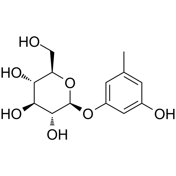
Orcinol glucoside is an orally active, blood-brain barrier permeable osteoblast proliferation promoter that targets the Nrf2/Keap1, mTOR and p38 signaling pathways. Orcinol glucoside promotes Nrf2 nuclear translocation, upregulates antioxidant enzyme levels, enhances the phosphorylation of mTOR and p70S6K, and inhibits the enzymatic activity of HAS2 as well as the nuclear translocation of GR. Orcinol glucoside also alleviates oxidative stress, inhibits autophagic flux, osteoclastogenesis and TGF-β1-induced M2 polarization, while reducing collagen deposition and effectively promoting the proliferation, differentiation and mineralization of osteoblasts. Orcinol glucoside also exhibits anti-pulmonary fibrosis, anxiolytic and antidepressant activities. Orcinol glucoside can be used in the research of senile and glucocorticoid-induced osteoporosis, idiopathic pulmonary fibrosis (IPF), anxiety and other related diseases.
For research use only. We do not sell to patients.
- Purity : 98.92%
- CAS No.: 21082-33-7
- Formula: C13H18O7
- Molecular Weight:286.28
-
Storage:
4°C, protect from light
* In solvent : -80°C, 6 months; -20°C, 1 month (protect from light)

To place orders, for customer services and technical support, please contact: MedChemExpress USA
Tel: 609-228-6898 E-mail: [email protected] [email protected]


-
 Biological Activity
-
Chemical Information
-
Solvent & Solubility
- Protocols
- Purity & Documentation
- References
-
 Help & FAQs


-
HY-N4123
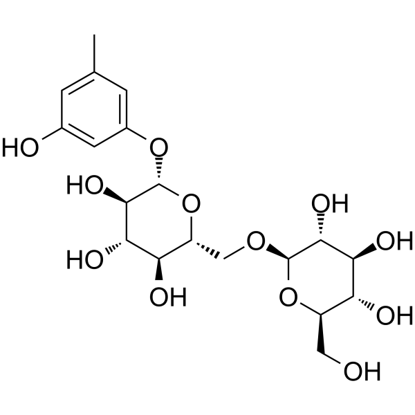
Orcinol gentiobioside -

HY-N0008R
Orcinol glucoside (Standard)Antidepressant -
HY-13764
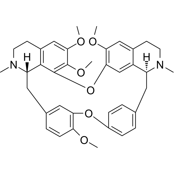
TetrandrineCalcium Channel Inhibitor -
HY-13076
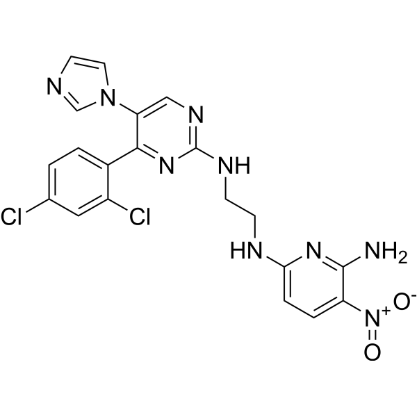
CHIR-98014GSK-3 Inhibitor -
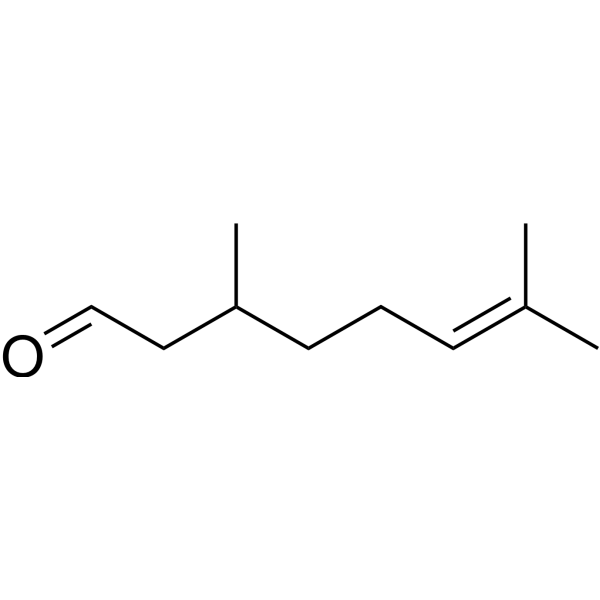
HY-N7126
CitronellalMonoterpenea -
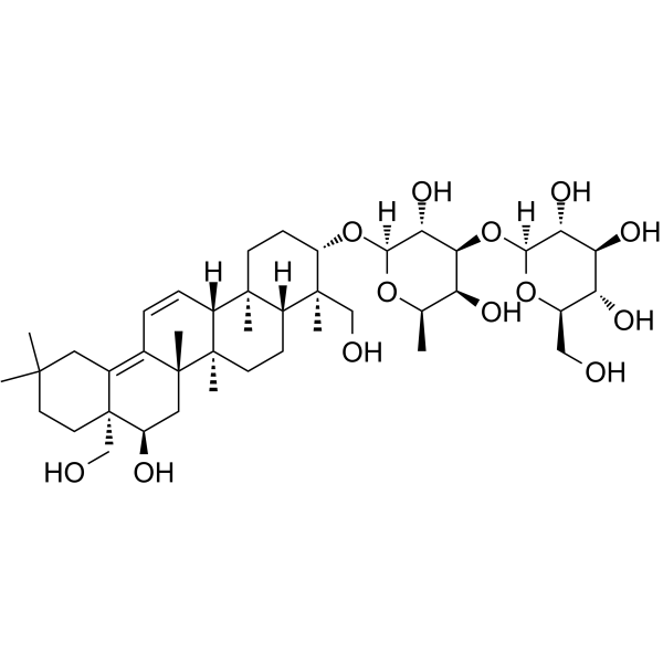
HY-N0248
Saikosaponin B2Solute Carrier Modulator, ATP-Binding Cassette Transporter Modulator -
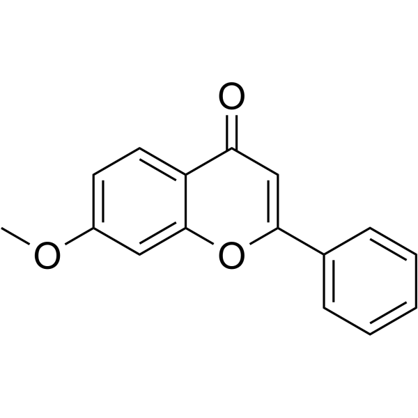
HY-N8487
7-MethoxyflavonePeripheral Analgesic -
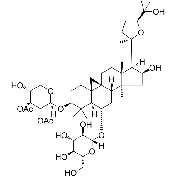
HY-N0432
Astragaloside IWnt/β-catenin Activator -

HY-13764R
Tetrandrine (Standard)Calcium Channel Inhibitor -

HY-N7126R
Citronellal (Standard)Monoterpenea -

HY-N8487R
7-Methoxyflavone (Standard)Peripheral Analgesic -

HY-N0248R
Saikosaponin B2 (Standard)HCV Inhibitor
Publications Citing Use of MedChemExpress (MCE) Orcinol glucoside
More

Biological Activity
Description
In Vitro
Orcinol glucoside (1-10 μM; 72 h) does not reduce the viability of H2O2-exposed RAW264.7 cells[1].
Orcinol glucoside (1-10 μM; 72 h) dose-dependently inhibits the differentiation and TRAP activity of RANKL- and H2O2-induced RAW264.7 osteoclasts[1].
Orcinol glucoside (5-10 μM; 48 h pre-incubation with RANKL before 4 h H2O2 exposure) activates the mTOR pathway in RANKL- and H2O2-induced RAW264.7 osteoclasts, increasing phosphorylation of mTOR and p70S6K[1].
Orcinol glucoside (10-100 nM; 72 h) promotes proliferation of primary mouse osteoblast cells in a dose-dependent manner at concentrations of 10, 50, and 100 nM over 72 h[2].
Orcinol glucoside (10-100 nM) upregulates p38 phosphorylation and the expression of osteogenic-related proteins (Collagen I, Runx2, Osx, Dlx5) in primary mouse osteoblast cells in vitro in a dose-dependent manner at concentrations of 10, 50, and 100 nM[2].
Orcinol glucoside (0-100 µM; 48 h) is non-toxic to NIH/3T3 mouse fibroblasts and HFL-1 human fibroblasts at concentrations ranging from 0 to 100 µM after 48 h incubation[3].
MedChemExpress (MCE) has not independently confirmed the accuracy of these methods. They are for reference only.
-
Cell Line:murine macrophage RAW264.7 cells
-
Concentration:1-10 μM
-
Incubation Time:72 h
-
Result:Exerted no cytotoxic effects on RAW264.7 cells, with cell viability remaining comparable to control groups.
-
Cell Line:RANKL- and H2O2-induced RAW264.7 cells
-
Concentration:1-10 μM
-
Incubation Time:72 h
-
Result:Decreased the number of TRAP-positive multinucleated osteoclasts and reduced TRAP activity in a dose-dependent manner compared to the H2O2-only treated group.
In Vivo
Orcinol glucoside (5-20 mg/kg, intragastric administration; daily dosing; for 8 consecutive weeks) dose-dependently increases BMD, BV/TV, Tb.Th and Tb.N in dexamethasone-induced osteoporotic mice by activating the p38 signaling pathway and upregulating downstream osteogenic proteins[2].
Orcinol glucoside (5 mg/kg, intragastric administration, once daily for 5 consecutive weeks) improves the bone microstructure and increases osteogenic biomarkers in dexamethasone-induced osteoporotic mice by activating p38, while p38 inhibitors block these effects[2].
Orcinol glucoside (25-100 mg/kg; p.o.; once daily; for 14 consecutive days) dose-dependently alleviates bleomycin-induced pulmonary fibrosis in male C57BL/6J mice by inhibiting the expression of fibrosis markers, HA accumulation, and TGF-β1 production, with an exposure-response relationship observed across different administration doses[3].
Orcinol glucoside (5-20 mg/kg; p.o.; single administration) exhibits anxiolytic activity in mice without inducing sedative effects[4].
MedChemExpress (MCE) has not independently confirmed the accuracy of these methods. They are for reference only.
-
Animal Model:C57BL/6 (male, 8 weeks old, dexamethasone-induced osteoporosis)[2]
-
Dosage:5 mg/kg; 10 mg/kg; 20 mg/kg
-
Administration:i.g.; daily; 8 weeks
-
Result:Increased bone mineral density (BMD), trabecular bone volume fraction (BV/TV), trabecular thickness (Tb.Th), and trabecular number (Tb.N) in a dose-dependent manner compared to the dexamethasone-only group.
Reversed dexamethasone-induced reductions in serum osteoblast-associated markers type I procollagen amino-terminal extension peptide (PINP) and alkaline phosphatase (ALP).
Increased femoral tissue expression of p38, phosphorylated p38 (p-p38), Collagen I, Runx2, Osx, Dlx5, glucocorticoid receptor (GR), and phosphorylated GR Ser226 (p-GR226).
-
Animal Model:CD-1 (male, 18-22 g)[4]
-
Dosage:5 mg/kg; 10 mg/kg; 20 mg/kg
-
Administration:p.o.; single dose
-
Result:Significantly increased the time spent in open arms and number of entries into open arms in the elevated plus-maze test compared to vehicle control.
Significantly increased the number of head-dips in the hole-board test compared to vehicle control (at 5 and 10 mg/kg).
Did not cause significant changes in mouse locomotor counts in the open-field test compared to vehicle control.
- Western Blot
- Collagen: Sirius Red Staining
- ROS/oxidative-stress fluorescent staining
- Connective Tissue: Masson's Trichrome/Collagen Trichrome Staining
- Cell Viability Determination by MTT Colorimetric Assay
- Kinase activity and phosphorylation assays
- Fibrosis/Collagen Morphometry
- Cytoplasmic-Nuclear Fractionated Protein Extraction
- Mesenchymal stromal/stem cell osteogenic differentiation
- Research Protocol for Endocrine Diseases
- Autophagy
- Protocol for Kinase activity and phosphorylation assays
- Macroautophagy Solutions
- Osteoclast differentiation from monocyte/macrophage precursors


Chemical Information
-
CAS No. 21082-33-7
-
Appearance Solid
-
Molecular Weight 286.28
-
Formula C13H18O7
-
Color White to off-white
-
SMILES
CC1=CC(O)=CC(O[C@@H]2O[C@@H]([C@@H](O)[C@H](O)[C@H]2O)CO)=C1
-
Structure Classification
-
Shipping
Room temperature in continental US; may vary elsewhere.
-
Storage
4°C, protect from light
* In solvent : -80°C, 6 months; -20°C, 1 month (protect from light)


Publications (1)
-
Journal Impact Factor
-
Most Recent


Solvent & Solubility
In Vitro:
DMSO : 125 mg/mL (436.64 mM; Need ultrasonic; Hygroscopic DMSO has a significant impact on the solubility of product, please use newly opened DMSO)
Please refer to the solubility information to select the appropriate solvent. Once prepared, please aliquot and store the solution to prevent product inactivation from repeated freeze-thaw cycles.
Storage method and period of stock solution: -80°C, 6 months; -20°C, 1 month (protect from light). When stored at -80°C, please use it within 6 months. When stored at -20°C, please use it within 1 month.
Please refer to the solubility information to select the appropriate solvent. Once prepared, please aliquot and store the solution to prevent product inactivation from repeated freeze-thaw cycles.
Storage method and period of stock solution: -80°C, 6 months; -20°C, 1 month (protect from light). When stored at -80°C, please use it within 6 months. When stored at -20°C, please use it within 1 month.
Concentration (start) × Volume (start) = Concentration (final) × Volume (final)
In Vivo:
Select the appropriate dissolution method based on your experimental animal and administration route.
- For the following dissolution methods, please ensure to first prepare a clear stock solution using an In Vitro approach and then sequentially add co-solvents:
- To ensure reliable experimental results, the clarified stock solution can be appropriately stored based on storage conditions. As for the working solution for In Vivo experiments, it is recommended to prepare freshly and use it on the same day.
- The percentages shown for the solvents indicate their volumetric ratio in the final prepared solution. If precipitation or phase separation occurs during preparation, heat and/or sonication can be used to aid dissolution.
Add each solvent one by one: 10% DMSO 40% PEG300 5% Tween-80 45% Saline
Solubility: ≥ 2.08 mg/mL (7.27 mM); Clear solution
This protocol yields a clear solution of ≥ 2.08 mg/mL (saturation unknown).
Taking 1 mL working solution as an example, add 100 μL DMSO stock solution (20.8 mg/mL) to 400 μL PEG300, and mix evenly; then add 50 μL Tween-80 and mix evenly; then add 450 μL Saline to adjust the volume to 1 mL.
Preparation of Saline: Dissolve 0.9 g sodium chloride in ddH₂O and dilute to 100 mL to obtain a clear Saline solution.
Add each solvent one by one: 10% DMSO 90% (20% SBE-β-CD in Saline)
Solubility: ≥ 2.08 mg/mL (7.27 mM); Clear solution
This protocol yields a clear solution of ≥ 2.08 mg/mL (saturation unknown).
Taking 1 mL working solution as an example, add 100 μL DMSO stock solution (20.8 mg/mL) to 900 μL 20% SBE-β-CD in Saline, and mix evenly.
Preparation of 20% SBE-β-CD in Saline (4°C, storage for one week): 2 g SBE-β-CD powder is dissolved in 10 mL Saline, completely dissolve until clear.
In Vivo Dissolution Calculator
Please enter the basic information of animal experiments:
-
-
-
-
Recommended: Prepare an additional quantity of animals to account for potential losses during experiments.
Please enter your animal formula composition:
-
%DMSO +
Recommended: Keep the proportion of DMSO in working solution below 2% if your animal is weak.
-
%+
-
+%Tween-80 + +
-
%Saline +
The co-solvents required include: DMSO, . All of co-solvents are available by MedChemExpress (MCE). , Tween 80. All of co-solvents are available by MedChemExpress (MCE).
Working solution concentration: 0.22 mg/mL
Method for preparing stock solution: mg drug dissolved in μL DMSO. Stock solution concentration: mg/mL. * In solvent : -80°C, 6 months; -20°C, 1 month (protect from light)
1. Take μL DMSO stock solution;
2. Add μL .
μL , mix evenly;
3. Then add μL Tween 80, mix evenly;
4. Then add μL
Please ensure that the stock solution in the first step is dissolved to a clear state, and add co-solvents in sequence. You can use ultrasonic heating (ultrasonic cleaner, recommended frequency 20-40 kHz), vortexing, etc. to assist dissolution.


Purity & Documentation
-
Data Sheet (283 KB)
-
SDS (393 KB)
- English - EN (393 KB)
- Français - FR (393 KB)
- Deutsch - DE (393 KB)
- Norwegian - NO (393 KB)
- Español - ES (393 KB)
- Swedish - SV (393 KB)
- Italian - IT (393 KB)
- Korean - KR (393 KB)
- Portuguese - PT (393 KB)
-
Handling Instructions (2659 KB)
References
[1]. Gong W, et al. Orcinol Glucoside Improves Senile Osteoporosis through Attenuating Oxidative Stress and Autophagy of Osteoclast via Activating Nrf2/Keap1 and mTOR Signaling Pathway. Oxid Med Cell Longev. 2022;2022:5410377. Published 2022 May 9. [Content Brief]
[2]. He XY, et al. Orcinol glucoside targeted p38 as an agonist to promote osteogenesis and protect glucocorticoid-induced osteoporosis. Phytomedicine. 2023;119:154953. [Content Brief]
[3]. Li C, et al. Orcinol glucoside ameliorates pulmonary fibrosis by suppressing hyaluronic acid synthesis and macrophage M2 polarization via targeting hyaluronic acid synthase 2. Int J Mol Med. 2026;57(4):93. [Content Brief]
[4]. Wang X, et al. Anxiolytic effects of orcinol glucoside and orcinol monohydrate in mice. Pharm Biol. 2015;53(6):876-881. [Content Brief]


Complete Stock Solution Preparation Table
Please refer to the solubility information to select the appropriate solvent. Once prepared, please aliquot and store the solution to prevent product inactivation from repeated freeze-thaw cycles.
Storage method and period of stock solution: -80°C, 6 months; -20°C, 1 month (protect from light). When stored at -80°C, please use it within 6 months. When stored at -20°C, please use it within 1 month.
| Optional Solvent | Concentration Solvent Mass | 1 mg | 5 mg | 10 mg | 25 mg |
|---|---|---|---|---|---|
| DMSO | 1 mM | 3.4931 mL | 17.4654 mL | 34.9308 mL | 87.3271 mL |
| 5 mM | 0.6986 mL | 3.4931 mL | 6.9862 mL | 17.4654 mL | |
| 10 mM | 0.3493 mL | 1.7465 mL | 3.4931 mL | 8.7327 mL | |
| 15 mM | 0.2329 mL | 1.1644 mL | 2.3287 mL | 5.8218 mL | |
| 20 mM | 0.1747 mL | 0.8733 mL | 1.7465 mL | 4.3664 mL | |
| 25 mM | 0.1397 mL | 0.6986 mL | 1.3972 mL | 3.4931 mL | |
| 30 mM | 0.1164 mL | 0.5822 mL | 1.1644 mL | 2.9109 mL | |
| 40 mM | 0.0873 mL | 0.4366 mL | 0.8733 mL | 2.1832 mL | |
| 50 mM | 0.0699 mL | 0.3493 mL | 0.6986 mL | 1.7465 mL | |
| 60 mM | 0.0582 mL | 0.2911 mL | 0.5822 mL | 1.4555 mL | |
| 80 mM | 0.0437 mL | 0.2183 mL | 0.4366 mL | 1.0916 mL | |
| 100 mM | 0.0349 mL | 0.1747 mL | 0.3493 mL | 0.8733 mL |


Orcinol glucoside Related Classifications
HY-N0008 Related Classifications
 Powered by Bioz