



PD146176
Based on 6 publication(s) in Google Scholar
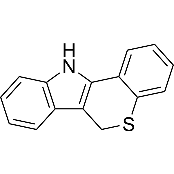
PD146176 (NSC168807), a 15-Lipoxygenase (15-LO) inhibitor, inhibits rabbit reticulocyte 15-LO (Ki=197 nM, IC50=0.54 μM). PD146176 reverses cognitive impairment, brain amyloidosis, and tau pathology by stimulating autophagy in aged triple transgenic mice.
For research use only. We do not sell to patients.
- Purity : 99.59%
- CAS No.: 4079-26-9
- Formula: C15H11NS
- Molecular Weight:237.32
-
Storage:Powder -20°C, 3 years ; In solvent -80°C, 6 months , -20°C, 1 month

To place orders, for customer services and technical support, please contact: MedChemExpress USA
Tel: 609-228-6898 E-mail: [email protected] [email protected]


-
 Biological Activity
-
Chemical Information
-
Solvent & Solubility
- Protocols
- Purity & Documentation
- References
-
 Help & FAQs


-
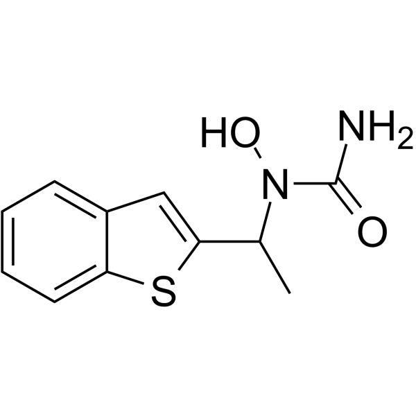
HY-14164
Zileuton5-Lipoxygenase Inhibitor -
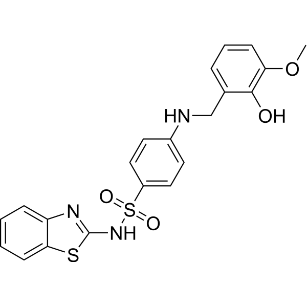
HY-12341
Onvoloxastat12-LOX Inhibitor -
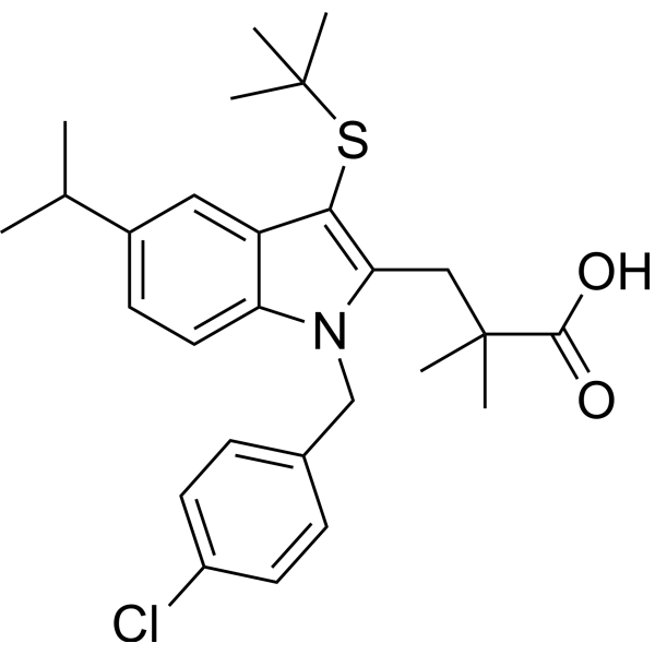
HY-14166
MK-886FLAP Inhibitor -

HY-100579
Ferrostatin-1Ferroptosis Inhibitor -
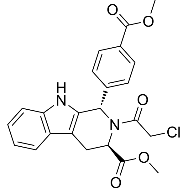
HY-100218A
RSL3GPX4 Inhibitor -
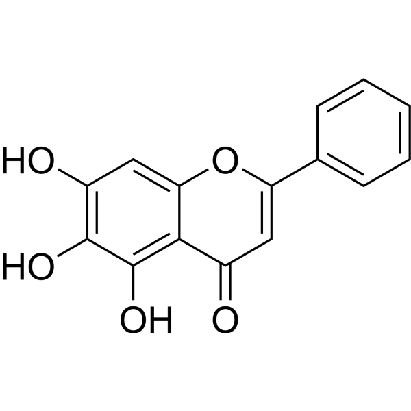
HY-N0196
BaicaleinXanthine Oxidase Inhibitor -
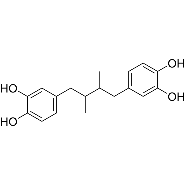
HY-N0198
Nordihydroguaiaretic acidAutophagy Inducer -

HY-15763
ErastinFerroptosis Inducer -
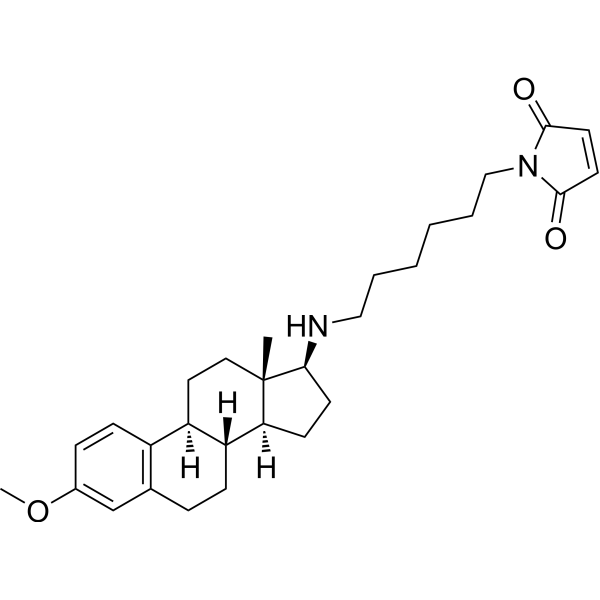
HY-13419
U-73122Phospholipase C Inhibitor -
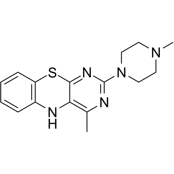
HY-118480
4-MMPB15-lipoxygenase Inhibitor -
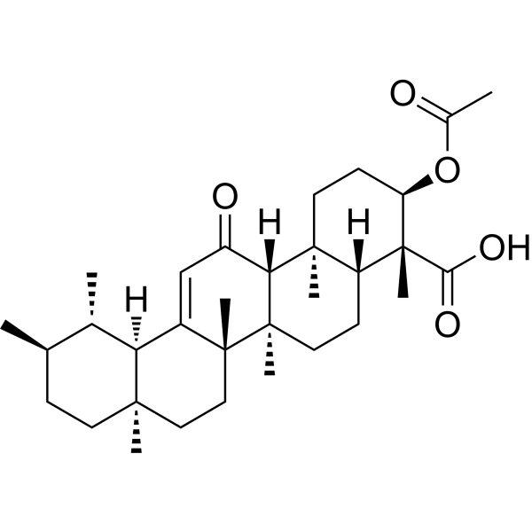
HY-N0892
AKBANrf2 Activator -

HY-103157R
PD146176 (Standard)15-LO Inhibitor
Publications Citing Use of MedChemExpress (MCE) PD146176
More Customer Validation & Images

-
In Vivo Efficacy Study

-
Histological Imaging/Staining

-
Cell Imaging/Staining

-
Flow Cytometry


Biological Activity
Description
IC50 & Target
Cellular Effect
|
Cell Line

|
Type | Value | Description | References |
|---|---|---|---|---|
| HEK293 | IC50 |
810 nM
Compound: PD-146176
|
Inhibition of human 12/15-LOX expressed in HEK293 cells after 30 mins by microplate reader analysis
Inhibition of human 12/15-LOX expressed in HEK293 cells after 30 mins by microplate reader analysis
|
[PMID: 26899595] |
In Vitro
In intact IC21 cells transfected with human 15-LO, PD146176 inhibits 13-HODE production with an IC50 of 0.81 μM[2].
MedChemExpress (MCE) has not independently confirmed the accuracy of these methods. They are for reference only.
In Vivo
MedChemExpress (MCE) has not independently confirmed the accuracy of these methods. They are for reference only.
-
Animal Model:Triple transgenic (3xTg) mice[3]
-
Dosage:80 mg/kg
-
Administration:Chowing; 12 weeks
-
Result:Significantly lower amyloid beta levels and deposition, less tau neuropathology, increased synaptic integrity, and autophagy activation.


Chemical Information
-
CAS No. 4079-26-9
-
Appearance Solid
-
Molecular Weight 237.32
-
Formula C15H11NS
-
Color Light yellow to yellow
-
SMILES
C1(N2)=C(CSC3=CC=CC=C31)C4=C2C=CC=C4
-
Synonyms
NSC168807
-
Shipping
Room temperature in continental US; may vary elsewhere.
-
Storage
Powder -20°C 3 years In solvent -80°C 6 months -20°C 1 month


Publications (6)
-
Journal Impact Factor
-
Most Recent
-
Nat Commun
Intrinsic temperature increase drives lipid metabolism towards ferroptosis evasion and chemotherapy resistance in pancreatic cancer. [Abstract]2024 Oct 2;15(1):8540. PMID: 39358362 -
Redox Biol
Neutrophil Irgm1 ameliorates sepsis-induced myocardial dysfunction by promoting Alox15 degradation. [Abstract]2026 May:92:104104. PMID: 41795420
PD146176 purchased from MedChemExpress. Usage Cited in: Redox Biol. 2026 May:92:104104. [Abstract]
The chart shows the serum 15-HETE levels in Irgm1-cKO mice after CLP surgery, when the mice were given either the carrier or PD146176 (10 mg/kg).
PD146176 purchased from MedChemExpress. Usage Cited in: Redox Biol. 2026 May:92:104104. [Abstract]
PD146176 (10 mg/kg). Representative images of H&E staining and quantitative analysis of myocardial injury.
-
J Biomed Sci
Exosomal PGE2 from M2 macrophages inhibits neutrophil recruitment and NET formation through lipid mediator class switching in sepsis. [Abstract]2023 Aug 2;30(1):62. PMID: 37533081
PD146176 purchased from MedChemExpress. Usage Cited in: J Biomed Sci. 2023 Aug 2;30(1):62. [Abstract]
PD146176 (1 μM). The chemotaxis of neutrophils to IL-8 was detected by Transwell assay.
PD146176 purchased from MedChemExpress. Usage Cited in: J Biomed Sci. 2023 Aug 2;30(1):62. [Abstract]
PD146176 (1 μM). Flow cytometry was used to detect the expression of CXCR2 in co-cultured PMN.
-
J Pineal Res
2024 Aug;76(5):e12987. PMID: 38975671 -
Biosci Trends
Hepatic stellate cell exosome-derived circWDR25 promotes the progression of hepatocellular carcinoma via the miRNA-4474-3P-ALOX-15 and EMT axes. [Abstract]2022 Sep 17;16(4):267-281. PMID: 35934785 -


Solvent & Solubility
In Vitro:
DMSO : 100 mg/mL (421.37 mM; Need ultrasonic; Hygroscopic DMSO has a significant impact on the solubility of product, please use newly opened DMSO)
Please refer to the solubility information to select the appropriate solvent. Once prepared, please aliquot and store the solution to prevent product inactivation from repeated freeze-thaw cycles.
Storage method and period of stock solution: -80°C, 6 months; -20°C, 1 month. When stored at -80°C, please use it within 6 months. When stored at -20°C, please use it within 1 month.
Please refer to the solubility information to select the appropriate solvent. Once prepared, please aliquot and store the solution to prevent product inactivation from repeated freeze-thaw cycles.
Storage method and period of stock solution: -80°C, 6 months; -20°C, 1 month. When stored at -80°C, please use it within 6 months. When stored at -20°C, please use it within 1 month.
Concentration (start) × Volume (start) = Concentration (final) × Volume (final)
In Vivo:
Select the appropriate dissolution method based on your experimental animal and administration route.
- For the following dissolution methods, please ensure to first prepare a clear stock solution using an In Vitro approach and then sequentially add co-solvents:
- To ensure reliable experimental results, the clarified stock solution can be appropriately stored based on storage conditions. As for the working solution for In Vivo experiments, it is recommended to prepare freshly and use it on the same day.
- The percentages shown for the solvents indicate their volumetric ratio in the final prepared solution. If precipitation or phase separation occurs during preparation, heat and/or sonication can be used to aid dissolution.
Add each solvent one by one: 10% DMSO 40% PEG300 5% Tween-80 45% Saline
Solubility: 2.5 mg/mL (10.53 mM); Suspended solution; Need ultrasonic
This protocol yields a suspended solution of 2.5 mg/mL. Suspended solution can be used for oral and intraperitoneal injection.
Taking 1 mL working solution as an example, add 100 μL DMSO stock solution (25.0 mg/mL) to 400 μL PEG300, and mix evenly; then add 50 μL Tween-80 and mix evenly; then add 450 μL Saline to adjust the volume to 1 mL.
Preparation of Saline: Dissolve 0.9 g sodium chloride in ddH₂O and dilute to 100 mL to obtain a clear Saline solution.
Add each solvent one by one: 10% DMSO 90% (20% SBE-β-CD in Saline)
Solubility: 2.5 mg/mL (10.53 mM); Suspended solution; Need ultrasonic
This protocol yields a suspended solution of 2.5 mg/mL. Suspended solution can be used for oral and intraperitoneal injection.
Taking 1 mL working solution as an example, add 100 μL DMSO stock solution (25.0 mg/mL) to 900 μL 20% SBE-β-CD in Saline, and mix evenly.
Preparation of 20% SBE-β-CD in Saline (4°C, storage for one week): 2 g SBE-β-CD powder is dissolved in 10 mL Saline, completely dissolve until clear.
In Vivo Dissolution Calculator
Please enter the basic information of animal experiments:
-
-
-
-
Recommended: Prepare an additional quantity of animals to account for potential losses during experiments.
Please enter your animal formula composition:
-
%DMSO +
Recommended: Keep the proportion of DMSO in working solution below 2% if your animal is weak.
-
%+
-
+%Tween-80 + +
-
%Saline +
The co-solvents required include: DMSO, . All of co-solvents are available by MedChemExpress (MCE). , Tween 80. All of co-solvents are available by MedChemExpress (MCE).
Working solution concentration: 0.22 mg/mL
Method for preparing stock solution: mg drug dissolved in μL DMSO. Stock solution concentration: mg/mL.
1. Take μL DMSO stock solution;
2. Add μL .
μL , mix evenly;
3. Then add μL Tween 80, mix evenly;
4. Then add μL
Please ensure that the stock solution in the first step is dissolved to a clear state, and add co-solvents in sequence. You can use ultrasonic heating (ultrasonic cleaner, recommended frequency 20-40 kHz), vortexing, etc. to assist dissolution.


Purity & Documentation
-
Data Sheet (279 KB)
-
SDS (393 KB)
- English - EN (393 KB)
- Français - FR (393 KB)
- Deutsch - DE (393 KB)
- Norwegian - NO (393 KB)
- Español - ES (393 KB)
- Swedish - SV (393 KB)
- Italian - IT (393 KB)
- Korean - KR (393 KB)
- Portuguese - PT (393 KB)
-
Handling Instructions (2659 KB)
References
[1]. Sendobry SM, et al. Attenuation of diet-induced atherosclerosis in rabbits with a highly selective 15-lipoxygenase inhibitor lacking significant antioxidant properties. Br J Pharmacol. 1997;120(7):1199-1206. [Content Brief]
[2]. Bocan TM, et al. A specific 15-lipoxygenase inhibitor limits the progression and monocyte-macrophage enrichment of hypercholesterolemia-induced atherosclerosis in the rabbit [published correction appears in Atherosclerosis 1998 Jul;139(1):201]. Atherosclerosis. 1998;136(2):203-216. [Content Brief]
[3]. Di Meco A, et al. 12/15-Lipoxygenase Inhibition Reverses Cognitive Impairment, Brain Amyloidosis, and Tau Pathology by Stimulating Autophagy in Aged Triple Transgenic Mice. Biol Psychiatry. 2017;81(2):92-100. [Content Brief]


Complete Stock Solution Preparation Table
Please refer to the solubility information to select the appropriate solvent. Once prepared, please aliquot and store the solution to prevent product inactivation from repeated freeze-thaw cycles.
Storage method and period of stock solution: -80°C, 6 months; -20°C, 1 month. When stored at -80°C, please use it within 6 months. When stored at -20°C, please use it within 1 month.
| Optional Solvent | Concentration Solvent Mass | 1 mg | 5 mg | 10 mg | 25 mg |
|---|---|---|---|---|---|
| DMSO | 1 mM | 4.2137 mL | 21.0686 mL | 42.1372 mL | 105.3430 mL |
| 5 mM | 0.8427 mL | 4.2137 mL | 8.4274 mL | 21.0686 mL | |
| 10 mM | 0.4214 mL | 2.1069 mL | 4.2137 mL | 10.5343 mL | |
| 15 mM | 0.2809 mL | 1.4046 mL | 2.8091 mL | 7.0229 mL | |
| 20 mM | 0.2107 mL | 1.0534 mL | 2.1069 mL | 5.2671 mL | |
| 25 mM | 0.1685 mL | 0.8427 mL | 1.6855 mL | 4.2137 mL | |
| 30 mM | 0.1405 mL | 0.7023 mL | 1.4046 mL | 3.5114 mL | |
| 40 mM | 0.1053 mL | 0.5267 mL | 1.0534 mL | 2.6336 mL | |
| 50 mM | 0.0843 mL | 0.4214 mL | 0.8427 mL | 2.1069 mL | |
| 60 mM | 0.0702 mL | 0.3511 mL | 0.7023 mL | 1.7557 mL | |
| 80 mM | 0.0527 mL | 0.2634 mL | 0.5267 mL | 1.3168 mL | |
| 100 mM | 0.0421 mL | 0.2107 mL | 0.4214 mL | 1.0534 mL |


PD146176 Related Classifications
HY-103157 Related Classifications
 Powered by Bioz