



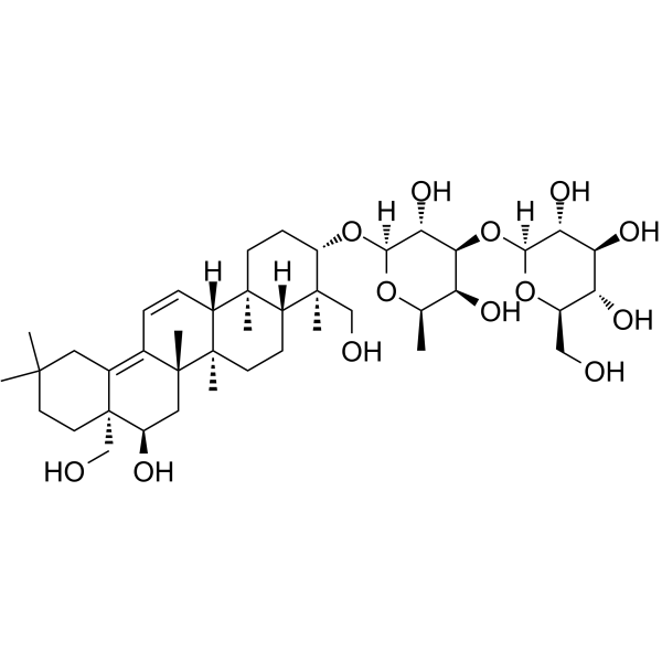
Saikosaponin B1
Based on 2 publication(s) in Google Scholar
Saikosaponin B1 is a bioactive constituent of Radix Bupleuri. Saikosaponin B1 is an agonist of the 5-HT2C receptor with an EC50 of 147.41 μM. Saikosaponin B1 inhibits the Hedgehog (Hh) signaling pathway by targeting the transmembrane protein SMO. Sailosaponin B1 can reduce liver fibrosis. Saikosaponin B1 has anti-cancer activities thus can be studies in research for cancers such as Medulloblastoma (MB).
Nur für Forschungszwecke. Wir verkaufen nicht an Patienten.
- Reinheit : 99.73%
- CAS. Nr.: 58558-08-0
- Formel: C42H68O13
- Molecular Weight:780.98
-
Speicherung:
4°C, protect from light
* In solvent : -80°C, 6 months; -20°C, 1 month (protect from light)

To place orders, for customer services and technical support, please contact: MedChemExpress USA
Tel: 609-228-6898 E-mail: [email protected] [email protected]


-
 Biologische Aktivität
-
Chemical Information
-
Lösungsmittel & Löslichkeit
- Protokoll
- Reinheit & Dokumentation
- Verweise
-
 Help & FAQs


-
HY-N0246
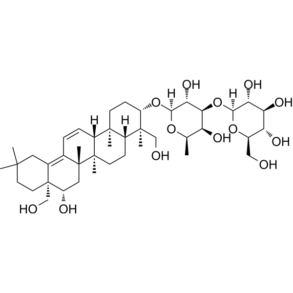
Saikosaponin AAnti-inflammatory Agent -
HY-N0249
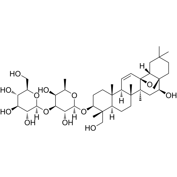
Saikosaponin CMMP-2 Inducer -
HY-N0248
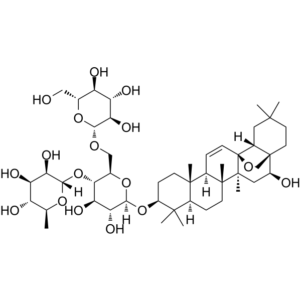
Saikosaponin B2Solute Carrier Modulator, ATP-Binding Cassette Transporter Modulator -
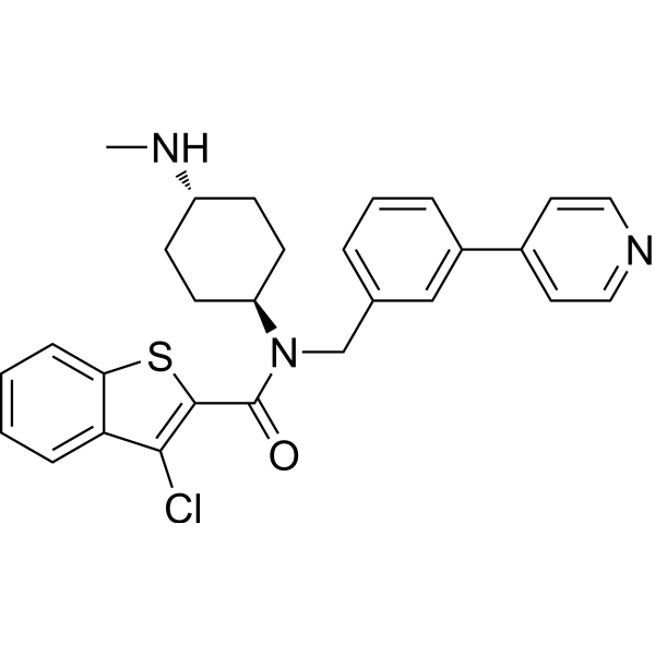
HY-12848
SAGSmoothened Agonist -

HY-N0247R
Saikosaponin B1 (Standard)SMO Inhibitor -
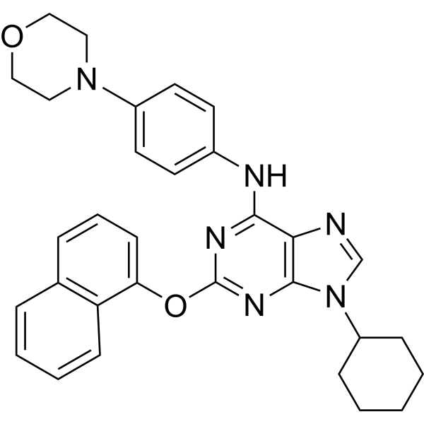
HY-15108
PurmorphamineSmoothened Receptor Agonist -
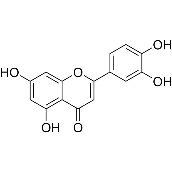
HY-N0162
LuteolinNrf2 Inhibitor -
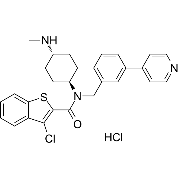
HY-12848B
SAG hydrochlorideSmoothened Agonist -
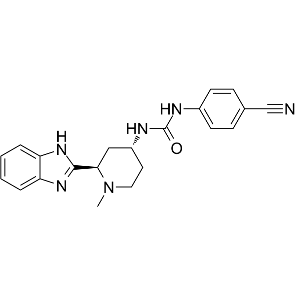
HY-16391
Glasdegib -
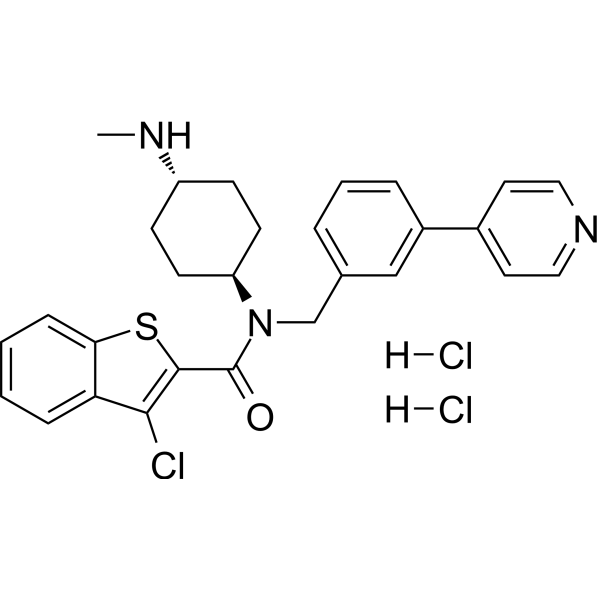
HY-12848C
SAG dihydrochlorideSmoothened Agonist -
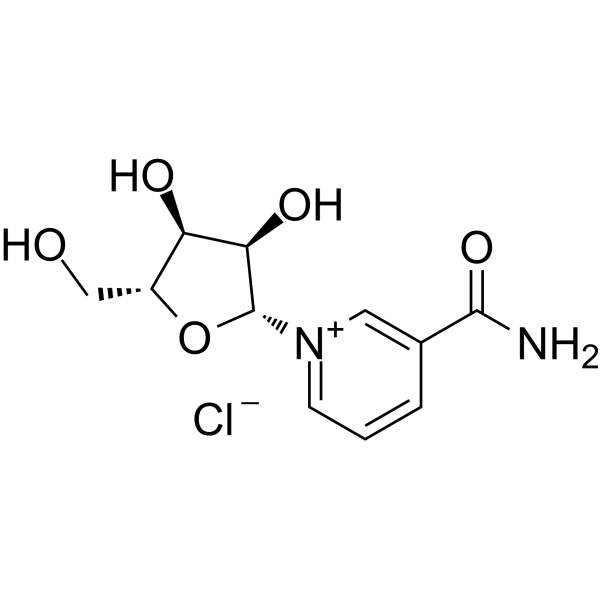
HY-123033A
Nicotinamide riboside chlorideEndogenous Metabolite -
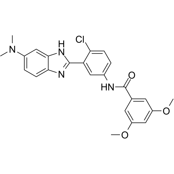
HY-15412
HhAntagSMO Antagonist
Publications Citing Use of MedChemExpress (MCE) Saikosaponin B1
MoreAlle TNF Receptor Isoform-spezifische Produkte anzeigen
More

Biologische Aktivität
Beschreibung
IC50 & Target
|
5-HT2C Receptor |
Cellular Effect
|
Cell Line

|
Type | Value | Description | References |
|---|---|---|---|---|
| A549 | IC50 |
>50 μM
Compound: 19
|
Cytotoxicity against human A549 cells assessed as induction of cell death incubated for 2 days by MTT assay
Cytotoxicity against human A549 cells assessed as induction of cell death incubated for 2 days by MTT assay
|
[PMID: 26259802] |
| Bcap37 | IC50 |
>50 μM
Compound: 19
|
Cytotoxicity against human Bcap37 cells assessed as induction of cell death incubated for 2 days by MTT assay
Cytotoxicity against human Bcap37 cells assessed as induction of cell death incubated for 2 days by MTT assay
|
[PMID: 26259802] |
| Hep 3B2 | IC50 |
>50 μM
Compound: 19
|
Cytotoxicity against human Hep3B cells assessed as induction of cell death incubated for 2 days by MTT assay
Cytotoxicity against human Hep3B cells assessed as induction of cell death incubated for 2 days by MTT assay
|
[PMID: 26259802] |
| HepG2 | IC50 |
>50 μM
Compound: 19
|
Cytotoxicity against human HepG2 cells assessed as induction of cell death incubated for 2 days by MTT assay
Cytotoxicity against human HepG2 cells assessed as induction of cell death incubated for 2 days by MTT assay
|
[PMID: 26259802] |
| MCF7 | IC50 |
>50 μM
Compound: 19
|
Cytotoxicity against human MCF7 cells assessed as induction of cell death incubated for 2 days by MTT assay
Cytotoxicity against human MCF7 cells assessed as induction of cell death incubated for 2 days by MTT assay
|
[PMID: 26259802] |
In Vitro
Saikosaponin B1 (10 nM-10 μM, 36 h) reduces the level of glioma-associated oncogene homolog (GLI)-luciferase activity in transfected Shh Light II cells with an IC50 of 241.8 nM[1].
Saikosaponin B1 (5 μM, 6-24 h) does not target GLI transcription factor and it downstream molecules, as Saikisaponin B1 shows no inhibition activity against PGE2 (HY-P3502B)-induced TCF/LEF and GLI activity in transfected LS174T cells, or TNF-α-induced NF-κB in HEK293T cell[1].
Saikosaponin B1 (3 μM, 36 h) fails to inhibit GLI-luciferase activity increased by limiting negative regulation from SUFU in SUFU-knockout Shh Light II cells, indicating Saikosaponin B1 targets Hh pathway via upstream components of SUFU[1].
Saikosaponin B1 (3 μM, 36 h) significantly prevents the increased of Gli1 mRNA expression induced by SAG (HY-12848) (a synthetic Hh pathway agonist binds to SMO) in Shh Light II cells with an IC50 of 3.64 μM[1].
Saikosaponin B1 (10 nM-10 μM, 72 h) significantly inhibits the proliferation of DAOY cell line (derived from nodular MB with GLI transcriptional activity)[1].
Saikisaponin B1 (1-10 μM, 0-24 h) decreases expression of TGF-β1-induced HSC activation biomarkers[2].
Saikisaponin B1 (0-10 μM) with biotin-label retains the inhibition activity of fibrotic protein expression in the activated HSC-T6 cells and shows the most remarkable binding to STAT3[2].
Saikisaponin B1 (50 μM) with biotin-label pulls down the STAT3 protein but no obvious interactions with JAK2 or IL-6 in HSC-T6 cells, LX-2 cells, and mouse liver tissues[2].
Saikisaponin B1 (1-10 μM) inhibits STAT3 phosphorylation in Tyr 705 but does not influence Ser 727 phosphorylation in HSC-t6 and LX-2 cells[2].
Saikisaponin B1 (5-10 μM, 24 h) disrupts STAT3 dimerization even in the presence of IL-6 (20 ng/mL) at 10 μM in HSC-T6 cells[2].
MedChemExpress (MCE) has not independently confirmed the accuracy of these methods. They are for reference only. Further protocols information, click here.
-
Cell Line:Shh Light II cells transfected with luciferase reporter plasmids and Renilla-TK construct
-
Concentration:10 nM-10 μM
-
Incubation Time:36 h
-
Result:Significantly reduced the level of Gli1 and Ptch 1 mRNA (two transcriptional targets of GLI), which further confirmed the inhibition of Hh signaling.
Showed little cytotoxicity in response to effective concentrations in Shh Light II cells.
-
Cell Line:Rat HSC-T6 cells; human LX-2 cells
-
Concentration:10 nM-10 μM
-
Incubation Time:0-24 h
-
Result:Inhibited the mRNA levels of the HSC activation biomarkers (α-SMA, collagen I, and vimentin) in TGF-β1-stimulated rat HSC-T6 cells.
Decreased expression of TGF-β1-induced α-SMA and Collagen I in human LX-2 cells and suggests that Saikisaponin B1 can reduce TGF-β1-induced HSC activation and matrix protein expression.
In Vivo
Saikosaponin B1 (200 mg/kg, i.p., three times a week from week 4 to week 7) ameliorates the Thioacetamide (TAA) (HY-Y0698)-induced morphological changes and liver damage with decreased collagen deposition in TAA-induced liver fibrosis model[2].
Saikosaponin B1 (10 mg/kg, s.c., twice a week for 4 weeks) attenuates the fibrosis histopathological characteristics of the liver tissues in CCl4-induced liver fibrotic mice[2].
Saikosaponin B1 (20 mg/kg, i.p., for 4 weeks) fails to further reduce liver fibrosis in response to CCl4 in CCl4mediated liver fibrosis STAT3 knockout mice model, suggesting STAT3 knockdown abrogated the antifibrosis effect of Saikosaponin B1[2].
Saikosaponin B1 (2.5-10 mg/kg, i.p., once per day for 7 consecutive days) inhibits pulmonary edema in LPS (HY-D1056)-induced ALI mice[3].
MedChemExpress (MCE) has not independently confirmed the accuracy of these methods. They are for reference only.
-
Animal Model:TAA (100 mg/kg, i.p., three times a week)-induced liver fibrosis male Balb/c mice model[2]
-
Dosage:5-20 μM, three times a week from week 4 to week 7
-
Administration:Intraperitoneal injection (i.p.)
-
Result:Decreased the liver/body weight ratio and the serum levels of ALT, AST, and TBIL compared to untreated model group.
Exerted a considerable effect on fibrosis alleviation with 10 mg/kg.
-
Animal Model:CCl4- (5 mg/kg of 40% CCl4 in the first week, 3 mg/kg of 40% CCl4 twice a week for another 6 weeks, s.c. )-induced liver fibrotic mice[2]
-
Dosage:10 mg/kg, twice a week for 4 weeks
-
Administration:Subcutaneous injection (s.c.)
-
Result:Exerted protective effect on liver function using the serum levels of ALT, AST, and TBIL.
Downregulated various proinflammatory cytokines (IL-6 and IL-1β) and fibrotic proteins (FN, collagen I , α-SMA and vimentin) in liver tissues.
Resulted in an obvious decrease in p-STAT3 and Gli1 expression levels.
Decreased STAT3 phosphorylation and Gli1 expression in the aHSCs and blocked interaction between STAT3 and Gli1.
-
Animal Model:CCl4- (5 mg/kg of 40% CCl4 in the first week, 3 mg/kg of 40% LPS (5 mg/kg, i.p.)-induced ALI mic
-
Dosage:2.5-10 mg/kg, once per day for 7 consecutive days
-
Administration:Intraperitoneal injection (i.p.)
-
Result:Significantly decreased the lung W/D ratios.
Significantly relieved the histological damage induced by LPS in mouse lungs.
Inhibited the expression of the inflammatory factors IL-6, IL-1β, and TNF-α in a dose-dependent manner.
Lowered TLR4 proteins and NF-κB proteins.


Chemical Information
-
CAS. Nr. 58558-08-0
-
Appearance Solid
-
Molecular Weight 780.98
-
Formel C42H68O13
-
Color White to off-white
-
SMILES
OC[C@@]1(C)[C@]2([H])CC[C@@]3(C)[C@]4(C)C[C@H](O)[C@@]5(CO)CCC(C)(C)CC5=C4C=C[C@]3([H])[C@@]2(C)CC[C@@H]1O[C@]6([H])O[C@H](C)[C@H](O)[C@H](O[C@]7([H])O[C@H](CO)[C@@H](O)[C@H](O)[C@H]7O)[C@H]6O
-
Structure Classification
-
Versand
Room temperature in continental US; may vary elsewhere.
-
Speicherung
4°C, protect from light
* In solvent : -80°C, 6 months; -20°C, 1 month (protect from light)


Publications (2)
-
Journal Impact Factor
-
Most Recent
-
Phytomedicine
CBX5 loss drives Pl3Kδ inhibitor resistance in mantle cell lymphoma and propolis restores sensitivity by inducing CBX5-mediated ferroptosis. [Abstract]2025 Jul 25:143:156911. PMID: 40466505 -
Biomater Res
Tryptamine-Functionalized Lipid Nanocarriers Co-delivering SMO/BRD4 Inhibitors for Synergistic Medulloblastoma Therapy. [Abstract]2025 Aug 8:29:0237. PMID: 40785845


Lösungsmittel & Löslichkeit
In Vitro:
DMSO : 100 mg/mL (128.04 mM; Need ultrasonic; Hygroscopic DMSO has a significant impact on the solubility of product, please use newly opened DMSO)
Please refer to the solubility information to select the appropriate solvent. Once prepared, please aliquot and store the solution to prevent product inactivation from repeated freeze-thaw cycles.
Storage method and period of stock solution: -80°C, 6 months; -20°C, 1 month (protect from light). When stored at -80°C, please use it within 6 months. When stored at -20°C, please use it within 1 month.
Please refer to the solubility information to select the appropriate solvent. Once prepared, please aliquot and store the solution to prevent product inactivation from repeated freeze-thaw cycles.
Storage method and period of stock solution: -80°C, 6 months; -20°C, 1 month (protect from light). When stored at -80°C, please use it within 6 months. When stored at -20°C, please use it within 1 month.
Konzentration (Stammlösung) × Volumen (Stammlösung) = Konzentration (Ziellösung) × Volumen (Ziellösung)
In Vivo:
Select the appropriate dissolution method based on your experimental animal and administration route.
- For the following dissolution methods, please ensure to first prepare a clear stock solution using an In Vitro approach and then sequentially add co-solvents:
- To ensure reliable experimental results, the clarified stock solution can be appropriately stored based on storage conditions. As for the working solution for In Vivo experiments, it is recommended to prepare freshly and use it on the same day.
- The percentages shown for the solvents indicate their volumetric ratio in the final prepared solution. If precipitation or phase separation occurs during preparation, heat and/or sonication can be used to aid dissolution.
Add each solvent one by one: 10% DMSO 40% PEG300 5% Tween-80 45% Saline
Solubility: 10 mg/mL (12.80 mM); Clear solution; Need ultrasonic
This protocol yields a clear solution of 10 mg/mL.
Taking 1 mL working solution as an example, add 100 μL DMSO stock solution (100.0 mg/mL) to 400 μL PEG300, and mix evenly; then add 50 μL Tween-80 and mix evenly; then add 450 μL Saline to adjust the volume to 1 mL.
Preparation of Saline: Dissolve 0.9 g sodium chloride in ddH₂O and dilute to 100 mL to obtain a clear Saline solution.
Add each solvent one by one: 10% DMSO 90% (20% SBE-β-CD in Saline)
Solubility: ≥ 10 mg/mL (12.80 mM); Clear solution
This protocol yields a clear solution of ≥ 10 mg/mL (saturation unknown).
Taking 1 mL working solution as an example, add 100 μL DMSO stock solution (100.0 mg/mL) to 900 μL 20% SBE-β-CD in Saline, and mix evenly.
Preparation of 20% SBE-β-CD in Saline (4°C, storage for one week): 2 g SBE-β-CD powder is dissolved in 10 mL Saline, completely dissolve until clear.
In Vivo Dissolution Calculator
Please enter the basic information of animal experiments:
-
-
-
-
Recommended: Prepare an additional quantity of animals to account for potential losses during experiments.
Please enter your animal formula composition:
-
%DMSO +
Recommended: Keep the proportion of DMSO in working solution below 2% if your animal is weak.
-
%+
-
+%Tween-80 + +
-
%Saline +
The co-solvents required include: DMSO, . All of co-solvents are available by MedChemExpress (MCE). , Tween 80. All of co-solvents are available by MedChemExpress (MCE).
Working solution concentration: 0.22 mg/mL
Method for preparing stock solution: mg drug dissolved in μL DMSO. Stock solution concentration: mg/mL. * In solvent : -80°C, 6 months; -20°C, 1 month (protect from light)
1. Take μL DMSO stock solution;
2. Add μL .
μL , mix evenly;
3. Then add μL Tween 80, mix evenly;
4. Then add μL
Please ensure that the stock solution in the first step is dissolved to a clear state, and add co-solvents in sequence. You can use ultrasonic heating (ultrasonic cleaner, recommended frequency 20-40 kHz), vortexing, etc. to assist dissolution.

Protokoll
-
Fibrosis/Collagen Morphometry
Fibrosis and collagen morphometry is based on the quantitative visualization of fibrillar collagen deposition in tissue sections using histochemical stains such as Sirius Red (Picrosirius Red) or Masson's trichrome, followed by image-based or polarization-enhanced analysis to estimate collagen proportional area as a surrogate of extracellular matrix accumulation during fibrotic remodeling. Sirius Red combined with polarized light microscopy enhances detection of collagen fibers due to birefringence properties, enabling more specific visualization of collagen type I and III fibrils compared to conventional bright-field histology, while whole-section or region-restricted digital morphometry reduces field-selection bias in fibrosis assessment. Alternative quantitative approaches include second harmonic generation (SHG) and two-photon excited fluorescence microscopy, which enable label-free detection of fibrillar collagen and have been validated against histological staining and biochemica
-
Collagen: Sirius Red Staining
Sirius Red or picrosirius red staining is a histochemical method for visualizing collagen-rich extracellular matrix in tissue sections, and collagen fibers are detected as red-stained structures under bright-field microscopy with enhanced birefringence under polarized light. Picrosirius red is useful for assessing total collagen organization, distribution, and fibrosis burden, but polarized color should not be interpreted as a definitive collagen type I versus type III readout because color is affected by fiber orientation, thickness, and packing.
-
Membrane Protein Extraction Using Detergents and Chaotropes
Membrane protein extraction with detergents and chaotropes solubilizes lipid-bilayer-associated proteins by disrupting protein-lipid and protein-protein interactions while maintaining proteins in a soluble state for downstream electrophoresis, purification, or mass spectrometry. Chaotropes such as urea and thiourea improve solubilization of difficult proteins, while nonionic and zwitterionic detergents such as CHAPS, ASB-14, SB 3-10, MEGA-10, dodecyl maltoside, and Triton X-100 differ in extraction efficiency depending on sample type and membrane protein properties.
-
Connective Tissue: Masson's Trichrome/Collagen Trichrome Staining
Masson’s Trichrome (collagen/trichrome staining) is a histological technique that differentially stains tissue compartments using sequential acidic dyes to distinguish collagen from muscle and cytoplasmic components based on dye affinity and tissue permeability differences, enabling visualization of fibrosis and connective tissue architecture in histological sections. The classical formulation typically uses Weigert's iron hematoxylin for nuclear staining, Biebrich scarlet-acid fuchsin for cytoplasm and muscle, and aniline blue (or light green variants) for collagen, producing a characteristic blue/green collagen signal contrasted against red cytoplasm and dark nuclei. The staining principle relies on selective displacement of smaller dye molecules by larger anionic dyes in collagen-rich regions under controlled acidified conditions, which enhances collagen-specific dye retention. This property makes the method widely used for fibrosis assessment in organs such as heart, liver, lung, a
-
Liver Histomorphometry
Liver histomorphometry is a quantitative histological approach used to measure structural alterations in hepatic tissue, including parenchymal loss, steatosis, fibrosis, and vascular remodeling, by combining stained tissue section analysis with stereological or computerized image-based measurements. Classical morphometric frameworks quantify volume fractions of liver compartments and fibrotic regions using systematic sampling and image analysis, enabling objective comparison of pathological changes across experimental groups. These approaches are widely applied in liver cirrhosis and fibrosis studies to reduce subjectivity in histological scoring and improve reproducibility of tissue evaluation. Recent methodological advances integrate automated image analysis and radiomics-based extraction of histological features from standard liver stains (e. g. , H&E and fibrotic stains), enabling quantitative correlation between morphometric features and fibrosis stages in non-alcoholic fatty live


Reinheit & Dokumentation
-
Data Sheet (288 KB)
-
SDS (394 KB)
- English - EN (394 KB)
- Français - FR (394 KB)
- Deutsch - DE (394 KB)
- Norwegian - NO (394 KB)
- Español - ES (394 KB)
- Swedish - SV (394 KB)
- Italian - IT (394 KB)
- Korean - KR (394 KB)
- Portuguese - PT (394 KB)
-
Handling Instructions (2659 KB)
Verweise
[1]. Luo J, et al., Saikosaponin B1 and Saikosaponin D inhibit tumor growth in medulloblastoma allograft mice via inhibiting the Hedgehog signaling pathway. J Nat Med. 2022 Jun;76(3):584-593. [Content Brief]
[3]. Peng D, et L., Saikosaponin A and Its Epimers Alleviate LPS-Induced Acute Lung Injury in Mice. Molecules. 2023 Jan 18;28(3):967. [Content Brief]
[4]. Sun, C. L., et al., (2017). Bioassay-guided isolation of saikosaponins with agonistic activity on 5-hydroxytryptamine 2C receptor from Bupleurum chinense and their potential use for the treatment of obesity. Chinese journal of natural medicines, 15(6), 467–473. [Content Brief]


Complete Stock Solution Preparation Table
Please refer to the solubility information to select the appropriate solvent. Once prepared, please aliquot and store the solution to prevent product inactivation from repeated freeze-thaw cycles.
Storage method and period of stock solution: -80°C, 6 months; -20°C, 1 month (protect from light). When stored at -80°C, please use it within 6 months. When stored at -20°C, please use it within 1 month.
| Optional Solvent | Concentration Solvent Mass | 1 mg | 5 mg | 10 mg | 25 mg |
|---|---|---|---|---|---|
| DMSO | 1 mM | 1.2804 mL | 6.4022 mL | 12.8044 mL | 32.0111 mL |
| 5 mM | 0.2561 mL | 1.2804 mL | 2.5609 mL | 6.4022 mL | |
| 10 mM | 0.1280 mL | 0.6402 mL | 1.2804 mL | 3.2011 mL | |
| 15 mM | 0.0854 mL | 0.4268 mL | 0.8536 mL | 2.1341 mL | |
| 20 mM | 0.0640 mL | 0.3201 mL | 0.6402 mL | 1.6006 mL | |
| 25 mM | 0.0512 mL | 0.2561 mL | 0.5122 mL | 1.2804 mL | |
| 30 mM | 0.0427 mL | 0.2134 mL | 0.4268 mL | 1.0670 mL | |
| 40 mM | 0.0320 mL | 0.1601 mL | 0.3201 mL | 0.8003 mL | |
| 50 mM | 0.0256 mL | 0.1280 mL | 0.2561 mL | 0.6402 mL | |
| 60 mM | 0.0213 mL | 0.1067 mL | 0.2134 mL | 0.5335 mL | |
| 80 mM | 0.0160 mL | 0.0800 mL | 0.1601 mL | 0.4001 mL | |
| 100 mM | 0.0128 mL | 0.0640 mL | 0.1280 mL | 0.3201 mL |


Saikosaponin B1 Related Classifications
HY-N0247 Related Classifications
 Powered by Bioz