



Tigogenin

Based on 1 Customer Validation
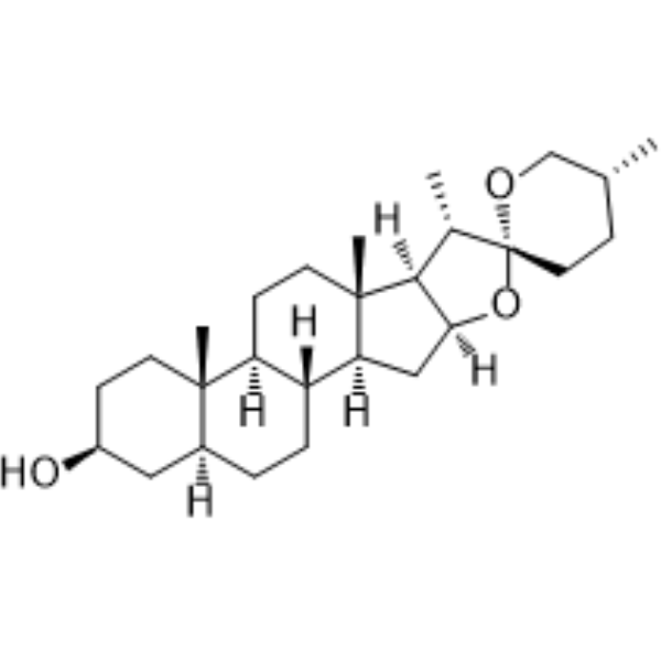
Tigogenin is a steroidal sapogenins. Tigogenin can inhibit adipocytic differentiation and induce osteoblastic differentiation in mouse bone marrow stromal cells. Tigogenin can inhibit cells proliferation and induce apoptosis. Tigogenin can be used for the researches of cancer, inflammation, immunology, metabolic and cardiovascular disease, such as mammary gland carcinoma, rheumatoid arthritis, osteoporosis and atherosclerosis.
For research use only. We do not sell to patients.
- Purity : 98.0%
- CAS No.: 77-60-1
- Formula: C27H44O3
- Molecular Weight:416.64
-
Storage:
4°C, protect from light
* In solvent : -80°C, 6 months; -20°C, 1 month (protect from light)

To place orders, for customer services and technical support, please contact: MedChemExpress USA
Tel: 609-228-6898 E-mail: [email protected] [email protected]


-
 Biological Activity
-
Chemical Information
-
Solvent & Solubility
- Protocols
- Purity & Documentation
- References
-
 Help & FAQs




Biological Activity
Description
In Vitro
Tigogenin (10-90 μM, 3 days) promotes bone marrow stromal cells (BMSCs) proliferation in a dose-dependent manner[1].
Tigogenin (10-90 μM, 18 days) decreases lipid accumulation and visfatin secretions in BMSCs[1].
Tigogenin (10-90 μM, 5-18 days) reduces PPAR mRNA and ap2 levels in BMSCs[1].
Tigogenin (10-90 μM, 5 days) increases levels of mRNA for Cbfa1, COL I, OCN and ALP activity in BMSCs[1].
Tigogenin (30 μM, 5-40 mins) activates the phosphorylation of p38 kinase in BMSCs[1].
Tigogenin (40 μM, 24-96 h) inhibits proliferation in rheumatoid arthritis (RA) synoviocytes[2].
Tigogenin (40 μM, 6-24 h) induces apoptosis in RA synoviocytes[2].
Tigogenin (40 μM, 1-24 h) increases P38, COX-2 and PEG2 levels in RA synoviocytes[2].
Tigogenin (20 μM, 24 h) reduces FXR, SHP, and BSEP levels companied with CDCA (HY-76847) in HepG2 cells[3].
Tigogenin (L–serine derivative of Tigogenin) (48 h) inhibits MCF-7 and MDA-MB-231 cells proliferation with IC50 values of 1.5 and 10.5 μM[4].
Tigogenin (L–serine derivative of Tigogenin) (2.8-7 μM, 18 h) induces apoptosis and increases caspase 3/7 activity in MCF-7 cells[4].
Tigogenin (L–serine derivative of Tigogenin) (5 μM, 6 h) reduces TNF-α, IL-1, IL-12, IL-10 and IL-4 levels in THP–1 cells[4].
MedChemExpress (MCE) has not independently confirmed the accuracy of these methods. They are for reference only. Further protocols information, click here.
-
Cell Line:BMSCs
-
Concentration:10, 30 and 90 μM
-
Incubation Time:3 days
-
Result:Promoted proliferation in a dose-dependent manner.
-
Cell Line:BMSCs
-
Concentration:10, 30 and 90 μM
-
Incubation Time:5 or 18 days
-
Result:Reduced PPAR mRNA and ap2 levels.
-
Cell Line:RA synoviocytes
-
Concentration:40 μM
-
Incubation Time:6 and 24 h
-
Result:Induced cell shrinkage, cytoplasm condensation and formation of cytoplasmic filaments.
Increased caspase-3, caspase-8 and caspase-9 activities.
Showed DNA fragmentation.


Chemical Information
-
CAS No. 77-60-1
-
Appearance Solid
-
Molecular Weight 416.64
-
Formula C27H44O3
-
Color White to off-white
-
SMILES
[H][C@]1(O[C@@]2(OC[C@H](C)CC2)[C@H]3C)C[C@@]4([H])[C@]5([H])CC[C@@]6([H])C[C@@H](O)CC[C@]6(C)[C@@]5([H])CC[C@]4(C)[C@]13[H]
-
Structure Classification
-
Initial Source
-
Shipping
Room temperature in continental US; may vary elsewhere.
-
Storage
4°C, protect from light
* In solvent : -80°C, 6 months; -20°C, 1 month (protect from light)


Solvent & Solubility
In Vitro:
Ethanol : 14.29 mg/mL (34.30 mM; Need ultrasonic)
Please refer to the solubility information to select the appropriate solvent. Once prepared, please aliquot and store the solution to prevent product inactivation from repeated freeze-thaw cycles.
Storage method and period of stock solution: -80°C, 6 months; -20°C, 1 month (protect from light). When stored at -80°C, please use it within 6 months. When stored at -20°C, please use it within 1 month.
Please refer to the solubility information to select the appropriate solvent. Once prepared, please aliquot and store the solution to prevent product inactivation from repeated freeze-thaw cycles.
Storage method and period of stock solution: -80°C, 6 months; -20°C, 1 month (protect from light). When stored at -80°C, please use it within 6 months. When stored at -20°C, please use it within 1 month.
Concentration (start) × Volume (start) = Concentration (final) × Volume (final)

Protocols
-
Collagen-Induced Arthritis
Collagen-induced arthritis (CIA) is an autoimmune murine model of rheumatoid arthritis in which immunization with type II collagen (CII) emulsified in an adjuvant induces a T cell- and autoantibody-driven inflammatory arthritis characterized by synovial hyperplasia, immune cell infiltration, and joint destruction. The model typically relies on genetically susceptible mouse strains (e. g. , DBA/1) and reproduces key features of human rheumatoid arthritis, including anti-collagen immune responses and progressive joint inflammation. Disease onset generally occurs within ~3-4 weeks after immunization, depending on antigen/adjuvant combinations and protocol variation. The immunopathology is driven by adaptive immune activation against CII, leading to systemic and local joint inflammation mediated by pro-inflammatory cytokines and effector immune cells, making CIA a standard preclinical platform for evaluating immunomodulatory and anti-arthritic interventions.
-
Apoptosis
Apoptosis, also called programmed cell death, is generally characterized by distinct morphological characteristics.
-
TUNEL staining for apoptotic DNA fragmentation
TUNEL staining detects DNA strand breaks by using terminal deoxynucleotidyl transferase to add labeled nucleotides to exposed 3′-OH DNA termini, generating either microscopic staining in fixed cells or tissue sections, or fluorescence/cytometric signal in cell suspensions. TUNEL positivity reflects DNA fragmentation but should not be interpreted alone as definitive apoptosis, because TUNEL can also label necrotic, autolytic, mechanically damaged, or DNA-repair-associated DNA breaks.
-
Annexin V plus membrane-impermeant dye apoptosis staining
Annexin V-based apoptosis assays rely on the detection of phosphatidylserine (PS) externalization from the inner leaflet of the plasma membrane to the outer leaflet, an early biochemical hallmark of apoptosis. Fluorescently labeled Annexin V binds PS in a calcium-dependent manner, enabling identification of early apoptotic cells by flow cytometry or fluorescence microscopy. When combined with a membrane-impermeant DNA-binding dye (e. g. , propidium iodide), this approach allows discrimination between viable (Annexin V−/dye−), early apoptotic (Annexin V+/dye−), and late apoptotic or necrotic (Annexin V+/dye+) cell populations by assessing membrane integrity and PS exposure.
-
Apoptosis Solutions
Apoptosis is a regulated, generally non-lytic cell-death pathway that removes unwanted, damaged, infected, or abnormal cells through coordinated morphological changes, caspase activation, DNA fragmentation, and membrane remodeling. The intrinsic apoptosis pathway is controlled mainly by mitochondrial outer membrane permeabilization, BCL-2 family proteins, cytochrome c release, apoptosome formation, caspase-9 activation, and downstream executioner caspase-3/7 activation. The extrinsic apoptosis pathway is initiated by death receptors such as Fas, TNFR, and TRAIL receptors, which recruit adaptor proteins and activate caspase-8 before engaging executioner caspases or mitochondrial amplification through BID cleavage. Apoptosis is linked to many phenotypes, including cancer cell killing, tissue homeostasis, immune regulation, neurodegeneration, infection response, and treatment-induced cytotoxicity; unresolved questions include how apoptosis interacts with necroptosis, pyroptosis, ferroptos
-
Research Protocol for Cardiovascular Diseases
Cardiovascular disease can be modeled as maladaptive cardiac remodeling, where ischemic injury or pressure overload activates inflammatory signaling, fibroblast activation, extracellular-matrix deposition, cardiomyocyte hypertrophy, vascular remodeling, and progressive ventricular dysfunction. The TGF-β/SMAD axis is a central profibrotic pathway after myocardial injury and pressure overload, while innate immune and cytokine pathways regulate leukocyte recruitment, scar formation, and adverse remodeling. Key unresolved questions include which inflammatory signals are reparative versus harmful, when fibrosis is protective versus maladaptive, and whether pathway inhibition improves function without weakening necessary infarct healing or compensatory remodeling.
-
Research Protocol for Inflammation-related Diseases
The NLRP3 inflammasome is a cytosolic innate immune signaling platform that integrates priming signals and danger-signal activation to promote caspase-1 activation, maturation of IL-1β and IL-18, and gasdermin D-mediated pyroptotic cell death. The core experimental logic is to determine whether inflammatory disease phenotypes are driven by increased NLRP3 expression, ASC-containing inflammasome assembly, caspase-1 cleavage, GSDMD cleavage, and extracellular release of IL-1β/IL-18 rather than by nonspecific cell injury alone. The pathway is strongly linked to inflammation-related disease phenotypes because monosodium urate crystals activate NALP3/NLRP3 inflammasome signaling in gout-like crystal inflammation, cholesterol crystals activate NLRP3 inflammasomes in atherogenesis models, and DSS-induced intestinal inflammation has been reported to involve NLRP3 inflammasome activity. However, experimental colitis studies also show context-dependent protective effects of NLRP3 inflammasome co
-
Cell Viability Determination by MTT Colorimetric Assay
The following protocol uses the MTT colorimetric assay as a classic literature-established method for assessing cell viability/metabolic activity in cultured mammalian cells. MTT[3-(4,5-dimethylthiazol-2-yl)-2,5-diphenyltetrazolium bromide] is reduced by metabolically active cells to a colored formazan product; the amount of formazan is quantified spectrophotometrically and provides an indirect measure of metabolically active viable cells. Importantly, MTT reduction reflects cellular oxidoreductase/metabolic activity rather than an absolute direct count of living cells, so changes in cellular metabolism can alter the signal independently of cell number.


Purity & Documentation
-
Data Sheet (277 KB)
-
SDS (393 KB)
- English - EN (393 KB)
- Français - FR (393 KB)
- Deutsch - DE (393 KB)
- Norwegian - NO (393 KB)
- Español - ES (393 KB)
- Swedish - SV (393 KB)
- Italian - IT (393 KB)
- Korean - KR (393 KB)
- Portuguese - PT (393 KB)
-
Handling Instructions (2659 KB)
References
[1]. Zhou H, et al. Tigogenin inhibits adipocytic differentiation and induces osteoblastic differentiation in mouse bone marrow stromal cells. Mol Cell Endocrinol. 2007 May 30;270(1-2):17-22. [Content Brief]
[2]. Liagre B, et al. Inhibition of human rheumatoid arthritis synovial cell survival by hecogenin and tigogenin is associated with increased apoptosis, p38 mitogen-activated protein kinase activity and upregulation of cyclooxygenase-2. Int J Mol Med. 2007 Oct;20(4):451-60. [Content Brief]
[3]. Bao R, et al. Dioscin Ameliorates Hyperuricemia-Induced Atherosclerosis by Modulating of Cholesterol Metabolism through FXR-Signaling Pathway. Nutrients. 2022 May 9;14(9):1983. [Content Brief]
[4]. Michalak O, et al. Synthesis and anti-tumour, immunomodulating activity of diosgenin and tigogenin conjugates. J Steroid Biochem Mol Biol. 2020 Apr;198:105573. [Content Brief]


Complete Stock Solution Preparation Table
Please refer to the solubility information to select the appropriate solvent. Once prepared, please aliquot and store the solution to prevent product inactivation from repeated freeze-thaw cycles.
Storage method and period of stock solution: -80°C, 6 months; -20°C, 1 month (protect from light). When stored at -80°C, please use it within 6 months. When stored at -20°C, please use it within 1 month.
| Optional Solvent | Concentration Solvent Mass | 1 mg | 5 mg | 10 mg | 25 mg |
|---|---|---|---|---|---|
| Ethanol | 1 mM | 2.4002 mL | 12.0008 mL | 24.0015 mL | 60.0038 mL |
| 5 mM | 0.4800 mL | 2.4002 mL | 4.8003 mL | 12.0008 mL | |
| 10 mM | 0.2400 mL | 1.2001 mL | 2.4002 mL | 6.0004 mL | |
| 15 mM | 0.1600 mL | 0.8001 mL | 1.6001 mL | 4.0003 mL | |
| 20 mM | 0.1200 mL | 0.6000 mL | 1.2001 mL | 3.0002 mL | |
| 25 mM | 0.0960 mL | 0.4800 mL | 0.9601 mL | 2.4002 mL | |
| 30 mM | 0.0800 mL | 0.4000 mL | 0.8001 mL | 2.0001 mL |


Tigogenin Related Classifications
HY-N1403 Related Classifications
 Powered by Bioz