



STAT3-IN-12
Based on 2 publication(s) in Google Scholar
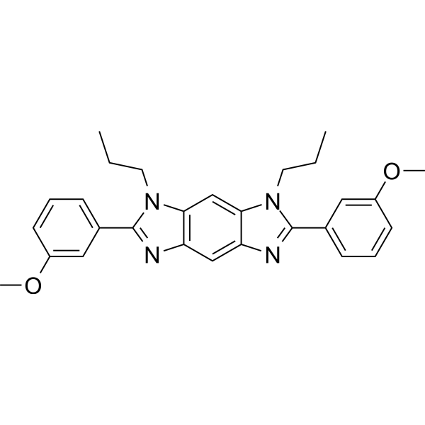
STAT3-IN-12 is a potent STAT3 signal inhibitor that can inhibit IL-6 induced JAK/STAT3 signalling pathway activation. STAT3-IN-12 inhibits cancer cell growth, migration, and induce cell apoptosis as well as cycle arrest. STAT3-IN-12 can be used in cancer-related research, such as hepatocellular carcinoma (HCC) and oesophageal carcinoma.
Nur für Forschungszwecke. Wir verkaufen nicht an Patienten.
- Reinheit : 99.87%
- CAS. Nr.: 2980758-31-2
- Formel: C28H30N4O2
- Molecular Weight:454.56
-
Speicherung:Powder -20°C, 3 years , 4°C, 2 years ; In solvent -80°C, 6 months , -20°C, 1 month

To place orders, for customer services and technical support, please contact: MedChemExpress USA
Tel: 609-228-6898 E-mail: [email protected] [email protected]


-
 Biologische Aktivität
-
Chemical Information
-
Lösungsmittel & Löslichkeit
- Protokoll
- Reinheit & Dokumentation
- Verweise
-
 Help & FAQs


-
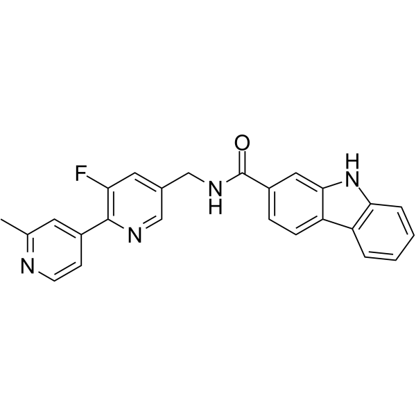
HY-111472
Porcn-IN-1Porcupine Inhibitor -
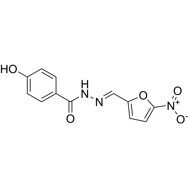
HY-B1436
NifuroxazideBacterial Inhibitor -
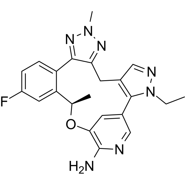
HY-152292
ZidesamtinibROS1 Inhibitor -
HY-N2116
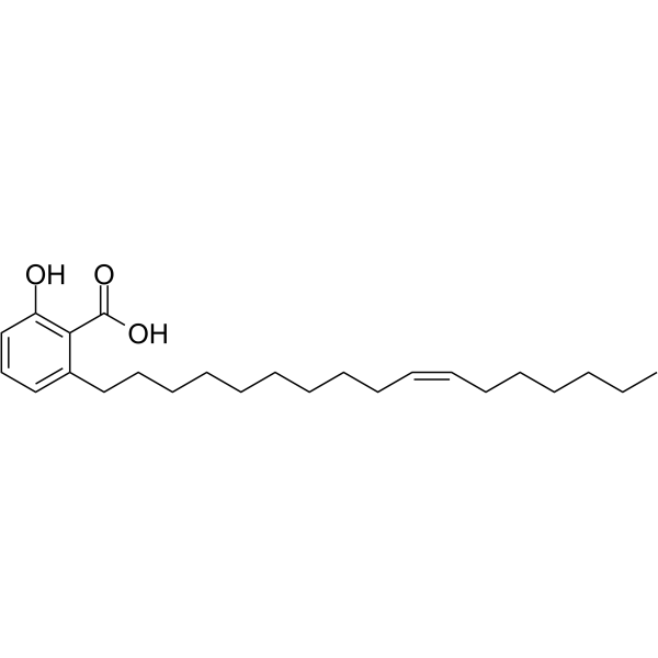
Ginkgolic acid C17:1Antitumor Agent -

HY-B1436R
Nifuroxazide (Standard)Bacterial Inhibitor -
HY-16975
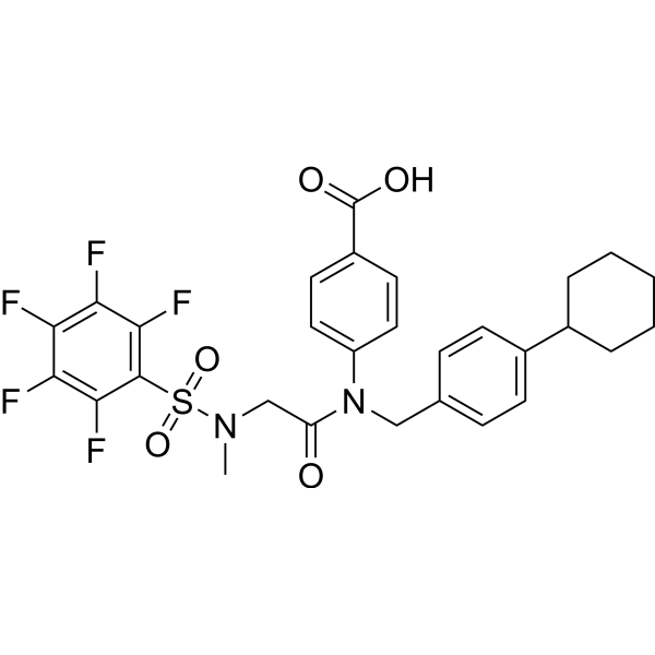
SH-4-54STAT3/5 Inhibitor -

HY-N2116R
Ginkgolic acid C17:1 (Standard)Antitumor Agent
Publications Citing Use of MedChemExpress (MCE) STAT3-IN-12
More

Biologische Aktivität
Beschreibung
IC50 & Target
[1]|
STAT3 |
Cellular Effect
|
Cell Line

|
Type | Value | Description | References |
|---|---|---|---|---|
| ECa-109 cell line | IC50 |
3.63 μM
Compound: 24
|
Antitumor activity against human EC109 cells assessed as reduction in cell viability incubated for 72 hrs by MTT assay
Antitumor activity against human EC109 cells assessed as reduction in cell viability incubated for 72 hrs by MTT assay
|
[PMID: 35504209] |
| HepG2 | IC50 |
4.3 μM
Compound: 24
|
Antitumor activity against human HepG2 cells assessed as reduction in cell viability incubated for 72 hrs by MTT assay
Antitumor activity against human HepG2 cells assessed as reduction in cell viability incubated for 72 hrs by MTT assay
|
[PMID: 35504209] |
In Vitro
STAT3-IN-12 (compound 24, 0-10 μM approximately, 72 h) inhibits cancer cell growth and migration in HepG2 and EC109 cells[1].
STAT3-IN-12 (0-20 μM, 16 h) binds to the STAT3 protein and inhibits IL-6-mediated STAT3 phosphorylation, also inhibts STAT3 nuclear localization and dimerization in EC109 and HepG2 cells[1].
STAT3-IN-12 (0-20 μM, 48 h) induces cell apoptosis as well as cycle arrest in HepG2 and EC109 cells[1].
MedChemExpress (MCE) has not independently confirmed the accuracy of these methods. They are for reference only.
-
Cell Line:HepG2 and EC109 cells
-
Concentration:0, 1.25, 2.5, 5 and 10 μM.
-
Incubation Time:72 h
-
Result:Inhibited cancer cell growth with IC50 values of 4.32 and 3.63 μM.
-
Cell Line:HepG2 and EC109 cells
-
Concentration:0-10 μM
-
Incubation Time:24 h
-
Result:Inhibited cancer cell migration.
-
Cell Line:HepG2 and EC109 cells
-
Concentration:0, 2.5, 5, 10 and 20 μM
-
Incubation Time:16 h
-
Result:Inhibited phosphorylation of STAT3 tyrosine 705 with high selectivity.
In Vivo
MedChemExpress (MCE) has not independently confirmed the accuracy of these methods. They are for reference only.
-
Animal Model:HepG2 cell xenograft tumor model[1]
-
Dosage:20, 40 mg/kg, daily for 24 days
-
Administration:Intraperitoneal injection
-
Result:Inhibited tumor growth without affecting the body weight.
- Detection of Apoptosis
- TUNEL staining for apoptotic DNA fragmentation
- Annexin V plus membrane-impermeant dye apoptosis staining
- Protocol for Cell Counting and Cell Density Analysis
- Cell migration
- Somatic Cell Culture
- Apoptosis Solutions
- CCK-8/WST-8 Cell Proliferation Assay
- Cell Counting-Based Growth Curve Assay
- Liver Cancer Modeling
- MTT Cell Proliferation Assay
- Cell Viability Determination by MTT Colorimetric Assay


Chemical Information
-
CAS. Nr. 2980758-31-2
-
Appearance Solid
-
Molecular Weight 454.56
-
Formel C28H30N4O2
-
Color Off-white to light yellow
-
SMILES
CCCN1C2=C(C=C3C(N(CCC)C(C4=CC=CC(OC)=C4)=N3)=C2)N=C1C5=CC=CC(OC)=C5
-
Versand
Room temperature in continental US; may vary elsewhere.
-
Speicherung
Powder -20°C 3 years 4°C 2 years In solvent -80°C 6 months -20°C 1 month


Publications (2)
-
Journal Impact Factor
-
Most Recent
-
Neurotherapeutics
Reticuline modulates astrocyte and microglial responses to enhance prognosis after traumatic brain injury. [Abstract]2025 Aug 5:e00709. PMID: 40769849 -
Microb Pathog
Extracellular vesicles derived from Cryptococcus neoformans promoted neutrophil extracellular traps. [Abstract]2026 Jul:216:108543. PMID: 42103018


Lösungsmittel & Löslichkeit
In Vitro:
DMSO : 100 mg/mL (219.99 mM; Need ultrasonic; Hygroscopic DMSO has a significant impact on the solubility of product, please use newly opened DMSO)
Please refer to the solubility information to select the appropriate solvent. Once prepared, please aliquot and store the solution to prevent product inactivation from repeated freeze-thaw cycles.
Storage method and period of stock solution: -80°C, 6 months; -20°C, 1 month. When stored at -80°C, please use it within 6 months. When stored at -20°C, please use it within 1 month.
Please refer to the solubility information to select the appropriate solvent. Once prepared, please aliquot and store the solution to prevent product inactivation from repeated freeze-thaw cycles.
Storage method and period of stock solution: -80°C, 6 months; -20°C, 1 month. When stored at -80°C, please use it within 6 months. When stored at -20°C, please use it within 1 month.
Konzentration (Stammlösung) × Volumen (Stammlösung) = Konzentration (Ziellösung) × Volumen (Ziellösung)


Reinheit & Dokumentation
-
Data Sheet (275 KB)
-
SDS (251 KB)
- English - EN (251 KB)
- Français - FR (251 KB)
- Deutsch - DE (251 KB)
- Norwegian - NO (251 KB)
- Español - ES (251 KB)
- Swedish - SV (251 KB)
- Italian - IT (251 KB)
- Korean - KR (251 KB)
- Portuguese - PT (251 KB)
-
Handling Instructions (2659 KB)
Verweise


Complete Stock Solution Preparation Table
Please refer to the solubility information to select the appropriate solvent. Once prepared, please aliquot and store the solution to prevent product inactivation from repeated freeze-thaw cycles.
Storage method and period of stock solution: -80°C, 6 months; -20°C, 1 month. When stored at -80°C, please use it within 6 months. When stored at -20°C, please use it within 1 month.
| Optional Solvent | Concentration Solvent Mass | 1 mg | 5 mg | 10 mg | 25 mg |
|---|---|---|---|---|---|
| DMSO | 1 mM | 2.1999 mL | 10.9996 mL | 21.9993 mL | 54.9982 mL |
| 5 mM | 0.4400 mL | 2.1999 mL | 4.3999 mL | 10.9996 mL | |
| 10 mM | 0.2200 mL | 1.1000 mL | 2.1999 mL | 5.4998 mL | |
| 15 mM | 0.1467 mL | 0.7333 mL | 1.4666 mL | 3.6665 mL | |
| 20 mM | 0.1100 mL | 0.5500 mL | 1.1000 mL | 2.7499 mL | |
| 25 mM | 0.0880 mL | 0.4400 mL | 0.8800 mL | 2.1999 mL | |
| 30 mM | 0.0733 mL | 0.3667 mL | 0.7333 mL | 1.8333 mL | |
| 40 mM | 0.0550 mL | 0.2750 mL | 0.5500 mL | 1.3750 mL | |
| 50 mM | 0.0440 mL | 0.2200 mL | 0.4400 mL | 1.1000 mL | |
| 60 mM | 0.0367 mL | 0.1833 mL | 0.3667 mL | 0.9166 mL | |
| 80 mM | 0.0275 mL | 0.1375 mL | 0.2750 mL | 0.6875 mL | |
| 100 mM | 0.0220 mL | 0.1100 mL | 0.2200 mL | 0.5500 mL |


STAT3-IN-12 Related Classifications
HY-150538 Related Classifications
 Powered by Bioz