



Suvecaltamide

Based on 1 Customer Validation
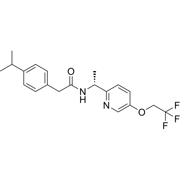
Suvecaltamide (MK-8998) is a selective T-type calcium channel inhibitor with oral efficacy. Suvecaltamide exhibits no cytotoxicity in myeloma cell lines and does not affect the antitumor efficacy of Bortezomib (BTZ). Suvecaltamide reverses BTZ-induced peripheral neuropathy (CIPN) in mouse and rat models, and helps inhibit myeloma growth.
For research use only. We do not sell to patients.
- Purity : 99.96%
- CAS No.: 953778-58-0
- Formula: C20H23F3N2O2
- Molecular Weight:380.40
-
Storage:Powder -20°C, 3 years , 4°C, 2 years ; In solvent -80°C, 2 years , -20°C, 1 year

To place orders, for customer services and technical support, please contact: MedChemExpress USA
Tel: 609-228-6898 E-mail: [email protected] [email protected]


-
 Biological Activity
-
Chemical Information
-
Solvent & Solubility
- Protocols
- Purity & Documentation
- References
-
 Help & FAQs



All Calcium Channel Isoforms
More

Biological Activity
Description
IC50 & Target
|
T-type calcium channel |
Cellular Effect
|
Cell Line

|
Type | Value | Description | References |
|---|---|---|---|---|
| HEK293 | IC50 |
1.2 nM
Compound: 1
|
Inhibition of T-type calcium channel Cav3.1 (unknown origin) expressed in HEK293 cells assessed as calcium influx by Fluo-4-AM dye-based FLIPR assay
Inhibition of T-type calcium channel Cav3.1 (unknown origin) expressed in HEK293 cells assessed as calcium influx by Fluo-4-AM dye-based FLIPR assay
|
[PMID: 27579577] |
| HEK293 | IC50 |
1.2 nM
Compound: 1; MK-8998
|
Inhibition of CaV 3.1 channel (unknown origin) expressed in HEK293 cells by FLEPR Ca2+ flux assay
Inhibition of CaV 3.1 channel (unknown origin) expressed in HEK293 cells by FLEPR Ca2+ flux assay
|
[PMID: 27933950] |
| HEK293 | IC50 |
1.5 nM
Compound: 1
|
Inhibition of T-type calcium channel Cav3.3 (unknown origin) expressed in HEK293 cells assessed as calcium influx by Fluo-4-AM dye-based FLIPR assay
Inhibition of T-type calcium channel Cav3.3 (unknown origin) expressed in HEK293 cells assessed as calcium influx by Fluo-4-AM dye-based FLIPR assay
|
[PMID: 27579577] |
| HEK293 | IC50 |
1.5 nM
Compound: 1; MK-8998
|
Inhibition of CaV 3.3 channel (unknown origin) expressed in HEK293 cells by FLEPR Ca2+ flux assay
Inhibition of CaV 3.3 channel (unknown origin) expressed in HEK293 cells by FLEPR Ca2+ flux assay
|
[PMID: 27933950] |
| HEK293 | IC50 |
8.2 nM
Compound: 1
|
Inhibition of recombinant T-type calcium channel Cav3.2 (unknown origin) expressed in HEK293 cells assessed as inhibition of CaCl2-induced calcium influx preincubated for 3 mins prior CaCl2 addition by Fluo-4-AM dye-based FLIPR assay
Inhibition of recombinant T-type calcium channel Cav3.2 (unknown origin) expressed in HEK293 cells assessed as inhibition of CaCl2-induced calcium influx preincubated for 3 mins prior CaCl2 addition by Fluo-4-AM dye-based FLIPR assay
|
[PMID: 27579577] |
| HEK293 | IC50 |
8.2 nM
Compound: 1; MK-8998
|
Inhibition of CaV 3.2 channel (unknown origin) expressed in HEK293 cells by FLEPR Ca2+ flux assay
Inhibition of CaV 3.2 channel (unknown origin) expressed in HEK293 cells by FLEPR Ca2+ flux assay
|
[PMID: 27933950] |
In Vitro
Suvecaltamide (10-1000 nM, 72 h) does not affect the inhibitory effect of Bortezomib (BTZ, HY-10227) on MM.1S, RPMI 8226, and U266B1 cells.
MedChemExpress (MCE) has not independently confirmed the accuracy of these methods. They are for reference only. Further protocols information, click here.
-
Cell Line:Bone marrow cancer cell lines MM.1S, RPMI 8226, U266B1 (cultured for 72 hours with 0.05-250nM BTZ)
-
Concentration:10, 30, 100, 300, 1000 nM
-
Incubation Time:72 h
-
Result:Did not affect the cytotoxicity of BTZ on cells, and using it alone did not impact cell survival.
In Vivo
Suvecaltamide (30 mg/kg, once daily for 28 days, orally) reduces tumor volume in a xenograft mouse model of human multiple myeloma without affecting the anti-cancer activity of BTZ[1].
MedChemExpress (MCE) has not independently confirmed the accuracy of these methods. They are for reference only.
-
Animal Model:BTZ-Induced CIPN Rat Model[1]
-
Dosage:3, 10, 30 mg/kg; once daily for 28 days
-
Administration:orally
-
Result:Increased raised the nerve conduction velocity (NCV) levels of the tail nerve and sciatic nerve by dose-dependent.
Reduced the β-tubulin polymerization caused by BTZ and increased the number of cutaneous unmyelinated fiber in the hindpaw (IENF) at 30 mg/kg, without affecting proteasome activity.
-
Animal Model:a START cell-based xenograft athymic nude mouse tumor model of human myeloma[1]
-
Dosage:30 mg/kg, once daily for 28 days
-
Administration:orally
-
Result:Reduced the size of the tumor without affecting the anti-cancer activity of BTZ or the weight loss effects.
Clinical Trial
| NCT Number | Sponsor | Condition | Start Date |
Phase
|
|---|---|---|---|---|
| NCT01329991 | Plexxikon| | 2011-05 | PHASE1 |


Chemical Information
-
CAS No. 953778-58-0
-
Appearance Solid
-
Molecular Weight 380.40
-
Formula C20H23F3N2O2
-
Color White to off-white
-
SMILES
O=C(N[C@@H](C1=NC=C(OCC(F)(F)F)C=C1)C)CC2=CC=C(C(C)C)C=C2
-
Synonyms
MK-8998; CX-8998; JZP385
-
Shipping
Room temperature in continental US; may vary elsewhere.
-
Storage
Powder -20°C 3 years 4°C 2 years In solvent -80°C 2 years -20°C 1 year


Solvent & Solubility
In Vitro:
DMSO : 100 mg/mL (262.88 mM; Need ultrasonic; Hygroscopic DMSO has a significant impact on the solubility of product, please use newly opened DMSO)
Please refer to the solubility information to select the appropriate solvent. Once prepared, please aliquot and store the solution to prevent product inactivation from repeated freeze-thaw cycles.
Storage method and period of stock solution: -80°C, 2 years; -20°C, 1 year. When stored at -80°C, please use it within 2 years. When stored at -20°C, please use it within 1 year.
Please refer to the solubility information to select the appropriate solvent. Once prepared, please aliquot and store the solution to prevent product inactivation from repeated freeze-thaw cycles.
Storage method and period of stock solution: -80°C, 2 years; -20°C, 1 year. When stored at -80°C, please use it within 2 years. When stored at -20°C, please use it within 1 year.
Concentration (start) × Volume (start) = Concentration (final) × Volume (final)
In Vivo:
Select the appropriate dissolution method based on your experimental animal and administration route.
- For the following dissolution methods, please ensure to first prepare a clear stock solution using an In Vitro approach and then sequentially add co-solvents:
- To ensure reliable experimental results, the clarified stock solution can be appropriately stored based on storage conditions. As for the working solution for In Vivo experiments, it is recommended to prepare freshly and use it on the same day.
- The percentages shown for the solvents indicate their volumetric ratio in the final prepared solution. If precipitation or phase separation occurs during preparation, heat and/or sonication can be used to aid dissolution.
Add each solvent one by one: 10% DMSO 40% PEG300 5% Tween-80 45% Saline
Solubility: ≥ 2.5 mg/mL (6.57 mM); Clear solution
This protocol yields a clear solution of ≥ 2.5 mg/mL (saturation unknown).
Taking 1 mL working solution as an example, add 100 μL DMSO stock solution (25.0 mg/mL) to 400 μL PEG300, and mix evenly; then add 50 μL Tween-80 and mix evenly; then add 450 μL Saline to adjust the volume to 1 mL.
Preparation of Saline: Dissolve 0.9 g sodium chloride in ddH₂O and dilute to 100 mL to obtain a clear Saline solution.
In Vivo Dissolution Calculator
Please enter the basic information of animal experiments:
-
-
-
-
Recommended: Prepare an additional quantity of animals to account for potential losses during experiments.
Please enter your animal formula composition:
-
%DMSO +
Recommended: Keep the proportion of DMSO in working solution below 2% if your animal is weak.
-
%+
-
+%Tween-80 + +
-
%Saline +
The co-solvents required include: DMSO, . All of co-solvents are available by MedChemExpress (MCE). , Tween 80. All of co-solvents are available by MedChemExpress (MCE).
Working solution concentration: 0.22 mg/mL
Method for preparing stock solution: mg drug dissolved in μL DMSO. Stock solution concentration: mg/mL.
1. Take μL DMSO stock solution;
2. Add μL .
μL , mix evenly;
3. Then add μL Tween 80, mix evenly;
4. Then add μL
Please ensure that the stock solution in the first step is dissolved to a clear state, and add co-solvents in sequence. You can use ultrasonic heating (ultrasonic cleaner, recommended frequency 20-40 kHz), vortexing, etc. to assist dissolution.

Protocols
-
Cell Cytotoxicity Assay
Cytotoxicity assays are usually based on the assessment of cell membrane damage, which can also be indirectly detected by measuring cell viability. Detection methods include MTT assay, CKK-8 assay, LDH assay and ATP assay, etc.
-
Mammalian live/dead viability and cytotoxicity staining
Live/dead viability and cytotoxicity staining assays are based on the simultaneous detection of intracellular esterase activity in metabolically active (viable) cells and membrane integrity loss in non-viable cells. In commonly used dual-staining approaches, membrane-permeant fluorogenic substrates are converted by intracellular esterases into fluorescent products in live cells, while impermeant DNA-binding dyes selectively enter cells with compromised plasma membranes and label nucleic acids in dead or dying cells, enabling discrimination between viable and non-viable populations by fluorescence microscopy or flow cytometry.
-
Patient-Derived Xenograft (PDX)
Patient-derived xenograft (PDX) models are generated by engrafting primary human tumor tissue directly into immunodeficient mice, allowing in vivo propagation of patient tumor biology without initial in vitro adaptation. These models are used to preserve key histopathological and molecular characteristics of the original tumor and enable assessment of tumor growth dynamics and therapeutic response in a living organism. The biological readout is tumor engraftment and subsequent growth in the murine host, which reflects the ability of human tumor cells to survive, vascularize, and expand in an immunocompromised microenvironment.
-
Cell Viability Determination by MTT Colorimetric Assay
The following protocol uses the MTT colorimetric assay as a classic literature-established method for assessing cell viability/metabolic activity in cultured mammalian cells. MTT[3-(4,5-dimethylthiazol-2-yl)-2,5-diphenyltetrazolium bromide] is reduced by metabolically active cells to a colored formazan product; the amount of formazan is quantified spectrophotometrically and provides an indirect measure of metabolically active viable cells. Importantly, MTT reduction reflects cellular oxidoreductase/metabolic activity rather than an absolute direct count of living cells, so changes in cellular metabolism can alter the signal independently of cell number.


Purity & Documentation
-
Data Sheet (271 KB)
-
SDS (396 KB)
- English - EN (396 KB)
- Français - FR (396 KB)
- Deutsch - DE (396 KB)
- Norwegian - NO (396 KB)
- Español - ES (396 KB)
- Swedish - SV (396 KB)
- Italian - IT (396 KB)
- Korean - KR (396 KB)
- Portuguese - PT (396 KB)
-
Handling Instructions (2659 KB)
References


Complete Stock Solution Preparation Table
Please refer to the solubility information to select the appropriate solvent. Once prepared, please aliquot and store the solution to prevent product inactivation from repeated freeze-thaw cycles.
Storage method and period of stock solution: -80°C, 2 years; -20°C, 1 year. When stored at -80°C, please use it within 2 years. When stored at -20°C, please use it within 1 year.
| Optional Solvent | Concentration Solvent Mass | 1 mg | 5 mg | 10 mg | 25 mg |
|---|---|---|---|---|---|
| DMSO | 1 mM | 2.6288 mL | 13.1441 mL | 26.2881 mL | 65.7203 mL |
| 5 mM | 0.5258 mL | 2.6288 mL | 5.2576 mL | 13.1441 mL | |
| 10 mM | 0.2629 mL | 1.3144 mL | 2.6288 mL | 6.5720 mL | |
| 15 mM | 0.1753 mL | 0.8763 mL | 1.7525 mL | 4.3814 mL | |
| 20 mM | 0.1314 mL | 0.6572 mL | 1.3144 mL | 3.2860 mL | |
| 25 mM | 0.1052 mL | 0.5258 mL | 1.0515 mL | 2.6288 mL | |
| 30 mM | 0.0876 mL | 0.4381 mL | 0.8763 mL | 2.1907 mL | |
| 40 mM | 0.0657 mL | 0.3286 mL | 0.6572 mL | 1.6430 mL | |
| 50 mM | 0.0526 mL | 0.2629 mL | 0.5258 mL | 1.3144 mL | |
| 60 mM | 0.0438 mL | 0.2191 mL | 0.4381 mL | 1.0953 mL | |
| 80 mM | 0.0329 mL | 0.1643 mL | 0.3286 mL | 0.8215 mL | |
| 100 mM | 0.0263 mL | 0.1314 mL | 0.2629 mL | 0.6572 mL |


Suvecaltamide Related Classifications
HY-101096 Related Classifications
 Powered by Bioz