



1-Thioglycerol
Based on 1 publication(s) in Google Scholar
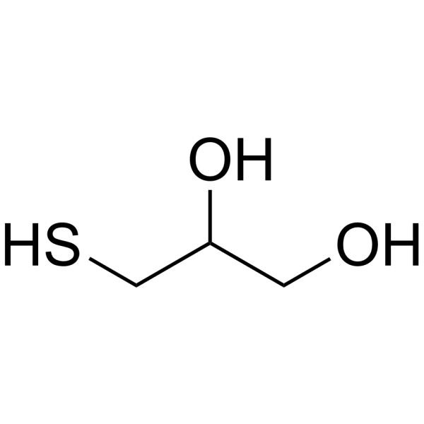
1-Thioglycerol, commonly used as a reducing agent in various biochemical and biophysical applications, especially in protein chemistry and molecular biology, it can protect proteins from oxidation and denaturation, and can reduce disulfide bonds to thiols base, which can then be modified or analyzed. In addition, 1-Thioglycerol has been investigated for potential medical applications, including as an inhibitor of cystic fibrosis, which may help improve the function of lung cells, and has also been studied for Used in the preparation of metal nanoparticles and as a stabilizer for certain pharmaceutical preparations.
For research use only. We do not sell to patients.
- Purity : 99.89%
- CAS No.: 96-27-5
- Formula: C3H8O2S
- Molecular Weight:108.16
-
Storage:
4°C, stored under nitrogen
* In solvent : -80°C, 6 months; -20°C, 1 month (stored under nitrogen)

To place orders, for customer services and technical support, please contact: MedChemExpress USA
Tel: 609-228-6898 E-mail: [email protected] [email protected]


-
 Biological Activity
-
Chemical Information
-
Solvent & Solubility
- Protocols
- Purity & Documentation
-
 Help & FAQs


-
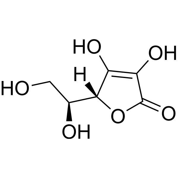
HY-B0166
L-Ascorbic acidAntioxidant -

HY-10182
LaduviglusibGSK-3 Inhibitor -
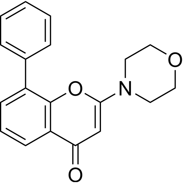
HY-10108
LY294002PI3K Inhibitor -

HY-10431
SB-431542TGF-β Receptor Kinase Inhibitor -

HY-10071
Y-27632ROCK Inhibitor -
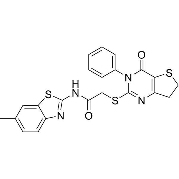
HY-13912
IWP-2Wnt Production Inhibitor -

HY-P0035
Insulin (human)Recombinant Human Insulin -
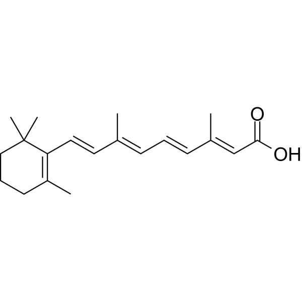
HY-14649
Retinoic acidRAR Agonist -

HY-14648
DexamethasoneGR Agonist -

HY-10583
Y-27632 dihydrochlorideROCK Inhibitor -

HY-K3019
Lipid Concentrate (Chemically Defined) -
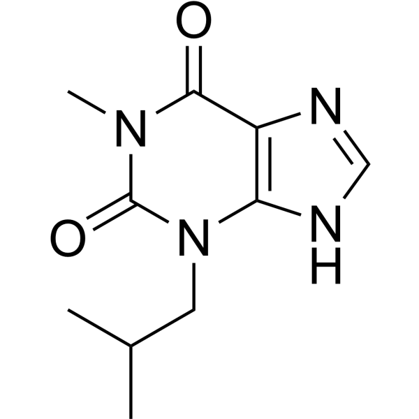
HY-12318
IBMXPDE Inhibitor
Publications Citing Use of MedChemExpress (MCE) 1-Thioglycerol
More

Biological Activity
Description
In Vitro
1-Thioglycerol is a biochemical reagent that can be used as a biological material or organic compound for life science related research.
MedChemExpress (MCE) has not independently confirmed the accuracy of these methods. They are for reference only. Further protocols information, click here.


Chemical Information
-
CAS No. 96-27-5
-
Appearance Liquid (Density: 1.25 g/cm3)
-
Molecular Weight 108.16
-
Formula C3H8O2S
-
Color Colorless to light yellow
-
SMILES
SCC(CO)O
-
Synonyms
alpha-Thioglycerol
-
Shipping
Room temperature in continental US; may vary elsewhere.
-
Storage
4°C, stored under nitrogen
* In solvent : -80°C, 6 months; -20°C, 1 month (stored under nitrogen)


Publications (1)
-
Journal Impact Factor
-
Most Recent


Solvent & Solubility
In Vitro:
DMSO : 100 mg/mL (924.56 mM; Need ultrasonic; Hygroscopic DMSO has a significant impact on the solubility of product, please use newly opened DMSO)
H2O : 50 mg/mL (462.28 mM; Need ultrasonic)
Please refer to the solubility information to select the appropriate solvent. Once prepared, please aliquot and store the solution to prevent product inactivation from repeated freeze-thaw cycles.
Storage method and period of stock solution: -80°C, 6 months; -20°C, 1 month (stored under nitrogen). When stored at -80°C, please use it within 6 months. When stored at -20°C, please use it within 1 month.
* Note: If you choose water as the stock solution, please dilute it to the working solution, then filter and sterilize it with a 0.22 μm filter before use.
Please refer to the solubility information to select the appropriate solvent. Once prepared, please aliquot and store the solution to prevent product inactivation from repeated freeze-thaw cycles.
Storage method and period of stock solution: -80°C, 6 months; -20°C, 1 month (stored under nitrogen). When stored at -80°C, please use it within 6 months. When stored at -20°C, please use it within 1 month.
* Note: If you choose water as the stock solution, please dilute it to the working solution, then filter and sterilize it with a 0.22 μm filter before use.
Concentration (start) × Volume (start) = Concentration (final) × Volume (final)
In Vivo:
Select the appropriate dissolution method based on your experimental animal and administration route.
- For the following dissolution methods, please ensure to first prepare a clear stock solution using an In Vitro approach and then sequentially add co-solvents:
- To ensure reliable experimental results, the clarified stock solution can be appropriately stored based on storage conditions. As for the working solution for In Vivo experiments, it is recommended to prepare freshly and use it on the same day.
- The percentages shown for the solvents indicate their volumetric ratio in the final prepared solution. If precipitation or phase separation occurs during preparation, heat and/or sonication can be used to aid dissolution.
In Vivo Dissolution Calculator
Please enter the basic information of animal experiments:
-
-
-
-
Recommended: Prepare an additional quantity of animals to account for potential losses during experiments.
Working solution concentration: 0.22 mg/mL
This product has good water solubility, please refer to the measured solubility data in water/PBS/Saline for details.

Protocols
-
Fibrosis/Collagen Morphometry
Fibrosis and collagen morphometry is based on the quantitative visualization of fibrillar collagen deposition in tissue sections using histochemical stains such as Sirius Red (Picrosirius Red) or Masson's trichrome, followed by image-based or polarization-enhanced analysis to estimate collagen proportional area as a surrogate of extracellular matrix accumulation during fibrotic remodeling. Sirius Red combined with polarized light microscopy enhances detection of collagen fibers due to birefringence properties, enabling more specific visualization of collagen type I and III fibrils compared to conventional bright-field histology, while whole-section or region-restricted digital morphometry reduces field-selection bias in fibrosis assessment. Alternative quantitative approaches include second harmonic generation (SHG) and two-photon excited fluorescence microscopy, which enable label-free detection of fibrillar collagen and have been validated against histological staining and biochemica
-
Collagen: Sirius Red Staining
Sirius Red or picrosirius red staining is a histochemical method for visualizing collagen-rich extracellular matrix in tissue sections, and collagen fibers are detected as red-stained structures under bright-field microscopy with enhanced birefringence under polarized light. Picrosirius red is useful for assessing total collagen organization, distribution, and fibrosis burden, but polarized color should not be interpreted as a definitive collagen type I versus type III readout because color is affected by fiber orientation, thickness, and packing.
-
Connective Tissue: Masson's Trichrome/Collagen Trichrome Staining
Masson’s Trichrome (collagen/trichrome staining) is a histological technique that differentially stains tissue compartments using sequential acidic dyes to distinguish collagen from muscle and cytoplasmic components based on dye affinity and tissue permeability differences, enabling visualization of fibrosis and connective tissue architecture in histological sections. The classical formulation typically uses Weigert's iron hematoxylin for nuclear staining, Biebrich scarlet-acid fuchsin for cytoplasm and muscle, and aniline blue (or light green variants) for collagen, producing a characteristic blue/green collagen signal contrasted against red cytoplasm and dark nuclei. The staining principle relies on selective displacement of smaller dye molecules by larger anionic dyes in collagen-rich regions under controlled acidified conditions, which enhances collagen-specific dye retention. This property makes the method widely used for fibrosis assessment in organs such as heart, liver, lung, a


Purity & Documentation
-
Data Sheet (243 KB)
-
SDS (483 KB)
- English - EN (483 KB)
- Français - FR (483 KB)
- Deutsch - DE (483 KB)
- Norwegian - NO (483 KB)
- Español - ES (483 KB)
- Swedish - SV (483 KB)
- Italian - IT (483 KB)
- Korean - KR (483 KB)
- Portuguese - PT (483 KB)
-
Handling Instructions (2659 KB)


Complete Stock Solution Preparation Table
Please refer to the solubility information to select the appropriate solvent. Once prepared, please aliquot and store the solution to prevent product inactivation from repeated freeze-thaw cycles.
Storage method and period of stock solution: -80°C, 6 months; -20°C, 1 month (stored under nitrogen). When stored at -80°C, please use it within 6 months. When stored at -20°C, please use it within 1 month.
| Optional Solvent | Concentration Solvent Mass | 1 mg | 5 mg | 10 mg | 25 mg |
|---|---|---|---|---|---|
| H2O / DMSO | 1 mM | 9.2456 mL | 46.2278 mL | 92.4556 mL | 231.1390 mL |
| 5 mM | 1.8491 mL | 9.2456 mL | 18.4911 mL | 46.2278 mL | |
| 10 mM | 0.9246 mL | 4.6228 mL | 9.2456 mL | 23.1139 mL | |
| 15 mM | 0.6164 mL | 3.0819 mL | 6.1637 mL | 15.4093 mL | |
| 20 mM | 0.4623 mL | 2.3114 mL | 4.6228 mL | 11.5570 mL | |
| 25 mM | 0.3698 mL | 1.8491 mL | 3.6982 mL | 9.2456 mL | |
| 30 mM | 0.3082 mL | 1.5409 mL | 3.0819 mL | 7.7046 mL | |
| 40 mM | 0.2311 mL | 1.1557 mL | 2.3114 mL | 5.7785 mL | |
| 50 mM | 0.1849 mL | 0.9246 mL | 1.8491 mL | 4.6228 mL | |
| 60 mM | 0.1541 mL | 0.7705 mL | 1.5409 mL | 3.8523 mL | |
| 80 mM | 0.1156 mL | 0.5778 mL | 1.1557 mL | 2.8892 mL | |
| 100 mM | 0.0925 mL | 0.4623 mL | 0.9246 mL | 2.3114 mL |
* Note: If you choose water as the stock solution, please dilute it to the working solution, then filter and sterilize it with a 0.22 μm filter before use.


1-Thioglycerol Related Classifications
HY-W010572 Related Classifications
 Powered by Bioz