



5-Ethynyl-2'-deoxyuridine
Based on 40 publication(s) in Google Scholar
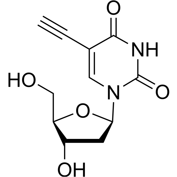
5-Ethynyl-2'-deoxyuridine (EdU), a thymidine analogue, is incorporated into cellular DNA during DNA replication and the subsequent reaction of EdU with a fluorescent azide in a “Click” reaction. EdU staining is a fast, sensitive and reproducible method to study cell proliferation. 5-Ethynyl-2'-deoxyuridine is an alkyl chain-based PROTAC linker that can be used in the synthesis of PROTACs. 5-Ethynyl-2'-deoxyuridine is a click chemistry reagent, it contains an Alkyne group and can undergo copper-catalyzed azide-alkyne cycloaddition (CuAAc) with molecules containing Azide groups.
For research use only. We do not sell to patients.
- Purity : 99.26%
- CAS No.: 61135-33-9
- Formula: C11H12N2O5
- Molecular Weight:252.23
-
Storage:
4°C, protect from light
* In solvent : -80°C, 6 months; -20°C, 1 month (protect from light)

To place orders, for customer services and technical support, please contact: MedChemExpress USA
Tel: 609-228-6898 E-mail: [email protected] [email protected]


-
 Biological Activity
-
Chemical Information
-
Solvent & Solubility
- Protocols
- Purity & Documentation
- References
-
 Help & FAQs


-
HY-15910
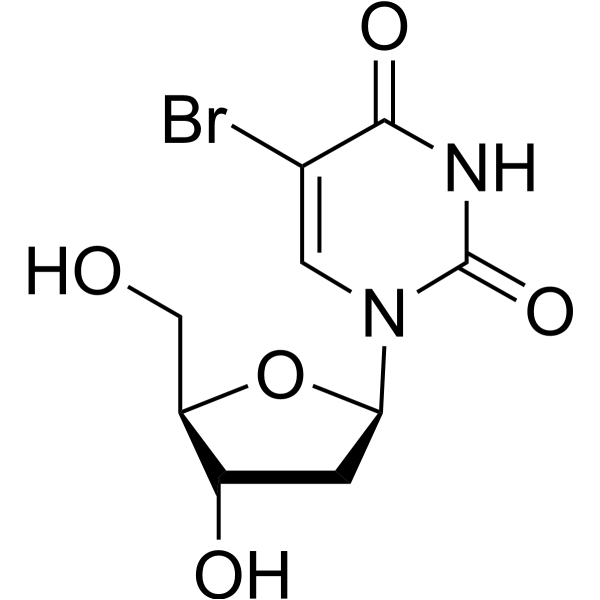
5-BrdUThymidine Analog -
HY-141140
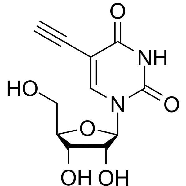
5-EthynyluridineCell-permeable Nucleoside -
HY-90006
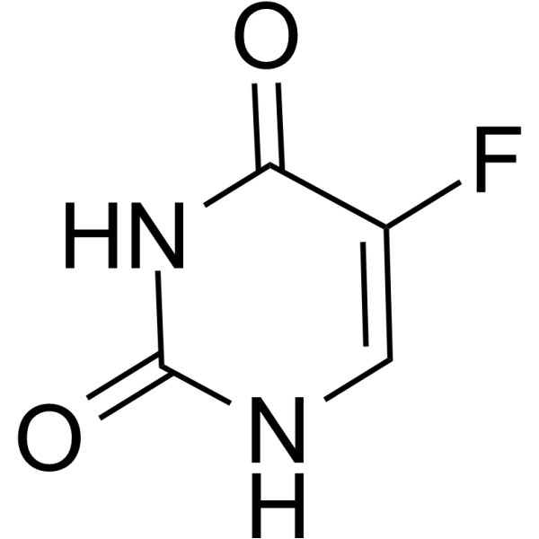
5-FluorouracilThymidylate Synthase Inhibitor -
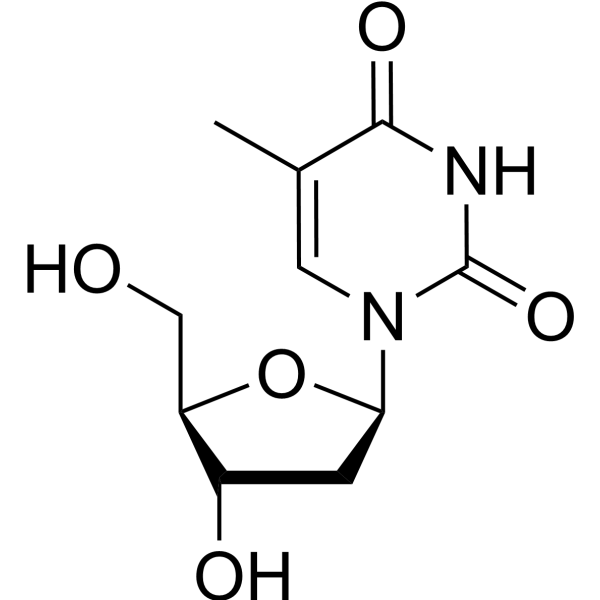
HY-N1150
ThymidineDNA Synthesis Inhibitor -
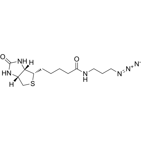
HY-129832
Biotin-azideClick Chemical Reagent -
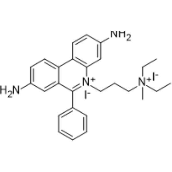
HY-D0815
Propidium IodideFluorochrome -
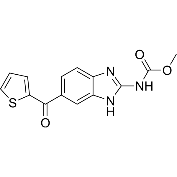
HY-13520
NocodazoleMicrotubule Inhibitor -
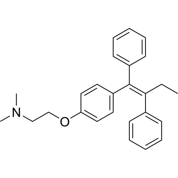
HY-13757A
TamoxifenEstrogen Receptor Modulator -
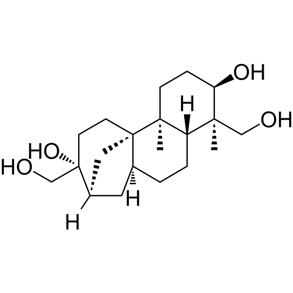
HY-N6733
AphidicolinDNA Polymerase Inhibitor -

HY-10583
Y-27632 dihydrochlorideROCK Inhibitor -
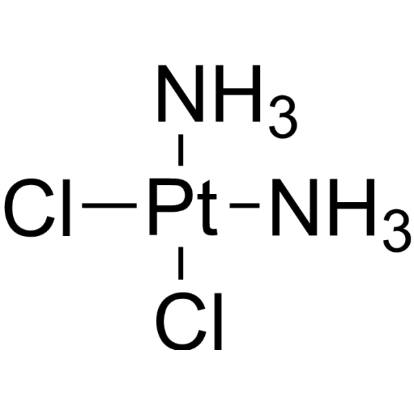
HY-17394
CisplatinDNA Alkylator -
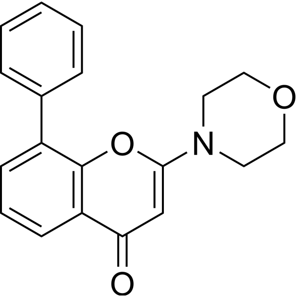
HY-10108
LY294002PI3K Inhibitor
Publications Citing Use of MedChemExpress (MCE) 5-Ethynyl-2'-deoxyuridine
More- Nat Genet. 2025 Dec;57(12):3050-3065. [Abstract]
- Nat Aging. 2025 Mar;5(3):401-418. [Abstract]
- Cell Stem Cell. 2022 Sep 1;29(9):1366-1381.e9. [Abstract]
- Cell Host Microbe. 2024 Feb 14;32(2):191-208.e9. [Abstract]
- Nat Commun. 2025 Nov 4;16(1):9664. [Abstract]
- J Am Chem Soc. 2026 Mar 11;148(9):9478-9493. [Abstract]
- J Clin Invest. 2023 Apr 3;133(7):e162324. [Abstract]
- Adv Sci (Weinh). 2026 Jul 9:e76479.
- Research (Wash D C). 2026 Jul 14:9:1360.
- NPJ Biofilms Microbiomes. 2026 Jan 8;12(1):38. [Abstract]
- J Cachexia Sarcopenia Muscle. 2024 Aug;15(4):1388-1403. [Abstract]
- Int J Biol Macromol. 2025 Jun;315(Pt 1):144297. [Abstract]
- NPJ Parkinsons Dis. 2026 May 26. [Abstract]
- Cell Rep. 2025 Nov 25;44(11):116496. [Abstract]
- Br J Pharmacol. 2025 Nov;182(21):5409-5429. [Abstract]
- PLoS Biol. 2022 Apr 27;20(4):e3001619 [Abstract]
- Neurosci Bull. 2025 May 2. [Abstract]
- JCI Insight. 2024 Dec 20;9(24):e183889. [Abstract]
- Inflamm Res. 2026 Jan 14;75(1):19. [Abstract]
- Front Endocrinol. 2023 Jan 9:13:1052487. [Abstract]
- Exp Neurol. 2026 May:399:115667. [Abstract]
- J Cell Mol Med. 2026 Mar 23;30(6):e71099.
- Reprod Biomed Online. 2022 May;44(5):803-816. [Abstract]
- Kaohsiung J Med Sci. 2022 Apr;38(4):357-366. [Abstract]
- Reprod Sci. 2022 Aug;29(8):2414-2427. [Abstract]
- Cell Microbiol. 2021 Dec;23(12):e13397. [Abstract]
- Nutr Res Pract. 2023 Dec;17(6):1070-1083. [Abstract]
- J Chemother. 2022 Oct;34(6):401-413. [Abstract]
- Lett Drug Des Discov. 2026 May 5.
- bioRxiv. 2026 Jul 13.
- bioRxiv. 2026 Jun 14.
- bioRxiv. 2026 May 26.
- bioRxiv. 2026 May 29.
- NAR Mol Med. 2026 Jan 23;3(1):ugag005. [Abstract]
- bioRxiv. 2025 Aug 1:2025.07.31.667081. [Abstract]
- SSRN. 2025 May 29.
- bioRxiv. 2025 Apr 28:2025.04.28.651040. [Abstract]
- Res Sq. 2024 Jun 03.
- Res Sq. 2024 May 20.
- bioRxiv. 2023 Jun 22.
 Customer Validation & Images

-
Histological Imaging/Staining
-
Histological Imaging/Staining
-
Histological Imaging/Staining
-
Histological Imaging/Staining
-
Histological Imaging/Staining
All PROTAC Linkers Isoforms
More

Biological Activity
Description
IC50 & Target
|
Alkyl-Chain |
Cellular Effect
|
Cell Line

|
Type | Value | Description | References |
|---|---|---|---|---|
| E6SM | EC50 |
0.1 μg/mL
Compound: 7, EDU
|
Antiviral activity against Vaccinia virus in E6SM cells assessed as reduction of virus-induced cytopathogenicity
Antiviral activity against Vaccinia virus in E6SM cells assessed as reduction of virus-induced cytopathogenicity
|
[PMID: 17518459] |
| E6SM | EC50 |
0.7 μg/mL
Compound: 7, EDU
|
Antiviral activity against HSV1 KOS in E6SM cells assessed as reduction of virus-induced cytopathogenicity
Antiviral activity against HSV1 KOS in E6SM cells assessed as reduction of virus-induced cytopathogenicity
|
[PMID: 17518459] |
| E6SM | EC50 |
0.7 μg/mL
Compound: 7, EDU
|
Antiviral activity against thymidine kinase-deficient HSV1 B2006 in E6SM cells assessed as reduction of virus-induced cytopathogenicity
Antiviral activity against thymidine kinase-deficient HSV1 B2006 in E6SM cells assessed as reduction of virus-induced cytopathogenicity
|
[PMID: 17518459] |
| E6SM | EC50 |
2 μg/mL
Compound: 7, EDU
|
Antiviral activity against HSV2 G in E6SM cells assessed as reduction of virus-induced cytopathogenicity
Antiviral activity against HSV2 G in E6SM cells assessed as reduction of virus-induced cytopathogenicity
|
[PMID: 17518459] |
| E6SM | EC50 |
4 μg/mL
Compound: 7, EDU
|
Antiviral activity against thymidine kinase deficient HSV1 VMW1837 in E6SM cells assessed as reduction of virus-induced cytopathogenicity
Antiviral activity against thymidine kinase deficient HSV1 VMW1837 in E6SM cells assessed as reduction of virus-induced cytopathogenicity
|
[PMID: 17518459] |
| FM3A | IC50 |
>=250 μM
Compound: 7, EDU
|
Inhibition of proliferation of thymidine kinase deficient mouse FM3A cells
Inhibition of proliferation of thymidine kinase deficient mouse FM3A cells
|
[PMID: 17518459] |
| FM3A | IC50 |
0.13 μM
Compound: 7, EDU
|
Inhibition of proliferation of thymidine kinase deficient mouse FM3A cells expressing HSV1 thymidine kinase
Inhibition of proliferation of thymidine kinase deficient mouse FM3A cells expressing HSV1 thymidine kinase
|
[PMID: 17518459] |
| FM3A | IC50 |
0.39 μM
Compound: 7, EDU
|
Inhibition of proliferation of thymidine kinase deficient mouse FM3A cells expressing HSV2 thymidine kinase
Inhibition of proliferation of thymidine kinase deficient mouse FM3A cells expressing HSV2 thymidine kinase
|
[PMID: 17518459] |
| HEL | CC50 |
2.5 μM
Compound: 7, EDU
|
Cytotoxicity against HEL cells
Cytotoxicity against HEL cells
|
[PMID: 17518459] |
| HEL | EC50 |
0.11 μM
Compound: 7, EDU
|
Antiviral activity against TK+ VZV YS in HEL cells assessed as reduction of virus plaque formation
Antiviral activity against TK+ VZV YS in HEL cells assessed as reduction of virus plaque formation
|
[PMID: 17518459] |
| HEL | EC50 |
0.16 μM
Compound: 7, EDU
|
Antiviral activity against TK+ VZV OKA in HEL cells assessed as reduction of virus plaque formation
Antiviral activity against TK+ VZV OKA in HEL cells assessed as reduction of virus plaque formation
|
[PMID: 17518459] |
| HEL | EC50 |
0.75 μM
Compound: 7, EDU
|
Antiviral activity against thymidine kinase deficient VZV YS in HEL cells assessed as reduction of virus plaque formation
Antiviral activity against thymidine kinase deficient VZV YS in HEL cells assessed as reduction of virus plaque formation
|
[PMID: 17518459] |
| HEL | EC50 |
0.81 μM
Compound: 7, EDU
|
Antiviral activity against thymidine kinase deficient VZV 07/1 in HEL cells assessed as reduction of virus plaque formation
Antiviral activity against thymidine kinase deficient VZV 07/1 in HEL cells assessed as reduction of virus plaque formation
|
[PMID: 17518459] |
| HEL | EC50 |
0.85 μM
Compound: 7, EDU
|
Antiviral activity against HCMV Davis in HEL cells assessed as reduction of virus plaque formation
Antiviral activity against HCMV Davis in HEL cells assessed as reduction of virus plaque formation
|
[PMID: 17518459] |
| HEL | EC50 |
1.2 μM
Compound: 7, EDU
|
Antiviral activity against HCMV AD169 in HEL cells assessed as reduction of virus plaque formation
Antiviral activity against HCMV AD169 in HEL cells assessed as reduction of virus plaque formation
|
[PMID: 17518459] |
| MCF7 | IC50 |
0.4 μM
Compound: 2h
|
Cytotoxicity against human MCF7 cells after 72 hrs
Cytotoxicity against human MCF7 cells after 72 hrs
|
[PMID: 17336074] |
| MDA-MB-231 | IC50 |
4.4 μM
Compound: 2h
|
Cytotoxicity against human MDA-MB-231 cells after 72 hrs
Cytotoxicity against human MDA-MB-231 cells after 72 hrs
|
[PMID: 17336074] |
| OST | IC50 |
23 μM
Compound: 7, EDU
|
Inhibition of proliferation of thymidine kinase deficient OST cells
Inhibition of proliferation of thymidine kinase deficient OST cells
|
[PMID: 17518459] |
| S49 | EC50 |
0.12 μM
Compound: EdUrd
|
Effective concentration to inhibit the growth of Wild-type S49 cells
Effective concentration to inhibit the growth of Wild-type S49 cells
|
[PMID: 6796687] |
In Vitro
5-Ethynyl-2'-deoxyuridine (EdU) staining is a fast, sensitive and reproducible method to study cell proliferation in the central nervous system[1].
MedChemExpress (MCE) has not independently confirmed the accuracy of these methods. They are for reference only. Further protocols information, click here.
In Vivo
MedChemExpress (MCE) has not independently confirmed the accuracy of these methods. They are for reference only.
-
Animal Model:Two month old female mice[1]
-
Dosage:10, 20, 50, 100 or 200 mg/kg
-
Administration:Single injection; intraperitoneally; 4 hours after EdU injection, brains were processed for EdU staining.
-
Result:EdU positive cell numbers slightly increased in a dose-dependent manner both in control and running mice.
Clinical Trial
| NCT Number | Sponsor | Condition | Start Date |
Phase
|
|---|---|---|---|---|
| NCT01329991 | Plexxikon| | 2011-05 | PHASE1 |


Chemical Information
-
CAS No. 61135-33-9
-
Appearance Solid
-
Molecular Weight 252.23
-
Formula C11H12N2O5
-
SMILES
O[C@H]1C[C@H](N2C(NC(C(C#C)=C2)=O)=O)O[C@@H]1CO
-
Synonyms
EdU
-
Shipping
Room temperature in continental US; may vary elsewhere.
-
Storage
4°C, protect from light
* In solvent : -80°C, 6 months; -20°C, 1 month (protect from light)


Publications (40)
-
Journal Impact Factor
-
Most Recent
-
Nat Genet
TGF-β builds a dual immune barrier in colorectal cancer by impairing T cell recruitment and instructing immunosuppressive SPP1+ macrophages. [Abstract]2025 Dec;57(12):3050-3065. PMID: 41203813 -
Nat Aging
2025 Mar;5(3):401-418. PMID: 39794469 -
Cell Stem Cell
Hepatic cytochrome P450 8B1 and cholic acid potentiate intestinal epithelial injury in colitis by suppressing intestinal stem cell renewal. [Abstract]2022 Sep 1;29(9):1366-1381.e9. PMID: 36055192
5-Ethynyl-2'-deoxyuridine purchased from MedChemExpress. Usage Cited in: Cell Stem Cell. 2022 Sep 1;29(9):1366-1381.e9. [Abstract]
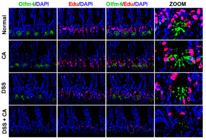
Representative Edu and Olfm4 staining and quantitation in intestine of CA-treated colitic mice. To measure the proliferation of intestinal epithelial cells, 5-ethynyl-2’-deoxyuridine (Edu) at 100 mg/kg was intraperitoneally injected into mice 2 h prior to killing.
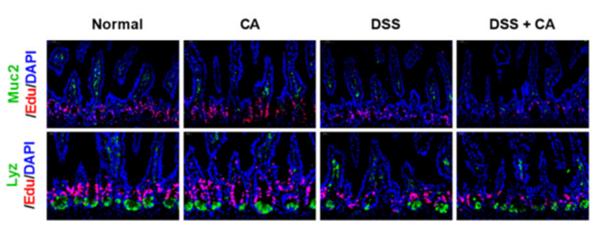
5-Ethynyl-2'-deoxyuridine purchased from MedChemExpress. Usage Cited in: Cell Stem Cell. 2022 Sep 1;29(9):1366-1381.e9. [Abstract]
Representative Muc2 or Lyz with Edu staining of intestine sections. To measure the proliferation of intestinal epithelial cells, 5-ethynyl-2’-deoxyuridine (Edu) at 100 mg/kg was intraperitoneally injected into mice 2 h prior to killing.
-
Cell Host Microbe
A gut microbiota-bile acid axis promotes intestinal homeostasis upon aspirin-mediated damage. [Abstract]2024 Feb 14;32(2):191-208.e9. PMID: 38237593
5-Ethynyl-2'-deoxyuridine purchased from MedChemExpress. Usage Cited in: Cell Host Microbe. 2024 Feb 14;32(2):191-208.e9. [Abstract]
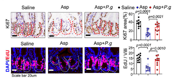
Ki67 and EdU staining of distal jejunum of mice treated. To measure the proliferation of intestinal epithelial cells, 5-ethynyl-20-deoxyuridine at 100 mg/kg was intraperitoneally injected into mice 12 h prior to sacrifice. P. g: Live P. goldsteinii. Red/white dotted line marks single intestinal crypt. Ten random points of one sample were picked and the average value for each mouse was calculated as one single point. Red: EdU; Blue: DAPI. n=10 replicates/group.
5-Ethynyl-2'-deoxyuridine purchased from MedChemExpress. Usage Cited in: Cell Host Microbe. 2024 Feb 14;32(2):191-208.e9. [Abstract]
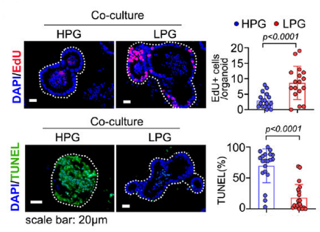
EdU (upper set) and TUNEL (bottom set) staining of organoids after being cocultured with HPG or LPG. White dotted line marks a single organoid. To measure the proliferation of intestinal epithelial cells, 5-ethynyl-20-deoxyuridine at 100 mg/kg was intraperitoneally injected into mice 12 h prior to sacrifice. Scale bars: 20 μm. Red: EdU; Green: TUNEL; Blue: DAPI. n=20 biological replicates/group.
5-Ethynyl-2'-deoxyuridine purchased from MedChemExpress. Usage Cited in: Cell Host Microbe. 2024 Feb 14;32(2):191-208.e9. [Abstract]
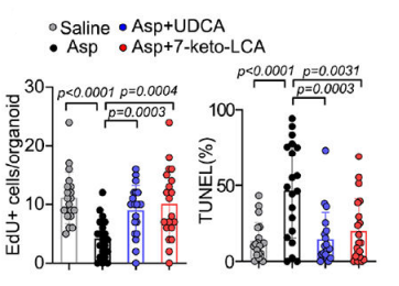
Statistical analysis of EdU (Left) and TUNEL (right) staining of organoids generated from mice treated. To measure the proliferation of intestinal epithelial cells, 5-ethynyl-20-deoxyuridine at 100 mg/kg was intraperitoneally injected into mice 12 h prior to sacrifice. n=20 biological replicates/group.
-
Nat Commun
Regenerating liver uses ammonia to support de novo pyrimidine synthesis and cell proliferation. [Abstract]2025 Nov 4;16(1):9664. PMID: 41188238 -
J Am Chem Soc
2026 Mar 11;148(9):9478-9493. PMID: 41739600 -
J Clin Invest
Coordinated activation of c-Src and FOXM1 drives tumor cell proliferation and breast cancer progression. [Abstract]2023 Apr 3;133(7):e162324. PMID: 36795481 -
-
-
NPJ Biofilms Microbiomes
Disruption of bile acid homeostasis potentiates Paneth cell ablation by activating the intestinal Farnesoid X receptor in necrotizing enterocolitis. [Abstract]2026 Jan 8;12(1):38. PMID: 41507219 -
J Cachexia Sarcopenia Muscle
Single-cell RNA-seq reveals novel interaction between muscle satellite cells and fibro-adipogenic progenitors mediated with FGF7 signalling. [Abstract]2024 Aug;15(4):1388-1403. PMID: 38751367 -
Int J Biol Macromol
Ionized Angelica polysaccharides derivatives slow tumor growth in vivo and induce the apoptosis of HepG2 cells via mitochondria death receptor-mediated pathways in vitro. [Abstract]2025 Jun;315(Pt 1):144297. PMID: 40383338 -
NPJ Parkinsons Dis
Cryopreservable dopaminergic progenitors derived from human iPSCs with accelerated loss of pluripotency and early functional restoration in Parkinsonian rats. [Abstract]2026 May 26. PMID: 42191734 -
Cell Rep
A generalized strategy to kill leukemic cells by targeting the regulatory systems governing mitochondrial membrane potential. [Abstract]2025 Nov 25;44(11):116496. PMID: 41166305 -
Br J Pharmacol
Cholinergic G protein-coupled bile acid receptor 1 (TGR5/GPBA) in the medial septal orchestrates adult hippocampal neurogenesis and cognition in Alzheimer's disease mice. [Abstract]2025 Nov;182(21):5409-5429. PMID: 40926398 -
PLoS Biol
Optineurin promotes myogenesis during muscle regeneration in mice by autophagic degradation of GSK3β. [Abstract]2022 Apr 27;20(4):e3001619 PMID: 35476671 -
Neurosci Bull
Endothelial Cell Integrin α6 Regulates Vascular Remodeling Through the PI3K/Akt-eNOS-VEGFA Axis After Stroke. [Abstract]2025 May 2. PMID: 40316875 -
JCI Insight
BPDCN MYB fusions regulate cell cycle genes, impair differentiation, and induce myeloid-dendritic cell leukemia. [Abstract]2024 Dec 20;9(24):e183889. PMID: 39499902 -
Inflamm Res
Erianin protects chondrocytes against IL-1β-induced oxidative stress and ferroptosis by activating GPX4/STING signaling in osteoarthritis. [Abstract]2026 Jan 14;75(1):19. PMID: 41533098 -
Front Endocrinol
Corticosterone induces obesity partly via promoting intestinal cell proliferation and survival. [Abstract]2023 Jan 9:13:1052487. PMID: 36699046 -
Exp Neurol
Exosomes-mediated delivery of miR-27a-3p antagomir alleviates white matter injury by regulating PPARγ/PRDX1/JNK pathway after subarachnoid hemorrhage in rats. [Abstract]2026 May:399:115667. PMID: 41581691 -
-
Reprod Biomed Online
MicroRNA let-7i inhibits granulosa-luteal cell proliferation and oestradiol biosynthesis by directly targeting IMP2. [Abstract]2022 May;44(5):803-816. PMID: 35339367 -
Kaohsiung J Med Sci
PAX9 functions as a tumor suppressor gene for cervical cancer via modulating cell proliferation and apoptosis. [Abstract]2022 Apr;38(4):357-366. PMID: 34931758 -
Reprod Sci
Identification of Circular RNA circ_0017068 as a Regulator of Proliferation and Apoptosis in Trophoblast Cells by miR-330-5p/XIAP Axis. [Abstract]2022 Aug;29(8):2414-2427. PMID: 34981461 -
Cell Microbiol
Chlamydia and HPV induce centrosome amplification in the host cell through additive mechanisms. [Abstract]2021 Dec;23(12):e13397. PMID: 34716742 -
Nutr Res Pract
Sanghuangporus sanghuang extract inhibits the proliferation and invasion of lung cancer cells in vitro and in vivo. [Abstract]2023 Dec;17(6):1070-1083. PMID: 38053828 -
J Chemother
Circ_SETD3 regulates gefitinib sensitivity and tumor progression by miR-873-5p-dependent regulation of APPBP2 in non-small cell lung cancer. [Abstract]2022 Oct;34(6):401-413. PMID: 34861803 -
-
-
-
-
-
NAR Mol Med
The fluoropyrimidine polymer CF10 synergizes with 5-ethynyl-2'-deoxyuridine by promoting telomere attrition and mitotic catastrophe. [Abstract]2026 Jan 23;3(1):ugag005. PMID: 41635782 -
bioRxiv
A small molecule inhibitor of NVL suppresses tumor growth by blocking ribosome biogenesis. [Abstract]2025 Aug 1:2025.07.31.667081. PMID: 40766499 -
-
bioRxiv
2025 Apr 28:2025.04.28.651040. PMID: 40492193 -
-
-


Solvent & Solubility
In Vitro:
DMSO : 50 mg/mL (198.23 mM; Need ultrasonic; Hygroscopic DMSO has a significant impact on the solubility of product, please use newly opened DMSO)
H2O : 25 mg/mL (99.12 mM; Need ultrasonic)
Please refer to the solubility information to select the appropriate solvent. Once prepared, please aliquot and store the solution to prevent product inactivation from repeated freeze-thaw cycles.
Storage method and period of stock solution: -80°C, 6 months; -20°C, 1 month (protect from light). When stored at -80°C, please use it within 6 months. When stored at -20°C, please use it within 1 month.
* Note: If you choose water as the stock solution, please dilute it to the working solution, then filter and sterilize it with a 0.22 μm filter before use.
Please refer to the solubility information to select the appropriate solvent. Once prepared, please aliquot and store the solution to prevent product inactivation from repeated freeze-thaw cycles.
Storage method and period of stock solution: -80°C, 6 months; -20°C, 1 month (protect from light). When stored at -80°C, please use it within 6 months. When stored at -20°C, please use it within 1 month.
* Note: If you choose water as the stock solution, please dilute it to the working solution, then filter and sterilize it with a 0.22 μm filter before use.
Concentration (start) × Volume (start) = Concentration (final) × Volume (final)
In Vivo:
For the following dissolution methods, please prepare the working solution directly:
It is recommended to prepare fresh solutions and use them promptly within a short period of time.
The percentages shown for the solvents indicate their volumetric ratio in the final prepared solution. If precipitation or phase separation occurs during preparation, heat and/or sonication can be used to aid dissolution.
Add each solvent one by one: PBS
Solubility: 4 mg/mL (15.86 mM); Clear solution; Need ultrasonic and warming and heat to 60°C

Protocols
-
BrdU Incorporation Assay
Bromodeoxyuridine (BrdU) incorporation assay is based on the principle that BrdU, a thymidine analog, is incorporated into newly synthesized DNA during the S phase of the cell cycle, thereby serving as a marker of DNA replication and cellular proliferation. Incorporated BrdU can be detected using anti-BrdU antibodies following DNA denaturation, enabling visualization or quantification of proliferating cells through immunochemical detection methods such as immunofluorescence or immunohistochemistry.
-
Somatic Cell Culture
A method of simulating the in vivo environment in vitro to maintain the cell growth, differentation and main functions.
-
CFSE Dye Dilution Proliferation Assay
The CFSE (carboxyfluorescein diacetate succinimidyl ester) dye dilution proliferation assay is based on the covalent labeling of intracellular proteins by a cell-permeant fluorescent dye that becomes fluorescent upon intracellular ester cleavage and then is stably retained within cells. As labeled cells divide, the dye is partitioned equally between daughter cells, resulting in a stepwise halving of fluorescence intensity that can be quantified by flow cytometry to determine the number of cell divisions undergone by each cell population. This fluorescence dilution approach enables quantitative tracking of lymphocyte proliferation at the single-cell level over multiple rounds of division. CFSE-based proliferation analysis has been widely applied to measure antigen-driven lymphocyte expansion in vitro, where discrete fluorescence peaks correspond to successive cell divisions and allow reconstruction of proliferative history within heterogeneous populations.
-
Dye-dilution cell tracking and proliferation staining
Dye-dilution cell tracking assays quantify cell proliferation by covalently labeling intracellular proteins with a stable fluorescent dye that is equally partitioned between daughter cells during mitosis, resulting in stepwise halving of fluorescence intensity with each cell division as measured by flow cytometry histograms. Carboxyfluorescein diacetate succinimidyl ester (CFSE) is a prototypical dye that diffuses into cells, is enzymatically converted into a fluorescent compound, and then covalently binds intracellular amine groups, producing long-lived fluorescence suitable for tracking multiple rounds of division in vitro and in vivo. Successive generations of dividing cells form discrete peaks of decreasing fluorescence intensity, enabling estimation of proliferation history, precursor frequency, and division index within heterogeneous populations. Alternative dyes such as CellTrace Violet (CTV) and far-red membrane dyes (e. g. , PKH26) follow the same dilution principle but differ
-
CCK-8/WST-8 Cell Proliferation Assay
The CCK-8/WST-8 assay is based on the reduction of the water-soluble tetrazolium salt WST-8 to a water-soluble formazan product by cellular dehydrogenases in metabolically active cells, where the generated formazan amount is proportional to the number of living cells and is quantified by measuring absorbance in the visible range, providing a colorimetric readout for cell viability and proliferation assessment. This class of tetrazolium-based assays improves upon earlier MTT-based systems by producing a water-soluble formazan, eliminating the need for organic solubilization steps and enabling direct spectrophotometric measurement in culture medium.
-
Protocol for Cell Cycle
Cell-cycle analysis by flow cytometry measures DNA content in single cells to estimate the fraction of cells in G0/G1, S, and G2/M phases. Propidium iodide intercalates into DNA, and after RNA removal with RNase, fluorescence intensity reflects cellular DNA content: 2N cells are assigned to G0/G1, cells between 2N and 4N to S phase, and 4N cells to G2/M. DNA-content analysis alone cannot reliably separate G0 from G1 or G2 from M. Ki-67 can distinguish quiescent G0 cells from cycling cells, EdU or BrdU incorporation marks active DNA synthesis in S phase, and phospho-histone H3 staining identifies mitotic cells within the 4N population.
-
Cell Counting-Based Growth Curve Assay
Cell counting-based growth curve assays quantify cell proliferation by directly measuring changes in viable cell number over time using manual or automated counting methods such as hemocytometer-based counting or instrument-assisted cell enumeration, enabling construction of growth curves that reflect population expansion dynamics in response to culture conditions. A widely used approach is trypan blue exclusion with hemocytometer counting, where membrane-compromised (non-viable) cells take up the dye, allowing discrimination between viable and non-viable cells while simultaneously enabling total cell number quantification. Repeated sampling across time points allows estimation of proliferation rate, growth phases, and comparative growth kinetics between experimental conditions.
-
Colony Formation (Clonogenic) Assay
The clonogenic (colony formation) assay measures the ability of a single cell to retain reproductive viability and form a macroscopic colony, typically defined as a cluster derived from one progenitor cell after a defined growth period. This assay is widely used to evaluate cell survival after exposure to ionizing radiation or cytotoxic treatments and is considered a standard method in radiation biology for generating dose-response relationships of reproductive cell death. Colony formation reflects long-term proliferative capacity rather than short-term metabolic activity, and survival is quantified by comparing treated versus untreated conditions based on colony number and derived survival fractions.
-
Cell Viability Determination by MTT Colorimetric Assay
The following protocol uses the MTT colorimetric assay as a classic literature-established method for assessing cell viability/metabolic activity in cultured mammalian cells. MTT[3-(4,5-dimethylthiazol-2-yl)-2,5-diphenyltetrazolium bromide] is reduced by metabolically active cells to a colored formazan product; the amount of formazan is quantified spectrophotometrically and provides an indirect measure of metabolically active viable cells. Importantly, MTT reduction reflects cellular oxidoreductase/metabolic activity rather than an absolute direct count of living cells, so changes in cellular metabolism can alter the signal independently of cell number.
-
EdU Incorporation Assay (Click Chemistry-Based DNA Synthesis Measurement)
The EdU incorporation assay measures DNA synthesis by adding the thymidine analog 5-ethynyl-2′-deoxyuridine to cells or tissues, where it is incorporated into newly synthesized DNA during S phase. Incorporated EdU is detected by copper-catalyzed azide-alkyne cycloaddition, in which a fluorescent azide covalently reacts with the ethynyl group on EdU, allowing S-phase cells to be detected by fluorescence microscopy, flow cytometry, or high-content imaging. EdU detection does not require DNA denaturation or anti-BrdU antibody access, which preserves sample structure and improves compatibility with immunostaining and multiparameter cytometry compared with BrdU-based detection. EdU can be cytotoxic in a cell-type- and exposure-dependent manner, so pulse duration, concentration, and continuous-labeling designs should be validated for each cell type.
-
Ki-67 Immunostaining Proliferation Assay
Ki-67 immunostaining measures the growth fraction of a cell population by detecting Ki-67, a nuclear antigen present in proliferating cells and absent in quiescent G0 cells. The readout is the percentage of Ki-67-positive nuclei among total counted cells, commonly called the Ki-67 labeling index or proliferation index.
-
PCNA Immunodetection Proliferation Assay
PCNA immunodetection measures proliferative activity by detecting proliferating cell nuclear antigen, a nuclear protein associated with DNA polymerase δ function and DNA replication. The assay readout is the proportion of PCNA-positive nuclei among total counted cells, but PCNA labeling is not identical to BrdU labeling because PCNA can mark late G1/early S-associated replication competence and may persist beyond active DNA synthesis depending on fixation and extraction conditions.
-
Protocol for Cell Counting and Cell Density Analysis
Cell counting and cell-density analysis estimate the number of cells in a known volume or field area. Manual hemocytometer counting uses a chamber of defined geometry to convert counted cells into cells/mL, while automated counters and image-analysis workflows detect cell objects from optical, brightfield, fluorescence, impedance, or digital-image features. Trypan blue viability counting is based on dye exclusion: viable cells with intact membranes exclude dye, while non-viable cells with compromised membranes stain blue. The readout is total cell density, viable-cell density, dead-cell density, and percent viability. Cell density can also be estimated from microscopy images by counting objects per image area, from flow cytometry using calibrated volume or reference particles, or from in situ microscopy in bioreactors after calibration against reference methods such as hemocytometer or flow cytometry.
-
MTT Cell Proliferation Assay
The MTT assay is a colorimetric endpoint assay for estimating viable cell number, cell growth, cytotoxicity, or cell activation in cultured mammalian cells. Living cells reduce the yellow tetrazolium salt MTT into purple/blue formazan, while dead cells do not generate the same signal; the resulting color can be quantified with a multiwell spectrophotometer. MTT reduction is commonly interpreted as a readout of metabolic activity that often correlates with viable cell number, but it should not be treated as a direct cell-counting method unless the assay is optimized for the cell type and experimental condition. Studies show that MTT reduction can involve mitochondrial and non-mitochondrial reducing systems, and formazan may accumulate in intracellular lipid droplets rather than simply marking mitochondria.


Purity & Documentation
-
Data Sheet (278 KB)
-
SDS (419 KB)
- English - EN (419 KB)
- Français - FR (419 KB)
- Deutsch - DE (419 KB)
- Norwegian - NO (419 KB)
- Español - ES (419 KB)
- Swedish - SV (419 KB)
- Italian - IT (419 KB)
- Korean - KR (419 KB)
- Portuguese - PT (419 KB)
-
Handling Instructions (2659 KB)
References
[1]. Chenbo Zeng, et al. Evaluation of 5-ethynyl-2'-deoxyuridine staining as a sensitive and reliable method for studying cell proliferation in the adult nervous system. Brain Res. 2010 Mar 10;1319:21-32. [Content Brief]
[2]. An S, et al. Small-molecule PROTACs: An emerging and promising approach for the development of targeted therapy drugs. EBioMedicine. 2018 Oct;36:553-562 [Content Brief]


Complete Stock Solution Preparation Table
Please refer to the solubility information to select the appropriate solvent. Once prepared, please aliquot and store the solution to prevent product inactivation from repeated freeze-thaw cycles.
Storage method and period of stock solution: -80°C, 6 months; -20°C, 1 month (protect from light). When stored at -80°C, please use it within 6 months. When stored at -20°C, please use it within 1 month.
| Optional Solvent | Concentration Solvent Mass | 1 mg | 5 mg | 10 mg | 25 mg |
|---|---|---|---|---|---|
| H2O / DMSO | 1 mM | 3.9646 mL | 19.8232 mL | 39.6464 mL | 99.1159 mL |
| 5 mM | 0.7929 mL | 3.9646 mL | 7.9293 mL | 19.8232 mL | |
| 10 mM | 0.3965 mL | 1.9823 mL | 3.9646 mL | 9.9116 mL | |
| 15 mM | 0.2643 mL | 1.3215 mL | 2.6431 mL | 6.6077 mL | |
| 20 mM | 0.1982 mL | 0.9912 mL | 1.9823 mL | 4.9558 mL | |
| 25 mM | 0.1586 mL | 0.7929 mL | 1.5859 mL | 3.9646 mL | |
| 30 mM | 0.1322 mL | 0.6608 mL | 1.3215 mL | 3.3039 mL | |
| 40 mM | 0.0991 mL | 0.4956 mL | 0.9912 mL | 2.4779 mL | |
| 50 mM | 0.0793 mL | 0.3965 mL | 0.7929 mL | 1.9823 mL | |
| 60 mM | 0.0661 mL | 0.3304 mL | 0.6608 mL | 1.6519 mL | |
| 80 mM | 0.0496 mL | 0.2478 mL | 0.4956 mL | 1.2389 mL | |
| DMSO | 100 mM | 0.0396 mL | 0.1982 mL | 0.3965 mL | 0.9912 mL |
* Note: If you choose water as the stock solution, please dilute it to the working solution, then filter and sterilize it with a 0.22 μm filter before use.


5-Ethynyl-2'-deoxyuridine Related Classifications
HY-118411 Related Classifications
 Powered by Bioz