



Di-8-ANEPPS
Based on 1 publication(s) in Google Scholar
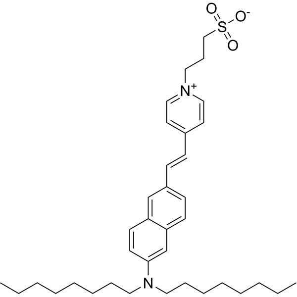
Di-8-ANEPPS is a naphthylstyryl voltage-sensitive dye, shifting both their fluorescence excitation and emission spectra upon changes in Vm .
For research use only. We do not sell to patients.
- Purity : 99.84%
- CAS No.: 157134-53-7
- Formula: C36H52N2O3S
- Molecular Weight:592.87
-
Storage:
-20°C, protect from light
* The compound is unstable in solutions, freshly prepared is recommended.

To place orders, for customer services and technical support, please contact: MedChemExpress USA
Tel: 609-228-6898 E-mail: [email protected] [email protected]


-
 Biological Activity
-
Chemical Information
-
Solvent & Solubility
- Protocols
- Purity & Documentation
- References
-
 Help & FAQs


-
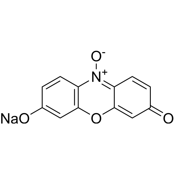
HY-111391
Resazurin sodiumBacterial Inhibitor -
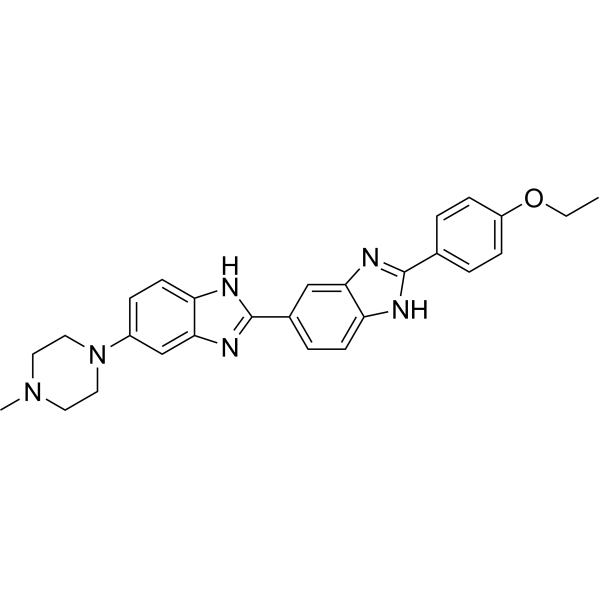
HY-15559
Hoechst 33342DNA Fluorescent Stain -
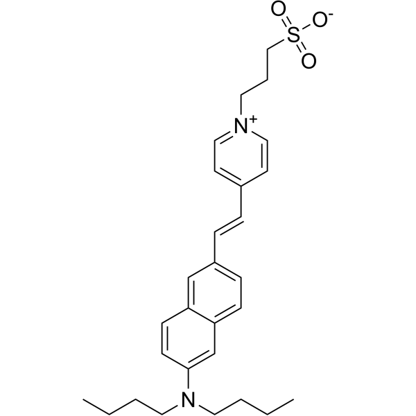
HY-129763
Di-4-ANEPPSVoltage-Sensitive Dye -
HY-W090090
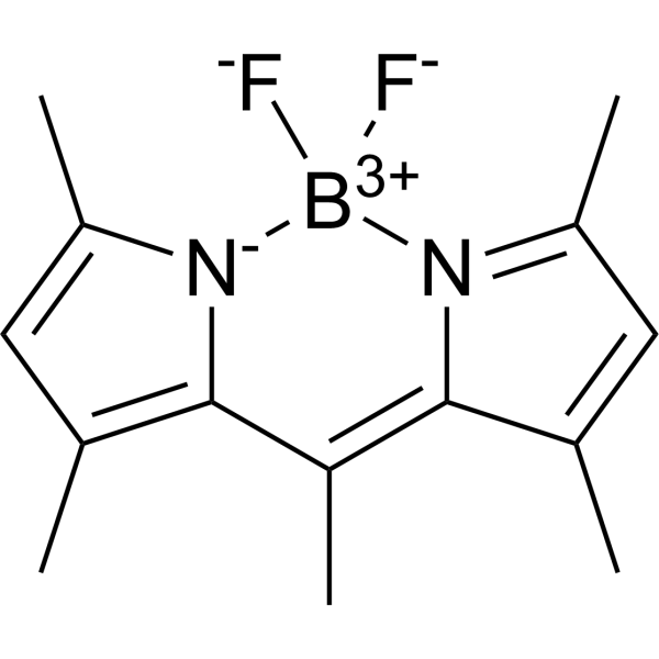
BODIPY 493/503Neutral Lipid Dye -
HY-129905A
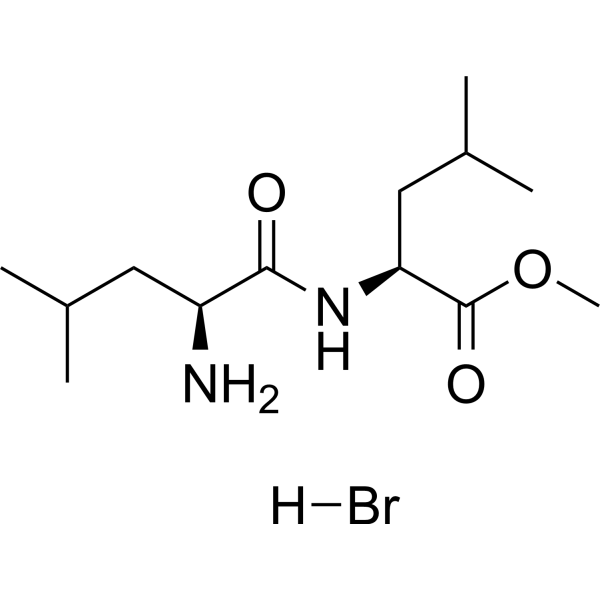
L-Leucyl-L-Leucine methyl ester hydrobromideDipeptide condensation product -
HY-103469
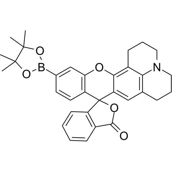
Peroxy Orange 1H2O2 probe -
HY-D0083
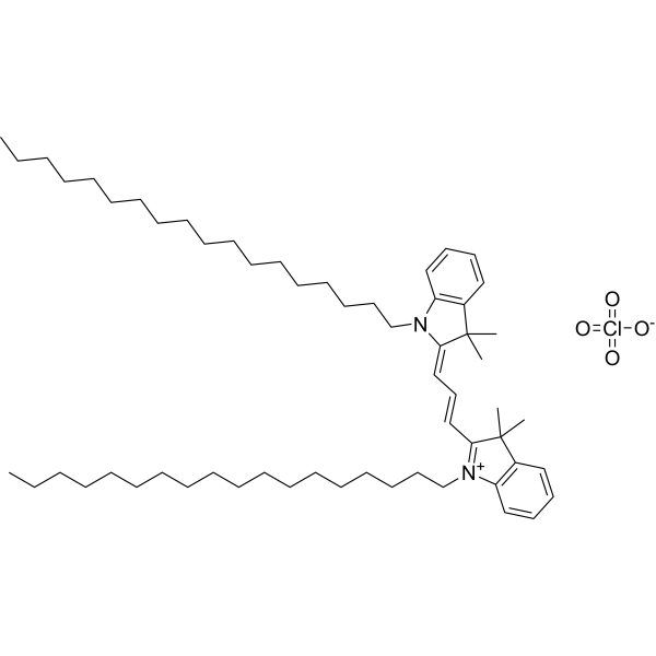
DiIFluorescent Dye -
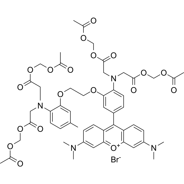
HY-D0989
Rhod-2 AMCa(2+) Indicator -

HY-DY1012
DiI (solution)Fluorescent Dye -

HY-DY1020
Rhod-2 AM (solution)Ca(2+) Indicator
Publications Citing Use of MedChemExpress (MCE) Di-8-ANEPPS
More

Biological Activity
Description
In Vitro
A mouse cell staining with Di-8-ANEPPS is applied voltage clamp pulses and immersed in Na+-containing solution. The Di-8-ANEPPS signal, which largely reflects t system voltage, has an asymmetrical positive component upon application of depolarizing pulses. This is interpreted as reflecting a propagating action potential, escaping from the voltage-clamped plasma membrane. Its temporal properties are not unlike those of the "center" signal. Notably, the peak of the signal occurrs 0.3 ms after the leading edge of the depolarizing pulse[1].
MedChemExpress (MCE) has not independently confirmed the accuracy of these methods. They are for reference only. Further protocols information, click here.
Emission (Em)
635
Excitation (Ex)
465


Chemical Information
-
CAS No. 157134-53-7
-
Appearance Solid
-
Molecular Weight 592.87
-
Formula C36H52N2O3S
-
Color Brown to red
-
SMILES
O=S(CCC[N+]1=CC=C(/C=C/C2=CC=C3C=C(N(CCCCCCCC)CCCCCCCC)C=CC3=C2)C=C1)([O-])=O
-
Shipping
Room temperature in continental US; may vary elsewhere.
-
Storage
-20°C, protect from light
* The compound is unstable in solutions, freshly prepared is recommended.


Publications (1)
-
Journal Impact Factor
-
Most Recent
-
Food Funct
2024 Sep 30;15(19):9863-9879. PMID: 39246047


Solvent & Solubility
In Vitro:
DMSO : 1 mg/mL (1.69 mM; ultrasonic and warming and heat to 60°C; Hygroscopic DMSO has a significant impact on the solubility of product, please use newly opened DMSO)
Please refer to the solubility information to select the appropriate solvent. The compound is unstable in solutions, freshly prepared is recommended.
Please refer to the solubility information to select the appropriate solvent. The compound is unstable in solutions, freshly prepared is recommended.
Concentration (start) × Volume (start) = Concentration (final) × Volume (final)

Protocols
-
Cardiac voltage-sensitive optical mapping
Cardiac voltage-sensitive optical mapping records changes in transmembrane potential from cardiac tissue by staining the preparation with a voltage-sensitive dye and imaging fluorescence changes during electrical activation; the resulting optical action potentials can be used to map activation time, action potential duration, conduction velocity, wavefront propagation, and arrhythmia dynamics. The optical signal represents a relative fluorescence change from a tissue volume rather than a single-cell intracellular recording, so spatial resolution, sampling rate, voltage resolution, optical magnification, light penetration, and motion control must be considered together when interpreting optical action potentials.
-
Neuronal voltage-sensitive dye imaging
Neuronal voltage-sensitive dye imaging detects membrane-potential-dependent optical changes from dyes associated with neuronal membranes, enabling optical recording of electrical activity from single neurons, dendrites, axons, spines, or neuronal populations in brain slices and cultured neurons. VSD signals are typically reported as fractional fluorescence or absorbance changes over baseline, such as ΔF/F or ΔI/I, and published protocols use high-speed cameras or photodiode arrays because neuronal voltage signals occur on millisecond time scales. Fast VSD imaging can be applied at two common scales: bulk staining of brain slices to measure circuit-level spatiotemporal activity, and single-cell loading or biolistic delivery to record membrane-potential transients from individual neuronal compartments. Optical signals should be interpreted as membrane-potential-related readouts, and validation by simultaneous electrophysiology or pharmacological controls is recommended when the experimen
-
Fluorescent plasma-membrane potential dye assay
Fluorescent plasma-membrane potential dye assays measure changes in cell membrane potential using voltage-sensitive dyes whose fluorescence changes when cells depolarize or hyperpolarize. Anionic bis-oxonol dyes such as DiBAC4(3) enter depolarized cells more readily and show increased fluorescence after intracellular binding, while hyperpolarization reduces dye accumulation and fluorescence. FMP/FLIPR membrane-potential dyes are used for faster, homogeneous microplate assays of ion-channel or receptor-mediated membrane-potential changes.


Purity & Documentation
-
Data Sheet (273 KB)
-
SDS (252 KB)
- English - EN (252 KB)
- Français - FR (252 KB)
- Deutsch - DE (252 KB)
- Norwegian - NO (252 KB)
- Español - ES (252 KB)
- Swedish - SV (252 KB)
- Italian - IT (252 KB)
- Korean - KR (252 KB)
- Portuguese - PT (252 KB)
-
Handling Instructions (2659 KB)
References


Complete Stock Solution Preparation Table
Please refer to the solubility information to select the appropriate solvent. The compound is unstable in solutions, freshly prepared is recommended.
| Optional Solvent | Concentration Solvent Mass | 1 mg | 5 mg | 10 mg | 25 mg |
|---|---|---|---|---|---|
| DMSO | 1 mM | 1.6867 mL | 8.4336 mL | 16.8671 mL | 42.1678 mL |


Di-8-ANEPPS Related Classifications
HY-101891 Related Classifications
 Powered by Bioz