



Quin-2AM
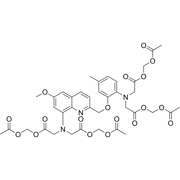
Quin-2AM is a fluorecent Ca2+ chelator, with high affinity for calcium. Quin-2AM can specifically identify intracellular calcium ions, with high sensitivity, low cytotoxicity, increased AM acetylmethyl ester can enter the cell well, after being sheared by the intracellular esterase stay in the cell to bind to calcium ions, produce strong fluorescence.
Nur für Forschungszwecke. Wir verkaufen nicht an Patienten.
- CAS. Nr.: 83104-85-2
- Formel: C38H43N3O18
- Molecular Weight:829.76
-
Speicherung:
Please store the product under the recommended conditions in the Certificate of Analysis.

To place orders, for customer services and technical support, please contact: MedChemExpress USA
Tel: 609-228-6898 E-mail: [email protected] [email protected]


-
 Biologische Aktivität
-
Chemical Information
- Protokoll
- Reinheit & Dokumentation
- Verweise
-
 Help & FAQs





Biologische Aktivität
Beschreibung
In Vitro
Guide (The following is our recommended protocol. This protocol is for reference only and should be modified according to your specific needs).
1. Preparation of Quin-2AM working solution
1.1 Preparation of stock solution
Dilute Quin-2AM with anhydrous DMSO to prepare a 10 mM stock solution.
Note: It is recommended to aliquot the Quin-2AM stock solution and store at -20°C or -80°C protected from light.
1.2 Preparation of working solution
Dilute the stock solution with pre-warmed HBSS to prepare a 1–10 μM Quin-2AM working solution.
Note: Adjust the concentration of Quin-2AM working solution according to actual needs and prepare fresh before use.
2. Cell staining (suspension cells)
2.1 Collect cells by centrifugation and wash twice with PBS, 5 min each time. Cell density: 1×10⁶/mL.
2.2 Add 1 mL Quin-2AM working solution and incubate at room temperature for 5–30 min.
2.3 Centrifuge at 400 g for 3–4 min and discard the supernatant.
2.4 Wash cells twice with PBS, 5 min each time.
2.5 Resuspend cells in 1 mL HBSS and observe using a fluorescence microscope or flow cytometer.
3. Cell staining (adherent cells)
3.1 Culture adherent cells on sterile coverslips.
3.2 Remove the coverslips from the culture medium and aspirate excess medium.
3.3 Add 100 μL dye working solution, gently shake to fully cover the cells, and incubate for 5–30 min.
3.4 Remove the dye working solution, wash 2–3 times with culture medium (5 min each), and observe using a fluorescence microscope.
MedChemExpress (MCE) has not independently confirmed the accuracy of these methods. They are for reference only.
Emission (Em)
492
Excitation (Ex)
339


Chemical Information
-
CAS. Nr. 83104-85-2
-
Molecular Weight 829.76
-
Formel C38H43N3O18
-
SMILES
O=C(OCOC(C)=O)CN(CC(OCOC(C)=O)=O)C1=CC=C(C)C=C1OCC2=NC3=C(N(CC(OCOC(C)=O)=O)CC(OCOC(C)=O)=O)C=C(OC)C=C3C=C2
-
Synonyms
Quin-2 acetoxymethyl ester
-
Versand
Room temperature in continental US; may vary elsewhere.
-
Speicherung
Please store the product under the recommended conditions in the Certificate of Analysis.


Reinheit & Dokumentation
Verweise
[1]. Chu AJ, et al. Possible role of Marcks in the cellular modulation of monocytic tissue factor-initiated hypercoagulation. Br J Haematol. 2002 Aug;118(2):569-76. [Content Brief]
[2]. Chu AJ, et al. Possible role of Marcks in the cellular modulation of monocytic tissue factor-initiated hypercoagulation. Br J Haematol. 2002 Aug;118(2):569-76. [Content Brief]

Calculators
Konzentration (Stammlösung) × Volumen (Stammlösung) = Konzentration (Ziellösung) × Volumen (Ziellösung)


Quin-2AM Related Classifications
HY-101902 Related Classifications
 Powered by Bioz