



Y15
Based on 31 publication(s) in Google Scholar
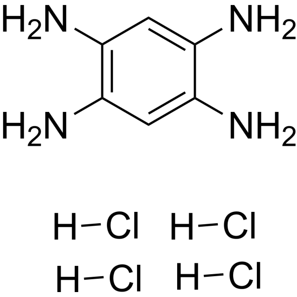
Y15 is a potent and specific inhibitor of focal adhesion kinase (FAK) that inhibits its autophosphorylation activity, decreases the viability of cancer cells, and blocks tumor growth.
For research use only. We do not sell to patients.
- Purity : 98.0%
- CAS No.: 4506-66-5
- Formula: C6H14Cl4N4
- Molecular Weight:284.01
-
Storage:
Store at room temperature, keep dry and cool.
In solvent -80°C, 1 year , -20°C, 6 months

To place orders, for customer services and technical support, please contact: MedChemExpress USA
Tel: 609-228-6898 E-mail: [email protected] [email protected]


-
 Biological Activity
-
Chemical Information
-
Solvent & Solubility
- Protocols
- Purity & Documentation
- References
-
 Help & FAQs


-
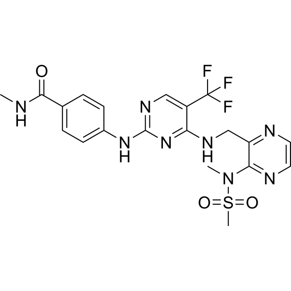
HY-12289
DefactinibFAK Inhibitor -
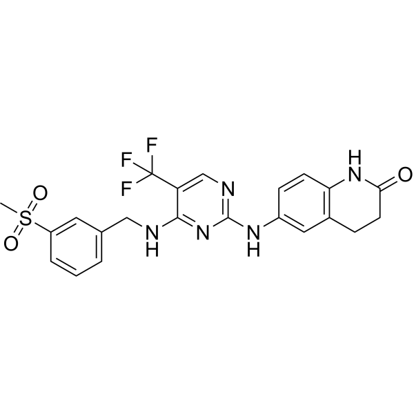
HY-10461
PF-573228FAK Inhibitor -
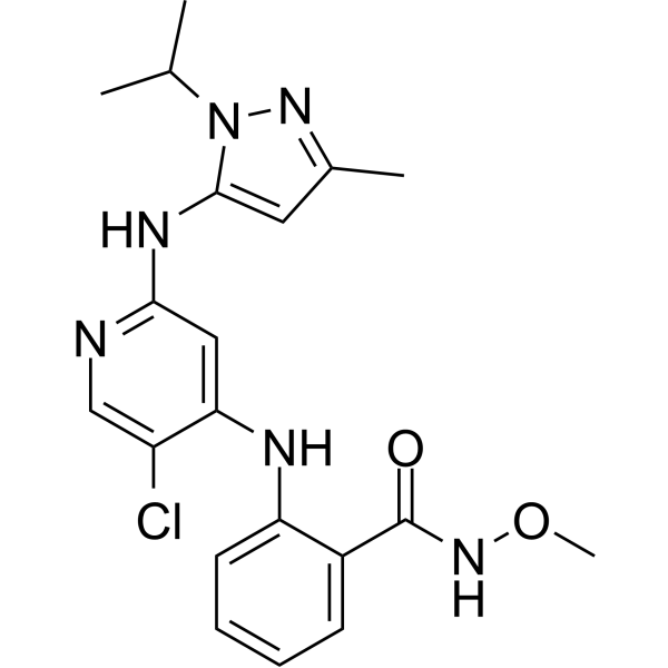
HY-100498
GSK2256098FAK Inhibitor -
HY-10181
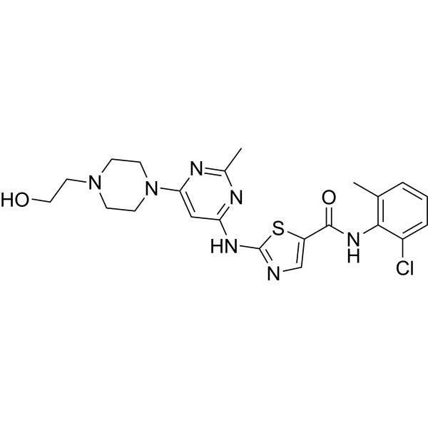
DasatinibSrc/Bcr-Abl Inhibitor -
HY-B0146
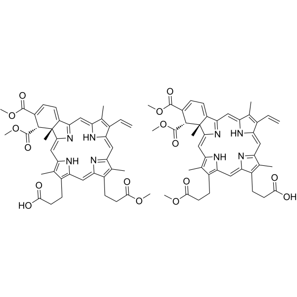
VerteporfinYAP Inhibitor -
HY-13966
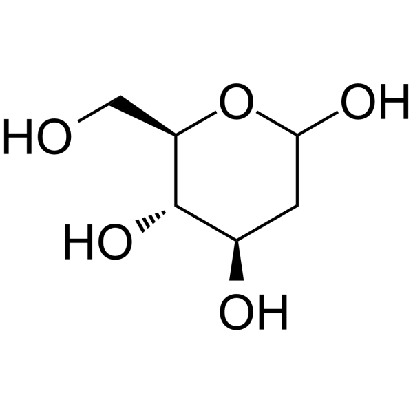
2-Deoxy-D-glucoseGlycolysis Inhibitor -
HY-134570
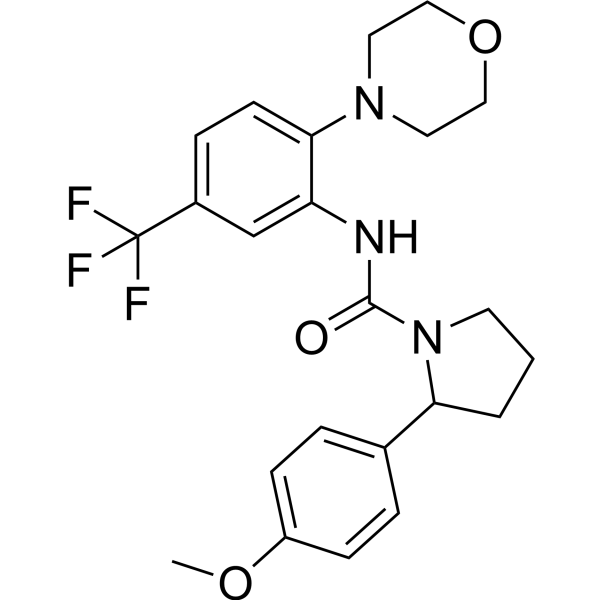
ZINC40099027FAK Activator -

HY-13259
MG-132Proteasome Inhibitor -
HY-10459
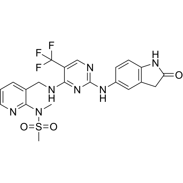
PF-562271FAK/Pyk2 Inhibitor -
HY-12028
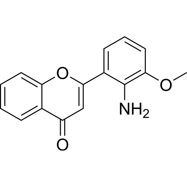
PD98059MEK Inhibitor -
HY-12041
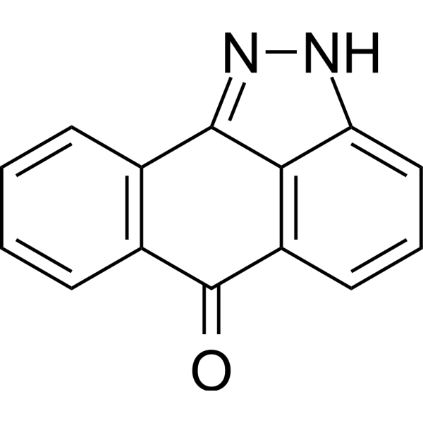
SP600125JNK Inhibitor -
HY-16578
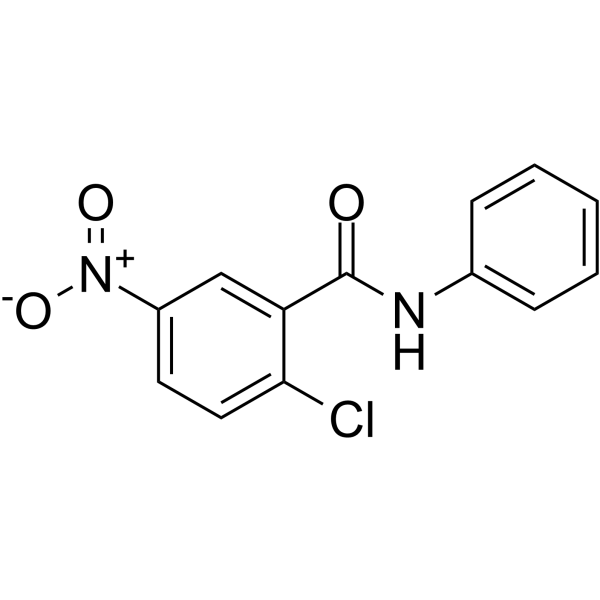
GW9662PPARγ Antagonist
Publications Citing Use of MedChemExpress (MCE) Y15
More- J Extracell Vesicles. 2025 Aug;14(8):e70146. [Abstract]
- Autophagy. 2025 Mar;21(3):664-673. [Abstract]
- Nat Commun. 2025 Mar 31;16(1):3093. [Abstract]
- J Adv Res. 2025 Nov 19:S2090-1232(25)00929-4. [Abstract]
- Adv Sci (Weinh). 2024 Dec;11(47):e2405705. [Abstract]
- Carbohydr Polym. 2026 Feb 15:374:124706. [Abstract]
- Neuro Oncol. 2024 Jan 5;26(1):137-152. [Abstract]
- NPJ Precis Oncol. 2023 Aug 11;7(1):74. [Abstract]
- Cell Mol Gastroenterol Hepatol. 2021;11(3):683-696. [Abstract]
- Stem Cell Res Ther. 2021 Oct 7;12(1):523. [Abstract]
- Atherosclerosis. 2023 Jul:377:43-59. [Abstract]
- Cell Biosci. 2025 Oct 31;15(1):150. [Abstract]
- ACS Biomater Sci Eng. 2024 Jul 8;10(7):4463-4479. [Abstract]
- J Hypertens. 2022 Sep 1;40(9):1666-1681. [Abstract]
- Front Cell Dev Biol. 2021 May 28:9:607001. [Abstract]
- Exp Neurol. 2024 Jun:376:114776. [Abstract]
- J Cell Mol Med. 2026 Jul;30(13):e71275.
- Acta Biochim Biophys Sin (Shanghai). 2018 Oct 1;50(10):976-983. [Abstract]
- FASEB J. 2024 May 31;38(10):e23698. [Abstract]
- J Cell Sci. 2026 Jul 1;139(13):jcs264323.
- Am J Cancer Res. 2023 May 15;13(5):1718-1743. [Abstract]
- Biomed Res Int. 2021 Mar 1:2021:6685493. [Abstract]
- Biochem Biophys Res Commun. 2025 May 12:763:151788. [Abstract]
- Comp Biochem Physiol A Mol Integr Physiol. 2020 Mar:241:110639. [Abstract]
- Exp Ther Med. 2020 Aug;20(2):1405-1414. [Abstract]
- Reprod Dev Med. October 27, 2022.
- bioRxiv. 2025 May 14:2025.05.12.653281. [Abstract]
- Patent. US20240100051A1
- bioRxiv. 2023 Feb 5.
- Patent. US20200383981A1.
- Oncotarget. 2017 Oct 5;8(56):95586-95595. [Abstract]
 Customer Validation & Images

-
Cell Proliferation/Viability Assay
-
Cell Imaging/Staining
-
WB
-
IP
-
WB


Biological Activity
Description
IC50 & Target
FAK[1]
In Vitro
Y15 directly blocks autophosphorylation activity of FAK. Y15 inhibits Y397 phosphorylation of FAK starting at 0.1 μM in Panc-1 cells. At a dose of 100 μM, Y15 has the same or better inhibition as TAE226. Of note, total FAK is downregulated at higher doses of Y15. Y15 also blocks phosphorylation of the FAK downstream substrate, paxillin. Total paxillin is decreased at higher doses similar to FAK. Thus, Y15 inhibits FAK phosphorylation in a dose-dependent manner[1]. MTS assay is completed using a range of Y15 doses on all cell lines (TT, K1, BCPAP, and TPC1, respectively).Y15 inhibited cell viability in a dose-dependent manner across all thyroid cell lines evaluated. IC50 is 2.05, 5.74, 9.99, and 17.54 μM for TT, TPC1, BCPAP, and K1, respectively[2].
In macrophages treated with Y15, the filopodia and lamellipodia remain mobile. The lawn of particles surrounding macrophages appeared larger in cells treated with Y15[4].
MedChemExpress (MCE) has not independently confirmed the accuracy of these methods. They are for reference only. Further protocols information, click here.
In Vivo
MedChemExpress (MCE) has not independently confirmed the accuracy of these methods. They are for reference only.


Chemical Information
-
CAS No. 4506-66-5
-
Appearance Solid
-
Molecular Weight 284.01
-
Formula C6H14Cl4N4
-
Color Off-white to gray
-
SMILES
[H]Cl.[H]Cl.[H]Cl.[H]Cl.NC1=CC(N)=C(N)C=C1N
-
Synonyms
FAK Inhibitor 14
-
Shipping
Room temperature in continental US; may vary elsewhere.
-
Storage
Store at room temperature, keep dry and cool
In solvent -80°C 1 year -20°C 6 months


Publications (31)
-
Journal Impact Factor
-
Most Recent
-
J Extracell Vesicles
Exomeres From Adventitial Fibroblasts of Spontaneously Hypertensive Rats Promote Vascular Remodelling via Transferring Osteopontin. [Abstract]2025 Aug;14(8):e70146. PMID: 40767027
Y15 purchased from MedChemExpress. Usage Cited in: J Extracell Vesicles. 2025 Aug;14(8):e70146. [Abstract]
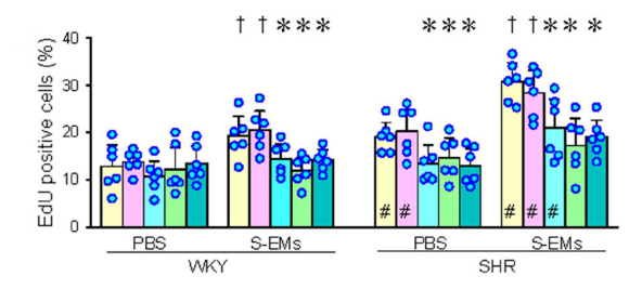
The effects of FAK inhibitor Y15 (2.5 µM; bar chart 3), PI3K inhibitor LY294002 (5 µM), or AKT inhibitor AKTi1/2 (0.5 µM) on S-EMs-induced VSMC proliferation and migration. S-EMs (40 µg/mL) were administered 2 hours after inhibitor application. VSMC proliferation and migration were assessed by the percentage of EdU-positive cells and Boyden chamber assays.
-
Autophagy
Deciphering melanophagy: role of the PTK2-ITCH-MLANA-OPTN cascade on melanophagy in melanocytes. [Abstract]2025 Mar;21(3):664-673. PMID: 39477686
Y15 purchased from MedChemExpress. Usage Cited in: Autophagy. 2025 Mar;21(3):664-673. [Abstract]
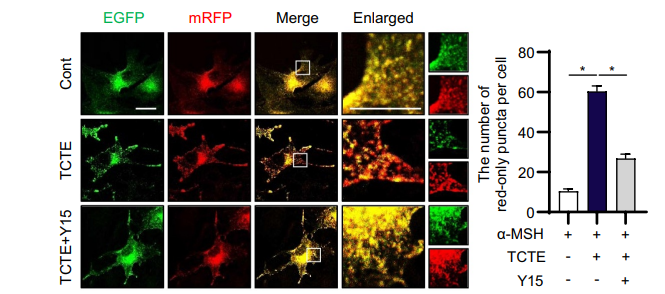
B16F1/TPC2-mRFP-EGFP cells stimulated with α-MSH were treated with TCTE (10 μg/ml) and Y15 (1.5 μM) for 24 hours. The cells were then fixed and imaged using a confocal microscope.
Y15 purchased from MedChemExpress. Usage Cited in: Autophagy. 2025 Mar;21(3):664-673. [Abstract]
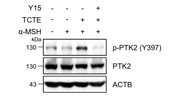
α-MSH-stimulated B16F1 cells were cotreated with TCTE (10 μg/ml) and PTK2 inhibitor Y15 (1.5 μM) for 24 h. Then, the cells were harvested and analyzed by western blotting with indicated antibodies.
Y15 purchased from MedChemExpress. Usage Cited in: Autophagy. 2025 Mar;21(3):664-673. [Abstract]
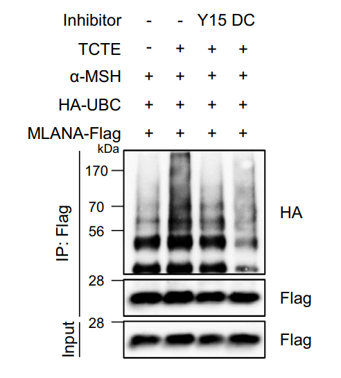
α-MSH-stimulated B16F1 cells were cotransfected with HA-UBC and MLANA-Flag. After 1 day, the cells were further treated with α-MSH (1 μM) for 48 h and exposed to TCTE (10 μg/ml) with Y15 (1.5 μM) or DC (25 μM) for 24 h. Then, the cells were further treated with MG132 (10 μM) for 4 h and pulled down with anti-Flag antibody conjugated with agarose beads. The immunoprecipitates were analyzed by western blotting with indicated antibodies.
-
Nat Commun
2025 Mar 31;16(1):3093. PMID: 40164597
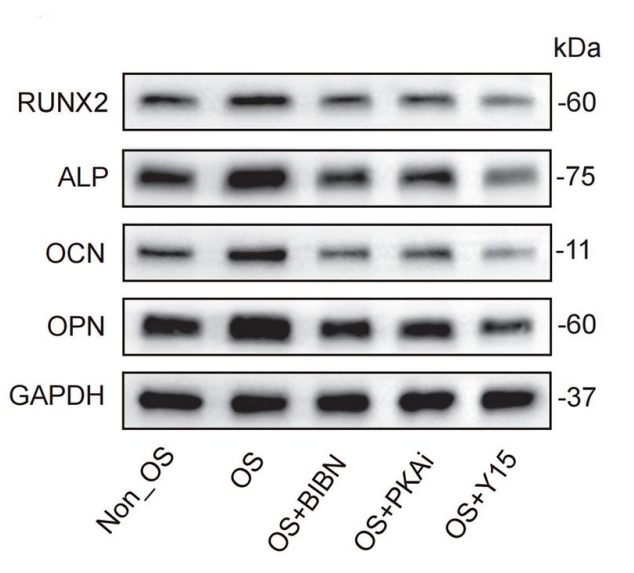
Y15 purchased from MedChemExpress. Usage Cited in: Nat Commun. 2025 Mar 31;16(1):3093. [Abstract]
Y15 (100 μg/kg). Expression levels of bone formation-related proteins (Runx2/ALP/OPN/OCN) in peri-implant bone tissue after 2 weeks of treatment with different antagonists/inhibitors.
-
J Adv Res
Dysregulated ITGA3/FAK/YAP axis mediates impaired alveolar type II epithelial cells function in COPD. [Abstract]2025 Nov 19:S2090-1232(25)00929-4. PMID: 41270956 -
Adv Sci (Weinh)
Piezo1 Regulates Stiffness-Dependent DRG Axon Regeneration via Modifying Cytoskeletal Dynamics. [Abstract]2024 Dec;11(47):e2405705. PMID: 39514408 -
Carbohydr Polym
Decoding the pharmacokinetics of intravenous lentinan: A multiscale journey from the blood to hepatic Kupffer cells' lysosomes. [Abstract]2026 Feb 15:374:124706. PMID: 41371850 -
Neuro Oncol
A disintegrin and metalloproteinase 22 activates integrin β1 through its disintegrin domain to promote the progression of pituitary adenoma. [Abstract]2024 Jan 5;26(1):137-152. PMID: 37555799 -
NPJ Precis Oncol
Chimeric antibody targeting unique epitope on onco-mucin16 reduces tumor burden in pancreatic and lung malignancies. [Abstract]2023 Aug 11;7(1):74. PMID: 37567918 -
Cell Mol Gastroenterol Hepatol
2021;11(3):683-696. PMID: 33075564 -
Stem Cell Res Ther
Regulation of the integrin αVβ3- actin filaments axis in early osteogenesis of human fibroblasts under cyclic tensile stress. [Abstract]2021 Oct 7;12(1):523. PMID: 34620239 -
Atherosclerosis
SIRT3-and FAK-mediated acetylation-phosphorylation crosstalk of NFATc1 regulates Nε-carboxymethyl-lysine-induced vascular calcification in diabetes mellitus. [Abstract]2023 Jul:377:43-59. PMID: 37392543 -
Cell Biosci
FAK-dependent activation of src family kinase member BLK contributed to endometrial fibrosis via endoplasmic reticulum stress. [Abstract]2025 Oct 31;15(1):150. PMID: 41174819 -
ACS Biomater Sci Eng
Hydroxyapatite Nanoparticles Promote the Development of Bone Microtissues for Accelerated Bone Regeneration by Activating the FAK/Akt Pathway. [Abstract]2024 Jul 8;10(7):4463-4479. PMID: 38848471 -
J Hypertens
WNT1-inducible signalling pathway protein 1 stabilizes atherosclerotic plaques in apolipoprotein-E-deficient mice via the focal adhesion kinase/mitogen-activated extracellular signal-regulated kinase/extracellular signal-regulated kinase pathway. [Abstract]2022 Sep 1;40(9):1666-1681. PMID: 35881419 -
Front Cell Dev Biol
Hepatoma Cell-Derived Extracellular Vesicles Promote Liver Cancer Metastasis by Inducing the Differentiation of Bone Marrow Stem Cells Through microRNA-181d-5p and the FAK/Src Pathway. [Abstract]2021 May 28:9:607001. PMID: 34124029 -
Exp Neurol
LAMC1 attenuates neuronal apoptosis via FAK/PI3K/AKT signaling pathway after subarachnoid hemorrhage. [Abstract]2024 Jun:376:114776. PMID: 38609046 -
-
Acta Biochim Biophys Sin (Shanghai)
Insulin-like growth factor 1 receptor signaling regulates embryonic epicardial cell proliferation through focal adhesion kinase pathway. [Abstract]2018 Oct 1;50(10):976-983. PMID: 30184089 -
FASEB J
FOXS1 acts as an oncogene and induces EMT through FAK/PI3K/AKT pathway by upregulating HILPDA in prostate cancer. [Abstract]2024 May 31;38(10):e23698. PMID: 38780613 -
-
Am J Cancer Res
Linc00662 m6A promotes the progression and metastasis of pancreatic cancer by activating focal adhesion through the GTF2B-ITGA1-FAK pathway. [Abstract]2023 May 15;13(5):1718-1743. PMID: 37293163 -
Biomed Res Int
Focal Adhesion Kinase Inhibitor Inhibits the Oxidative Damage Induced by Central Venous Catheter via Abolishing Focal Adhesion Kinase-Protein Kinase B Pathway Activation. [Abstract]2021 Mar 1:2021:6685493. PMID: 33748278 -
Biochem Biophys Res Commun
FAK regulates trophoblast functions of invasion and proliferation through Rap1 pathway in early-onset preeclampsia. [Abstract]2025 May 12:763:151788. PMID: 40220488 -
Comp Biochem Physiol A Mol Integr Physiol
Focal adhesion kinase and osmotic responses in ionocytes of Fundulus heteroclitus, a euryhaline teleost fish. [Abstract]2020 Mar:241:110639. PMID: 31863842 -
Exp Ther Med
Collagen triple helix repeat containing 1 promotes endometrial cancer cell migration by activating the focal adhesion kinase signaling pathway. [Abstract]2020 Aug;20(2):1405-1414. PMID: 32742375 -
-
bioRxiv
Epithelial eversion, a collective rearrangement from apical-in to apical-out polarity, is initiated by α6β4 integrins and sustained by increased cell proliferation and anchorage-independence. [Abstract]2025 May 14:2025.05.12.653281. PMID: 40463241 -
-
-
-
Oncotarget
Collagen I promotes hepatocellular carcinoma cell proliferation by regulating integrin β1/FAK signaling pathway in nonalcoholic fatty liver. [Abstract]2017 Oct 5;8(56):95586-95595. PMID: 29221151


Solvent & Solubility
In Vitro:
H2O : 50 mg/mL (176.05 mM; ultrasonic and warming and heat to 50°C)
DMSO : 25 mg/mL (88.03 mM; ultrasonic and warming and heat to 60°C; Hygroscopic DMSO has a significant impact on the solubility of product, please use newly opened DMSO)
Please refer to the solubility information to select the appropriate solvent. Once prepared, please aliquot and store the solution to prevent product inactivation from repeated freeze-thaw cycles.
Storage method and period of stock solution: -80°C, 1 year; -20°C, 6 months. When stored at -80°C, please use it within 1 year. When stored at -20°C, please use it within 6 months.
* Note: If you choose water as the stock solution, please dilute it to the working solution, then filter and sterilize it with a 0.22 μm filter before use.
Please refer to the solubility information to select the appropriate solvent. Once prepared, please aliquot and store the solution to prevent product inactivation from repeated freeze-thaw cycles.
Storage method and period of stock solution: -80°C, 1 year; -20°C, 6 months. When stored at -80°C, please use it within 1 year. When stored at -20°C, please use it within 6 months.
* Note: If you choose water as the stock solution, please dilute it to the working solution, then filter and sterilize it with a 0.22 μm filter before use.
Concentration (start) × Volume (start) = Concentration (final) × Volume (final)
In Vivo:
Select the appropriate dissolution method based on your experimental animal and administration route.
- For the following dissolution methods, please ensure to first prepare a clear stock solution using an In Vitro approach and then sequentially add co-solvents:
- To ensure reliable experimental results, the clarified stock solution can be appropriately stored based on storage conditions. As for the working solution for In Vivo experiments, it is recommended to prepare freshly and use it on the same day.
- The percentages shown for the solvents indicate their volumetric ratio in the final prepared solution. If precipitation or phase separation occurs during preparation, heat and/or sonication can be used to aid dissolution.
Add each solvent one by one: 10% DMSO 40% PEG300 5% Tween-80 45% Saline
Solubility: ≥ 2.5 mg/mL (8.80 mM); Clear solution
This protocol yields a clear solution of ≥ 2.5 mg/mL (saturation unknown).
Taking 1 mL working solution as an example, add 100 μL DMSO stock solution (25.0 mg/mL) to 400 μL PEG300, and mix evenly; then add 50 μL Tween-80 and mix evenly; then add 450 μL Saline to adjust the volume to 1 mL.
Preparation of Saline: Dissolve 0.9 g sodium chloride in ddH₂O and dilute to 100 mL to obtain a clear Saline solution.
Add each solvent one by one: 10% DMSO 90% (20% SBE-β-CD in Saline)
Solubility: ≥ 2.5 mg/mL (8.80 mM); Clear solution
This protocol yields a clear solution of ≥ 2.5 mg/mL (saturation unknown).
Taking 1 mL working solution as an example, add 100 μL DMSO stock solution (25.0 mg/mL) to 900 μL 20% SBE-β-CD in Saline, and mix evenly.
Preparation of 20% SBE-β-CD in Saline (4°C, storage for one week): 2 g SBE-β-CD powder is dissolved in 10 mL Saline, completely dissolve until clear.
For the following dissolution methods, please prepare the working solution directly:
It is recommended to prepare fresh solutions and use them promptly within a short period of time.
The percentages shown for the solvents indicate their volumetric ratio in the final prepared solution. If precipitation or phase separation occurs during preparation, heat and/or sonication can be used to aid dissolution.
Add each solvent one by one: PBS
Solubility: 10 mg/mL (35.21 mM); Clear solution; Need ultrasonic and warming and heat to 60°C
In Vivo Dissolution Calculator
Please enter the basic information of animal experiments:
-
-
-
-
Recommended: Prepare an additional quantity of animals to account for potential losses during experiments.
Working solution concentration: 0.22 mg/mL
This product has good water solubility, please refer to the measured solubility data in water/PBS/Saline for details.

Protocols
-
Patient-Derived Xenograft (PDX)
Patient-derived xenograft (PDX) models are generated by engrafting primary human tumor tissue directly into immunodeficient mice, allowing in vivo propagation of patient tumor biology without initial in vitro adaptation. These models are used to preserve key histopathological and molecular characteristics of the original tumor and enable assessment of tumor growth dynamics and therapeutic response in a living organism. The biological readout is tumor engraftment and subsequent growth in the murine host, which reflects the ability of human tumor cells to survive, vascularize, and expand in an immunocompromised microenvironment.
-
Patient-Derived Orthotopic Xenograft (PDOX)
Patient-derived orthotopic xenograft (PDOX) modeling implants fresh patient tumor tissue or patient-derived tumor cells into the anatomically corresponding organ or tissue site of immunodeficient mice, usually by surgical orthotopic implantation, to preserve patient tumor histology, local microenvironmental context, invasion, metastatic behavior, and treatment-response features better than subcutaneous implantation. PDOX readouts include tumor engraftment, orthotopic tumor growth, local invasion, metastasis, recurrence after resection, histologic similarity to the donor tumor, biomarker retention, molecular concordance, survival, and response or resistance to therapy. PDOX models are used for preclinical drug testing and individualized therapy evaluation, but engraftment success varies by tumor type and specimen quality.
-
Subcutaneous Cell-Line-Derived Xenograft
Subcutaneous cell-line-derived xenograft (CDX) models are established by implanting cultured human cancer cell lines into immunodeficient mice, where the injected cells form localized tumors that can be monitored in vivo as a measure of tumorigenic potential, growth kinetics, and treatment response. These models are widely used in oncology research because they allow reproducible tumor formation and enable comparative assessment of tumor growth between different cell lines or genetic manipulations in a controlled in vivo microenvironment. Subcutaneous implantation of cancer cells in immunodeficient mice is a standard approach for evaluating tumor growth behavior and therapeutic response across multiple cancer types, including prostate, esophageal, pancreatic, and colon cancer models.
-
Orthotopic Cell-Line Xenograft
Orthotopic cell-line xenograft models involve implantation of human cancer cell lines into the anatomically corresponding organ of immunodeficient mice to reproduce tumor growth within a native microenvironment, enabling more clinically relevant tumor behavior compared with subcutaneous models. These models are widely used because orthotopic placement better recapitulates tumor progression, including invasion and metastatic spread, which are often underrepresented in heterotopic implantation systems. Compared with conventional xenografts, orthotopic implantation is described as more technically complex but provides improved simulation of tumor-microenvironment interactions and metastatic behavior, making it particularly valuable for translational oncology research. Surgical orthotopic implantation approaches have been emphasized as enabling faithful reproduction of clinical cancer features, including metastasis and disease progression patterns that align with the tumor’s organ of origi
-
Cell Viability Determination by MTT Colorimetric Assay
The following protocol uses the MTT colorimetric assay as a classic literature-established method for assessing cell viability/metabolic activity in cultured mammalian cells. MTT[3-(4,5-dimethylthiazol-2-yl)-2,5-diphenyltetrazolium bromide] is reduced by metabolically active cells to a colored formazan product; the amount of formazan is quantified spectrophotometrically and provides an indirect measure of metabolically active viable cells. Importantly, MTT reduction reflects cellular oxidoreductase/metabolic activity rather than an absolute direct count of living cells, so changes in cellular metabolism can alter the signal independently of cell number.


Purity & Documentation
-
Data Sheet (286 KB)
-
SDS (396 KB)
- English - EN (396 KB)
- Français - FR (396 KB)
- Deutsch - DE (396 KB)
- Norwegian - NO (396 KB)
- Español - ES (396 KB)
- Swedish - SV (396 KB)
- Italian - IT (396 KB)
- Korean - KR (396 KB)
- Portuguese - PT (396 KB)
-
Handling Instructions (2659 KB)
References
[1]. Hochwald SN, et al. A novel small molecule inhibitor of FAK decreases growth of human pancreatic cancer. Cell Cycle. 2009 Aug;8(15):2435-43. [Content Brief]
[2]. O'Brien S, et al. FAK inhibition with small molecule inhibitor Y15 decreases viability, clonogenicity, and cell attachment in thyroid cancer cell lines and synergizes with targeted therapeutics. Oncotarget. 2014 Sep 15;5(17):7945-59. [Content Brief]
[3]. Zheng D, et al. A novel strategy to inhibit FAK and IGF-1R decreases growth of pancreatic cancer xenografts. Mol Carcinog. 2010 Feb;49(2):200-9. [Content Brief]
[4]. Xuchen Hu, et al. Release of cholesterol-rich particles from the macrophage plasma membrane during movement of filopodia and lamellipodia. Elife. 2019 Sep 5;8:e50231. [Content Brief]


Complete Stock Solution Preparation Table
Please refer to the solubility information to select the appropriate solvent. Once prepared, please aliquot and store the solution to prevent product inactivation from repeated freeze-thaw cycles.
Storage method and period of stock solution: -80°C, 1 year; -20°C, 6 months. When stored at -80°C, please use it within 1 year. When stored at -20°C, please use it within 6 months.
| Optional Solvent | Concentration Solvent Mass | 1 mg | 5 mg | 10 mg | 25 mg |
|---|---|---|---|---|---|
| DMSO / H2O | 1 mM | 3.5210 mL | 17.6050 mL | 35.2100 mL | 88.0251 mL |
| 5 mM | 0.7042 mL | 3.5210 mL | 7.0420 mL | 17.6050 mL | |
| 10 mM | 0.3521 mL | 1.7605 mL | 3.5210 mL | 8.8025 mL | |
| 15 mM | 0.2347 mL | 1.1737 mL | 2.3473 mL | 5.8683 mL | |
| 20 mM | 0.1761 mL | 0.8803 mL | 1.7605 mL | 4.4013 mL | |
| 25 mM | 0.1408 mL | 0.7042 mL | 1.4084 mL | 3.5210 mL | |
| 30 mM | 0.1174 mL | 0.5868 mL | 1.1737 mL | 2.9342 mL | |
| 40 mM | 0.0880 mL | 0.4401 mL | 0.8803 mL | 2.2006 mL | |
| 50 mM | 0.0704 mL | 0.3521 mL | 0.7042 mL | 1.7605 mL | |
| 60 mM | 0.0587 mL | 0.2934 mL | 0.5868 mL | 1.4671 mL | |
| 80 mM | 0.0440 mL | 0.2201 mL | 0.4401 mL | 1.1003 mL | |
| H2O | 100 mM | 0.0352 mL | 0.1761 mL | 0.3521 mL | 0.8803 mL |
* Note: If you choose water as the stock solution, please dilute it to the working solution, then filter and sterilize it with a 0.22 μm filter before use.


Y15 Related Classifications
HY-12444 Related Classifications
 Powered by Bioz