



β-Amyloid (12-28)
β-Amyloid (12-28) (Amyloid β-Protein (12-28)) is a peptide fragment of β-amyloid protein (β1-42). β1-42, a 42 amino acid protein , is the major component of senile plaque cores. β-Amyloid (12-28) shows aggregation properties. β-Amyloid (12-28) has the potential for Alzheimer’s disease research.
For research use only. We do not sell to patients.
- CAS No.: 107015-83-8
- Formula: C89H135N25O25
- Molecular Weight:1955.18
-
Storage:
Please store the product under the recommended conditions in the Certificate of Analysis.


To place orders, for customer services and technical support, please contact: MedChemExpress USA
Tel: 609-228-6898 E-mail: [email protected] [email protected]


-
 Biological Activity
-
Chemical Information
- Protocols
- Purity & Documentation
- References
-
 Help & FAQs


-
HY-P0128
β-Amyloid (25-35)Amyloid β Peptide -
HY-P1046
β-Amyloid (1-15)Peptide Fragment -

HY-P0128A
β-Amyloid (25-35), HFIP-treatedAmyloid β Peptide -

HY-P1363A
β-Amyloid (1-42), humanAmyloid β Peptide -

HY-P1363B
β-Amyloid (1-42), human, HFIP-treatedβ-Amyloid (1-42) Peptide -
HY-P1388
β-Amyloid (1-42), (rat/mouse)β-Amyloid (1-42) Peptide -
HY-P1362
β-Amyloid (42-1), humanInactive Form of Aβ(1-42) -
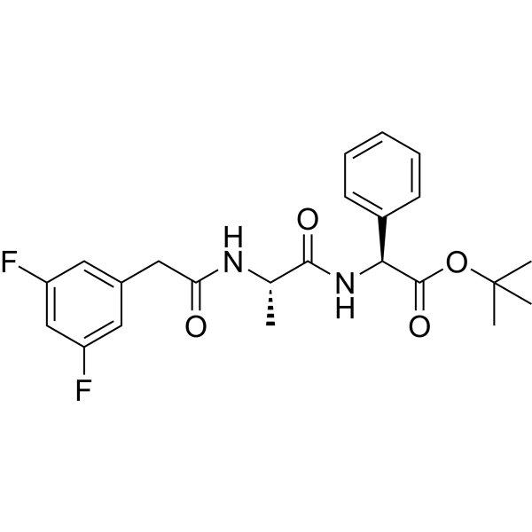
HY-13027
DAPTγ-secretase Inhibitor -

HY-P1061
ColivelinNeuroprotectant/STAT3 Activator -
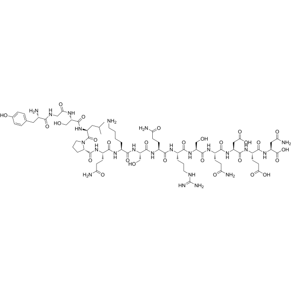
HY-P1048
Myelin Basic Protein (MBP) (68-82), guinea pigMBP Fragment -
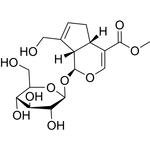
HY-N0009
GeniposideInfluenza Virus Inhibitor -
HY-13027G
DAPT (GMP)γ-Secretase Inhibitor


Biological Activity
Description
IC50 & Target
Amyloid-β[1]
In Vitro
β-amyloid (12-28) may exert dysregulation cognitive effects by means of defective coordination of potassium channel function in nerve, glia and endothelial cells[1].
β-Amyloid Aggregation Guidelines (Following is our recommended protocol. This protocol only provides a guideline, and should be modified according to your specific needs).
1. Solid Aβ peptide was dissolved in cold hexafluoro-2-propanol (HFIP). The peptide was incubated at room temperature for at least 1h to establish monomerization and randomization of structure.
2. The HFIP was removed by evaporation, and the resulting peptide was stored as a film at -20 or -80 °C.
3. The resulting film was dissolved in anhydrous DMSO at 5 mM and then diluted into the appropriate concentration and buffer (serum- and phenol red-free culture medium) with vortexing.
4. Next, the solution was age 48h at 4-8 °C. The sample was then centrifuged at 14000g for 10 min at 4-8 °C; the soluble oligomers were in the supernatant. The supernatant was diluted 10-200-fold for experiments.
Methods vary depends on the downstream applications.
MedChemExpress (MCE) has not independently confirmed the accuracy of these methods. They are for reference only. Further protocols information, click here.


Chemical Information
-
CAS No. 107015-83-8
-
Molecular Weight 1955.18
-
Formula C89H135N25O25
-
Synonyms
Amyloid β-Protein (12-28)
-
Sequence
Val-His-His-Gln-Lys-Leu-Val-Phe-Phe-Ala-Glu-Asp-Val-Gly-Ser-Asn-Lys
-
Sequence Shortening
VHHQKLVFFAEDVGSNK
-
Shipping
Room temperature in continental US; may vary elsewhere.
-
Storage
Please store the product under the recommended conditions in the Certificate of Analysis.

Protocols
-
Amyloid: Congo Red Amyloid Staining
Congo red amyloid staining is a histochemical method used to detect extracellular amyloid deposits in tissue sections based on the affinity of Congo red dye for β-pleated sheet-rich protein aggregates. When bound to amyloid, Congo red produces characteristic apple-green birefringence under polarized light microscopy, which is widely regarded as a diagnostic feature of amyloid deposition in histopathology. The diagnostic principle relies on the combination of dye binding (congophilia) and optical anisotropy under polarized illumination, which distinguishes amyloid from most non-amyloid eosinophilic extracellular deposits in routine histological evaluation. Amyloid identification by Congo red staining remains a cornerstone in diagnostic pathology despite the availability of adjunct methods such as immunohistochemistry and mass spectrometry, particularly because of its ability to localize deposits directly within tissue architecture. The specificity of Congo red-positive deposits is incre


Purity & Documentation
References
[1]. Rabanal F, et al. Structural, kinetic and cytotoxicity aspects of 12-28 beta-amyloid protein fragment: a reappraisal. J Pept Sci. 2002 Oct;8(10):578-88. [Content Brief]
[2]. Nikunj S Patel, et al. Potent anti-angiogenic motifs within the Alzheimer beta-amyloid peptide. Amyloid. 2008 Mar;15(1):5-19. [Content Brief]

Calculators
Concentration (start) × Volume (start) = Concentration (final) × Volume (final)


β-Amyloid (12-28) Related Classifications
HY-P1051 Related Classifications
 Powered by Bioz