



Anti-Mouse CD69 Antibody (CD69.2.2)
Anti-Mouse CD69 Antibody (CD69.2.2) is a mouse-derived IgG1 κ type antibody inhibitor, targeting to mouse CD69. Anti-Mouse CD69 Antibody (CD69.2.2) causes CD69 to be internalized but does not deplete CD69+ cells. Anti-Mouse CD69 Antibody (CD69.2.2) can be used for the researches of cancer, infection, inflammation and immunology, such as RMA-S tumor, arthritis and vaccinia virus (VACV) infection.
For research use only. We do not sell to patients.
- Molecular Weight:150 kDa
-
Storage:
Please store the product under the recommended conditions in the Certificate of Analysis.

To place orders, for customer services and technical support, please contact: MedChemExpress USA
Tel: 609-228-6898 E-mail: [email protected] [email protected]


-
 Biological Activity
-
Chemical Information
- Protocols
- Purity & Documentation
- References
-
 Help & FAQs




Biological Activity
Description
Isotype
Mouse IgG1 kappa
Recommend Isotype Controls
Species Reactivity
Mouse
IC50 & Target
CD69
In Vitro
Anti-Mouse CD69 Antibody (CD69.2.2) (10 μg/mL, 1 h) is readily internalized at 37°C in CD69+ cells[1].
Anti-Mouse CD69 Antibody (CD69.2.2) (10 μg/mL, 18 h) increases cytotoxicity of NK cells in splenocytes[1].
MedChemExpress (MCE) has not independently confirmed the accuracy of these methods. They are for reference only. Further protocols information, click here.
In Vivo
Anti-Mouse CD69 Antibody (CD69.2.2) (500 μg, i.p., at days 1 and 5) reduces tumor metastasis and prolongs survival in RM-1 tumor mice models[1].
Anti-Mouse CD69 Antibody (CD69.2.2) (200 μg, i.p., a single dose) consistently and significantly increases NK-dependent cytotoxic activity in RMA-S/RM-1/YAC-1 tumor mice models[1].
Anti-Mouse CD69 Antibody (CD69.2.2) (300 μg, i.p., at days 1 and 5) downregulates CD 69 expression and does not affect disease severity in collagen antibody-induced arthritis mice models[2].
Anti-Mouse CD69 Antibody (CD69.2.2) (500 μg, i.v., 24 h before infection) enhances the innate immune response in vaccinia virus (VACV)-infected mice models[3].
Anti-Mouse CD69 Antibody (CD69.2.2) (200 μg, i.v., twice a week) shows anti-infection in vaccinia virus (VACV)-infected Rag2-/- mice[3].
MedChemExpress (MCE) has not independently confirmed the accuracy of these methods. They are for reference only.
-
Animal Model:RM-1 tumor mice models[1]
-
Dosage:500 μg
-
Administration:Intraperitoneally injection, at days 1 and 5
-
Result:Drastically reduced tumor metastasis.
Increased survival rate of mice.
-
Animal Model:Vaccinia virus (VACV)-infected mice models[3]
-
Dosage:500 μg
-
Administration:Intraperitoneally injection, 24 h before infection
-
Result:Showed slightly lower ovarian viral titers.
Increased the numbers of TNF-α-producing NK, CD8+ T, and CD4+ T cells and IFN-γ-producing NK and CD4+ T cells.
Gene ID
Accession
Conjugated
Unconjugated
Reconsititution
The product can be reconstituted/diluted with sterile PBS or saline.
Format
-
Product Image
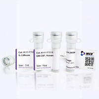
Application
in vivo down-regulation of CD69 expression; Functional assays


Chemical Information
-
Molecular Weight 150 kDa
-
SMILES
[Anti-Mouse CD69 Antibody (CD69.2.2)]
-
Formulation
Please refer to the lot-specific COA for specific buffer information.
-
Storage
Please store the product under the recommended conditions in the Certificate of Analysis.

Protocols
-
Research Protocol for Infectious Diseases
Infectious-disease experiments test how pathogens interact with host barriers, innate immune receptors, inflammatory signaling, pathogen replication, and tissue injury; pattern-recognition receptors such as TLRs, RIG-I-like receptors, NOD-like receptors, and inflammasomes detect microbial molecules and activate NF-κB, interferon, and cytokine responses. The central hypothesis is that infection severity reflects the balance between pathogen burden and host response: protective inflammation restricts pathogen growth, whereas excessive or mislocalized inflammation contributes to tissue damage and disease phenotype. Unresolved questions include which host pathways are protective versus pathogenic, why some infection models fail to translate to human disease, and which combined readouts best predict clinically relevant infection outcomes.
-
Collagen-Induced Arthritis
Collagen-induced arthritis (CIA) is an autoimmune murine model of rheumatoid arthritis in which immunization with type II collagen (CII) emulsified in an adjuvant induces a T cell- and autoantibody-driven inflammatory arthritis characterized by synovial hyperplasia, immune cell infiltration, and joint destruction. The model typically relies on genetically susceptible mouse strains (e. g. , DBA/1) and reproduces key features of human rheumatoid arthritis, including anti-collagen immune responses and progressive joint inflammation. Disease onset generally occurs within ~3-4 weeks after immunization, depending on antigen/adjuvant combinations and protocol variation. The immunopathology is driven by adaptive immune activation against CII, leading to systemic and local joint inflammation mediated by pro-inflammatory cytokines and effector immune cells, making CIA a standard preclinical platform for evaluating immunomodulatory and anti-arthritic interventions.
-
Research Protocol for Inflammation-related Diseases
The NLRP3 inflammasome is a cytosolic innate immune signaling platform that integrates priming signals and danger-signal activation to promote caspase-1 activation, maturation of IL-1β and IL-18, and gasdermin D-mediated pyroptotic cell death. The core experimental logic is to determine whether inflammatory disease phenotypes are driven by increased NLRP3 expression, ASC-containing inflammasome assembly, caspase-1 cleavage, GSDMD cleavage, and extracellular release of IL-1β/IL-18 rather than by nonspecific cell injury alone. The pathway is strongly linked to inflammation-related disease phenotypes because monosodium urate crystals activate NALP3/NLRP3 inflammasome signaling in gout-like crystal inflammation, cholesterol crystals activate NLRP3 inflammasomes in atherogenesis models, and DSS-induced intestinal inflammation has been reported to involve NLRP3 inflammasome activity. However, experimental colitis studies also show context-dependent protective effects of NLRP3 inflammasome co


Purity & Documentation
References
[1]. Esplugues E, et al. Induction of tumor NK-cell immunity by anti-CD69 antibody therapy. Blood. 2005 Jun 1;105(11):4399-406. [Content Brief]
[2]. Lamana A, et al. The role of CD69 in acute neutrophil-mediated inflammation. Eur J Immunol. 2006 Oct;36(10):2632-8. [Content Brief]
[3]. Notario L, et al. CD69 Targeting Enhances Anti-vaccinia Virus Immunity. J Virol. 2019 Sep 12;93(19):e00553-19. [Content Brief]

Calculators
Concentration (start) × Volume (start) = Concentration (final) × Volume (final)


Anti-Mouse CD69 Antibody (CD69.2.2) Related Classifications
HY-P990265 Related Classifications
 Powered by Bioz