



Bad Antibody (YA593)
(Synonyms: Bbc6, Bad, Bcl2-associated agonist of cell death, BAD, Bcl-2-binding component 6, Bcl-xL/Bcl-2-associated death promoter, Bcl2 antagonist of cell death)
Based on 1 Customer Validation
Bad Antibody (YA593) is a Rabbit-derived and non-conjugated IgG monoclonal antibody, targeting to Bad.
-
Host:
Rabbit
-
Isotype:
IgG
-
Application:
WB, ICC/IF, IHC-P, FC, IP
-
Reactivity :
Human
-
Formulation:
Supplied in 1*TBS (pH7.4), 0.05% BSA and 40% Glycerol. Preservative: 0.05% Sodium Azide.
-
Conjugation:
Non-conjugated





To place orders, for customer services and technical support, please contact: MedChemExpress USA
Tel: 609-228-6898 E-mail: [email protected] [email protected]





Applications
| Application |
WB

WB: Western Blot
|
ICC/IF
ICC/IF: Immunocytochemistry/
Immunofluorescence |
IHC-P
IHC-P: Immunohistochemistry-Paraffin
|
FC
FC: Flow Cytometry
|
IP
IP: Immunoprecipitation
|
|---|---|---|---|---|---|
| Dilution Ratio | 1:500 | 1:50 | 1:50-1:200 | 1:50 | Use at an assay dependent concentration. |


Product Details
Bad Antibody (YA593) is a Rabbit-derived and non-conjugated IgG monoclonal antibody, targeting to Bad.
-
Host Rabbit

-
Clonality Recombinant,Monoclonal

-
Species ReactivityHuman
-
Observed Molecular WeightObserved band size: 23 kDa
Note: Due to possible protein modifications or aggregation, the molecular weight should be confirmed by actual measurement, and the predicted value is for reference only.

-
Calculated Molecular Weight Predicted band size: 18 kDa

Synthetic peptide corresponding to Human Bad.AA range:1-50.
Endogenous
Protein A affinity purified.
Non-conjugated
Unmodified
IgG


Product Properties
-
Appearance
Solution
-
Formulation
Supplied in 1*TBS (pH7.4), 0.05% BSA and 40% Glycerol. Preservative: 0.05% Sodium Azide.
-
Concentration
Batch-dependent, Please check the COA for the concentration of each lot. Check Lot Concentration
-
Storage & Stability
Stored at -20°C for 1 year. Avoid repeated freeze / thaw cycles.
-
Shipping
Shipping with blue ice.


Verification Images
-
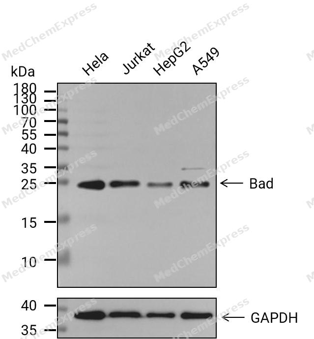 Western blot analysis of extracts from Hela(lane 1(20μg)) 、Jurkat(lane 2(20μg)) 、HepG2(lane 3(20μg))and A549(lane 4(20μg) using Bad Antibody(HY-P80025). Proteins were transferred to a PVDF membrane and blocked with 5%BSA in TBST for 1.5 hour at room temperature. The primary antibody ( 1/500) and Loading control antibody (GAPDH, 1/1000) was used in 5% BSA in TBST at 4℃ overnight. Goat Anti-Rabbit IgG-HRP Secondary Antibody (1/10,000) was used for 1 hour at room temperature.
 -
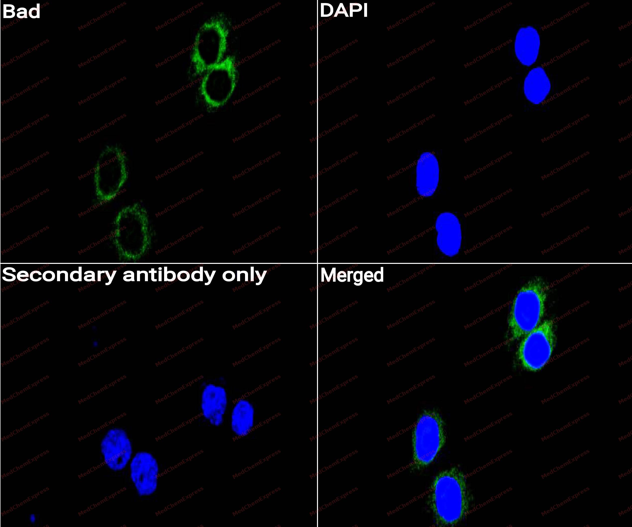 Immunocytochemistry analysis of Hela cells labeling Bad with Bad Antibody (HY-P80025) at 1/50 dilution. Cells were fixed in 4% paraformaldehyde for 15 minutes at room temperature, permeabilized with 0.1% Triton X-100 for 10 minutes at room temperature, then blocked with QuickBlock™ Blocking Buffer for Immunol Staining for 10 min at room temperature. Cells were then incubated with Bad Antibody (HY-P80025) at 1/50 dilution in QuickBlock™ Blocking Buffer for Immunol Staining at 4 ℃. Alexa Fluor® 488-conjugated AffiniPure Goat Anti-Rabbit IgG H&L(HY-P8002, Green) was used as the secondary antibody at 1/1,000 dilution. PBS instead of the primary antibody was used as the secondary antibody only control. The Nuclear counterstain was DAPI (Blue).
 -
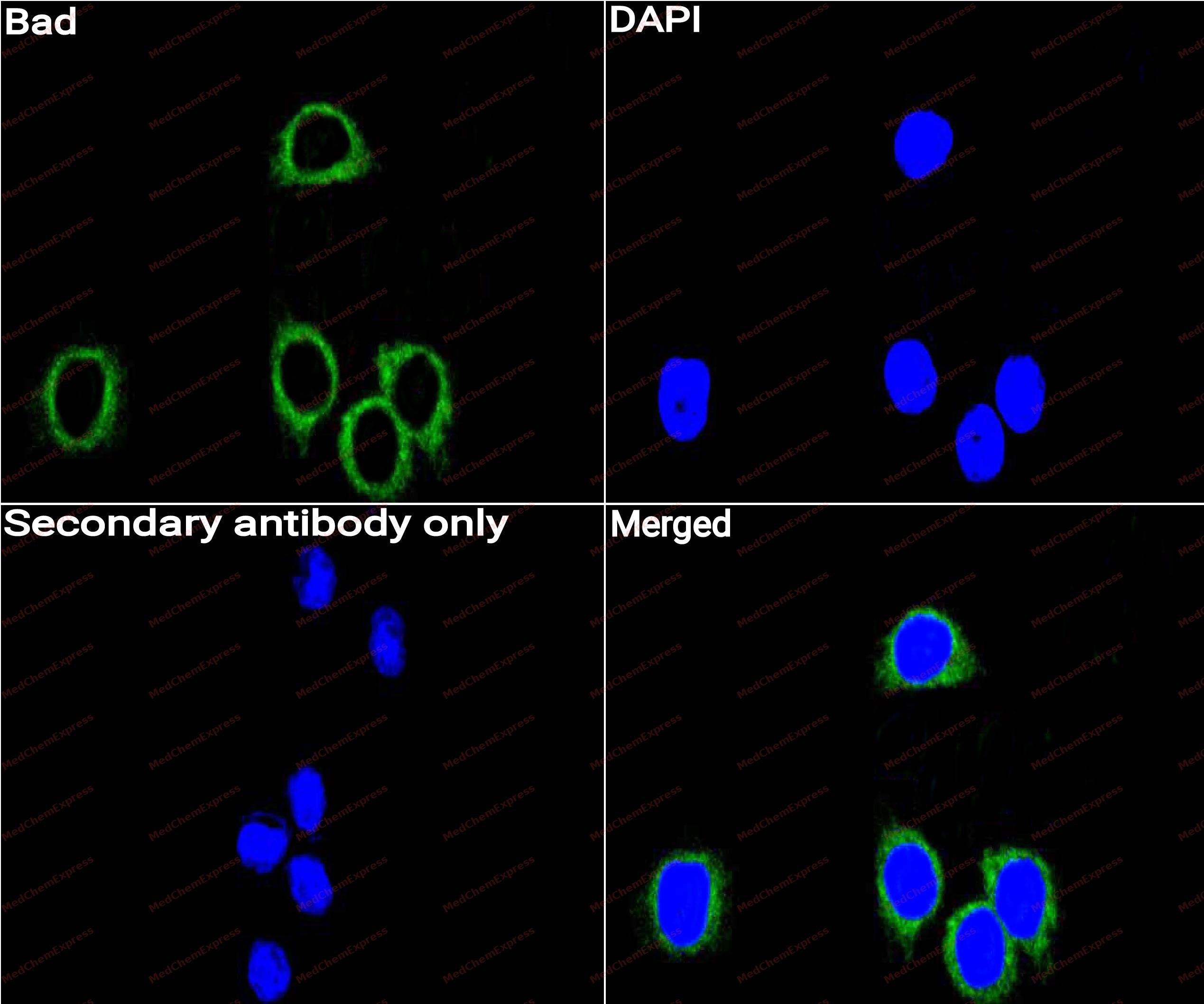 Immunocytochemistry analysis of HepG2 cells labeling Bad with Bad Antibody (HY-P80025) at 1/50 dilution. Cells were fixed in 4% paraformaldehyde for 15 minutes at room temperature, permeabilized with 0.1% Triton X-100 for 10 minutes at room temperature, then blocked with QuickBlock™ Blocking Buffer for Immunol Staining for 10 min at room temperature. Cells were then incubated with Bad Antibody (HY-P80025)at 1/50 dilution in QuickBlock™ Blocking Buffer for Immunol Staining at 4 ℃. Alexa Fluor® 488-conjugated AffiniPure Goat Anti-Rabbit IgG H&L(HY-P8002,Green) was used as the secondary antibody at 1/1,000 dilution. PBS instead of the primary antibody was used as the secondary antibody only control. The Nuclear counterstain was DAPI (Blue).
 -
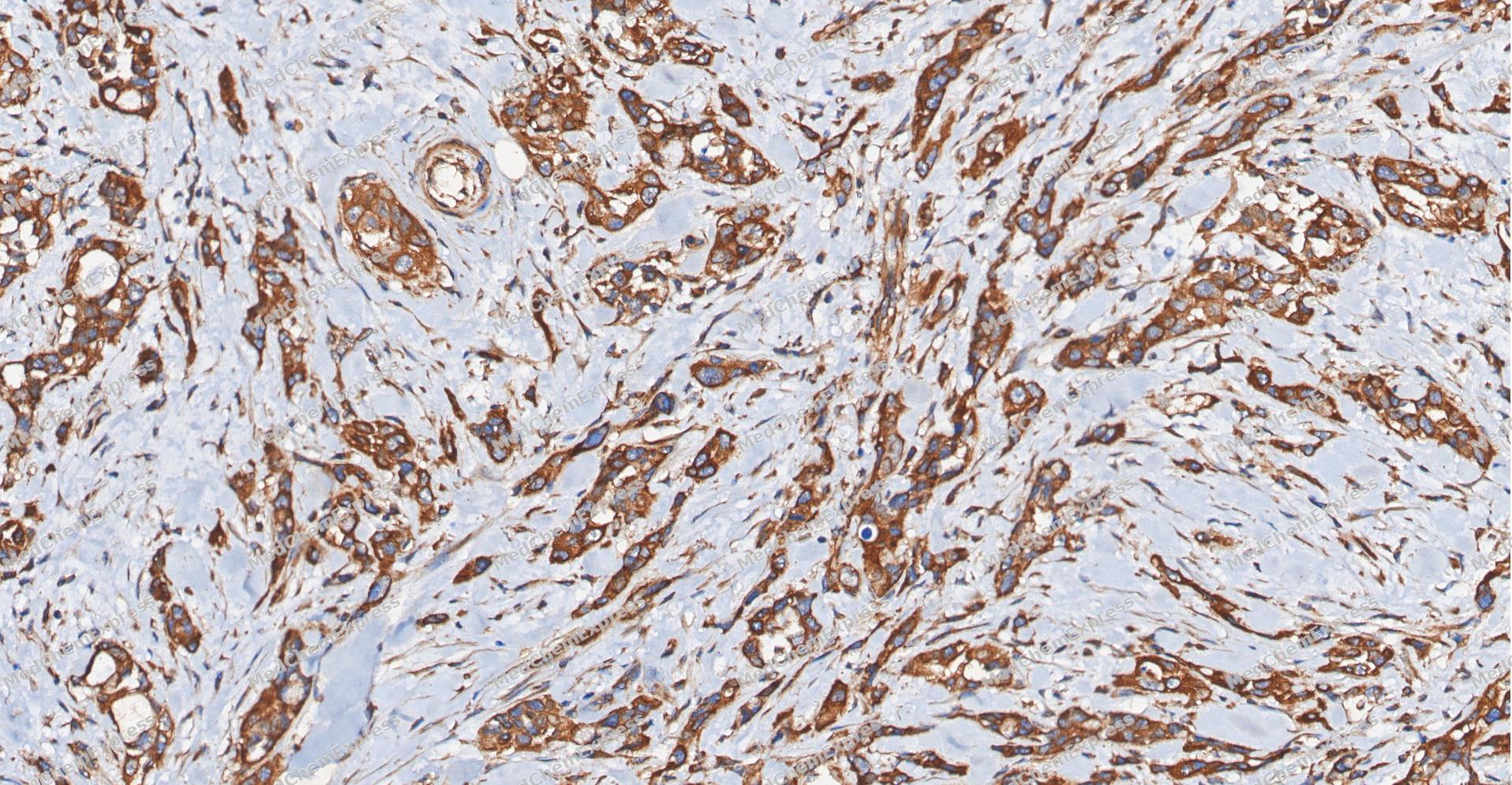 Immunohistochemical analysis of paraffin-embedded human Pancreatic carcinoma tissue using Bad antibody was performed. The section was pretreated using high-temperature and high-pressure mediated EDTA antigen retrieval buffer (pH 9.0), for 5 minutes. The tissues were incubated with primary antibody (HY-P80025, 1:100 dilution) at room temperature for 60 minutes. Detection was performed using an HRP conjugated compact polymer system. DAB was used as the chromogen. The tissues were counterstained with hematoxylin and mounted with neutral balsam mounting medium.
 -
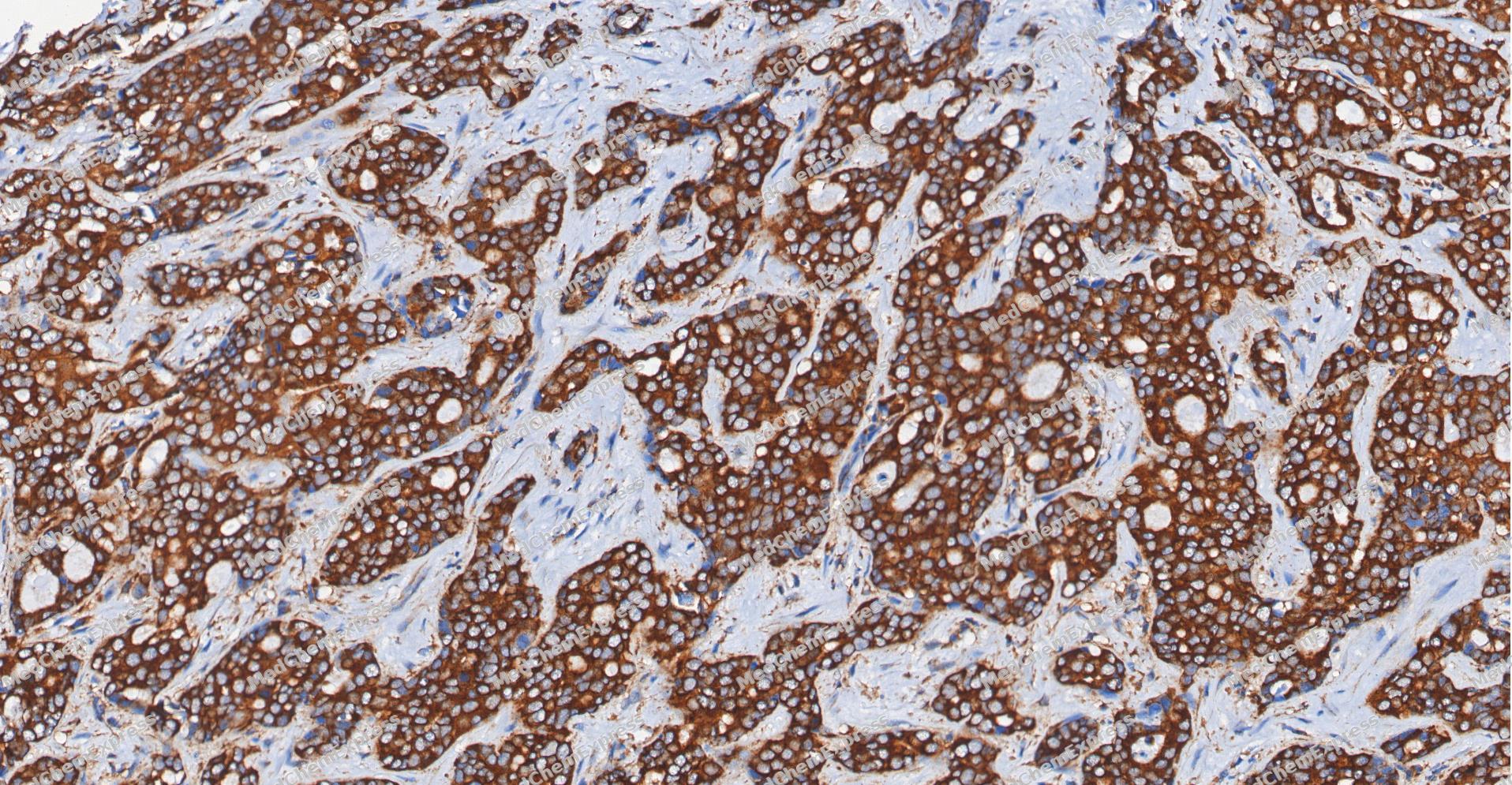 Immunohistochemical analysis of paraffin-embedded human cholangiocarcinoma tissue using Bad antibody was performed. The section was pretreated using high-temperature and high-pressure mediated EDTA antigen retrieval buffer (pH 9.0), for 5 minutes. The tissues were incubated with primary antibody (HY-P80025, 1:100 dilution) at room temperature for 60 minutes. Detection was performed using an HRP conjugated compact polymer system. DAB was used as the chromogen. The tissues were counterstained with hematoxylin and mounted with neutral balsam mounting medium.
 -
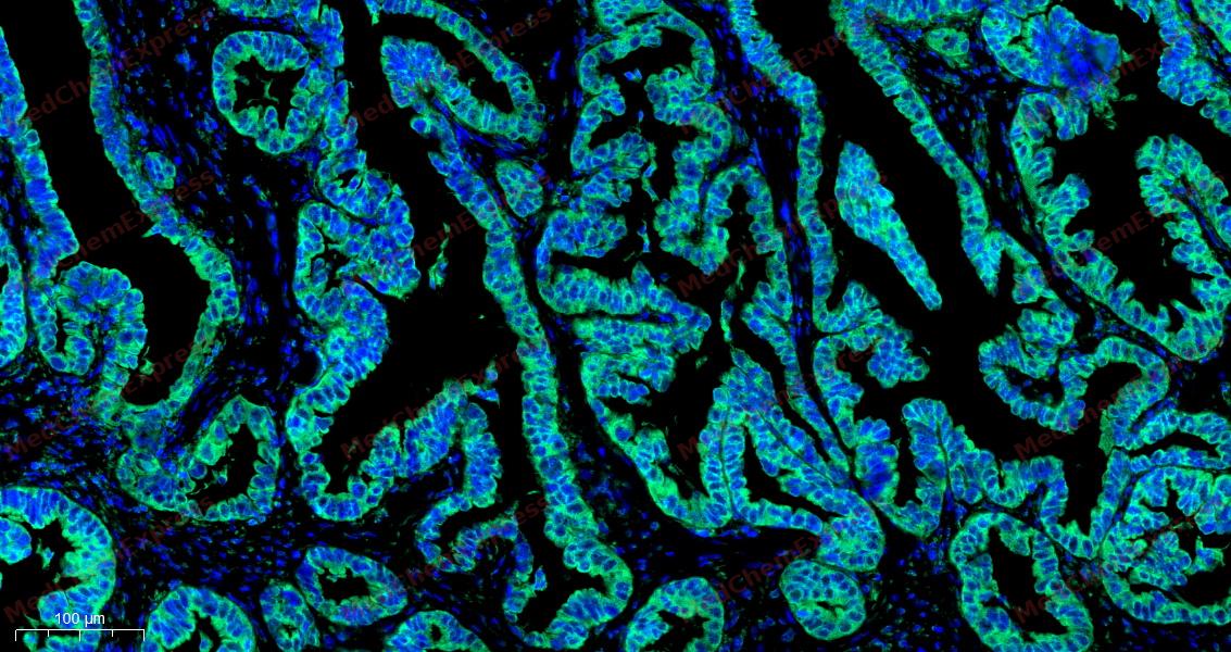
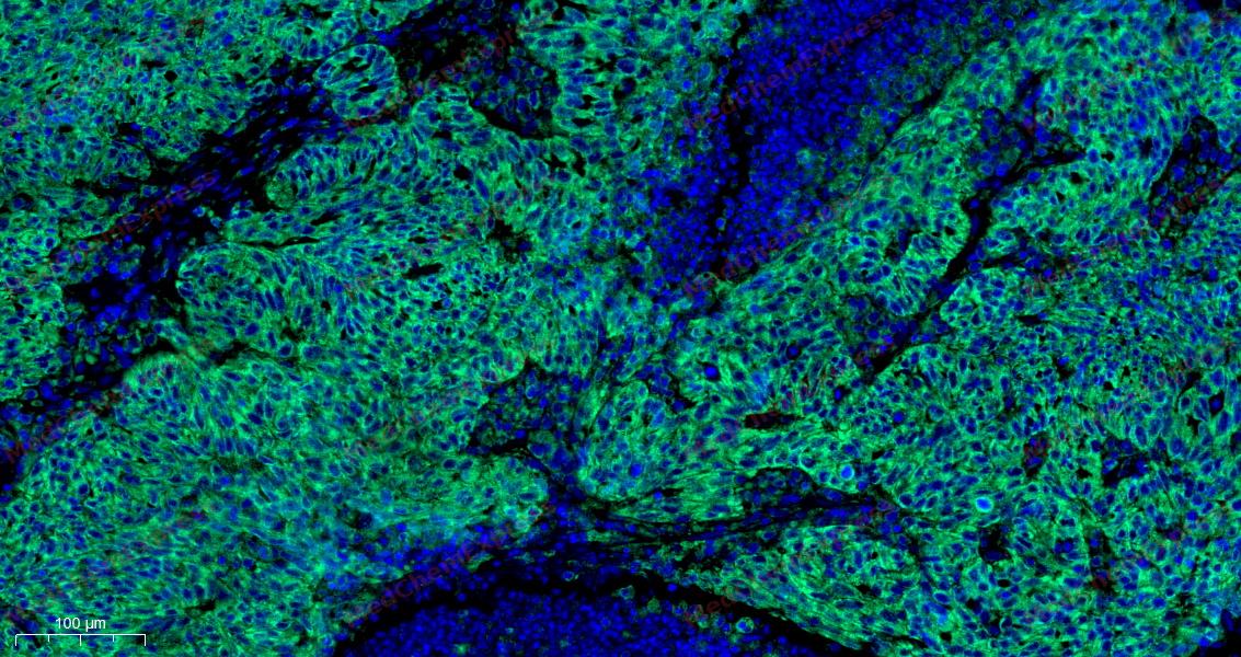 Tyramide signaling amplification based immunofluorescence was performed on paraffin-embedded human ovarian carcinoma (sample 1) tissue using Bad antibody. Antigen retrieval was performed in EDTA buffer pH 9.0 (95 °C, 20 min) followed by cooling to RT. Then incubated with primary antibody (HY-P80025, 1:100 dilution) at room temperature for 60 minutes and HRP conjugated secondary antibody for 10 minutes. Fluorescence was then developed with TSA520 . The tissues were counterstained with DAPI and mounted with Anti-fade mounting medium.
 -
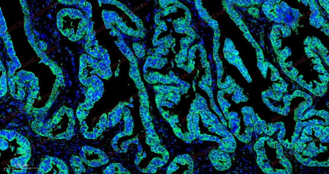 Tyramide signaling amplification based immunofluorescence was performed on paraffin-embedded human ovarian carcinoma (sample 2) tissue using Bad antibody. Antigen retrieval was performed in EDTA buffer pH 9.0 (95 °C, 20 min) followed by cooling to RT. Then incubated with primary antibody (HY-P80025, 1:100 dilution) at room temperature for 60 minutes and HRP conjugated secondary antibody for 10 minutes. Fluorescence was then developed with TSA520 . The tissues were counterstained with DAPI and mounted with Anti-fade mounting medium.
 -
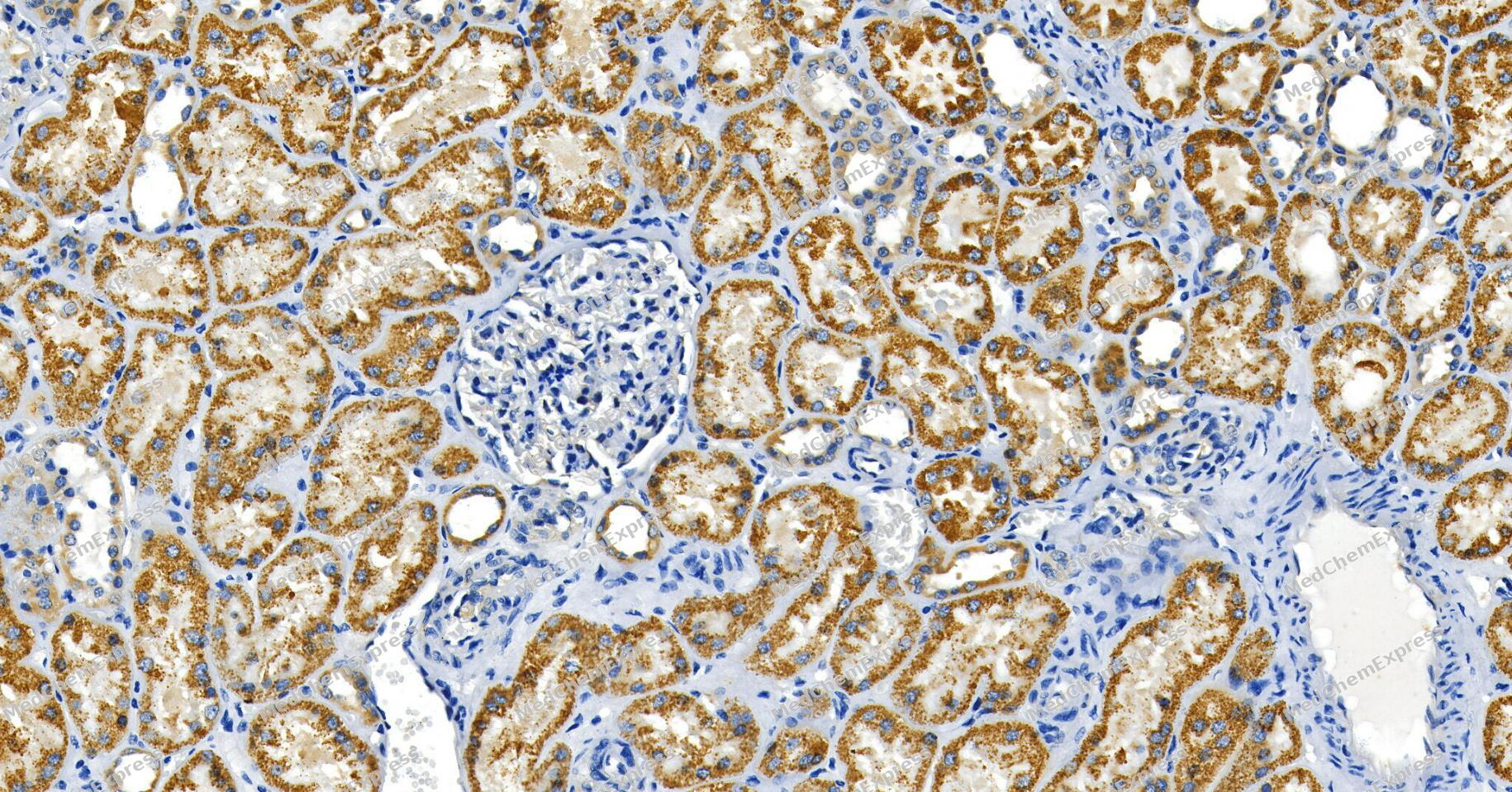 Immunohistochemical analysis of paraffin-embedded human kidney tissue using Bad Antibody (YA593). The section was pre-treated using heat mediated antigen retrieval with Tris/EDTA buffer (pH 9.0) for 20 minutes. The tissues were probed with the primary antibody (HY-P80025, 1/100) overnight at 4℃. The detection was performed using an HRP conjugated compact polymer system. DAB was used as the chromogen. Tissues were counterstained with hematoxylin and mounted with neutral balsam.
 -
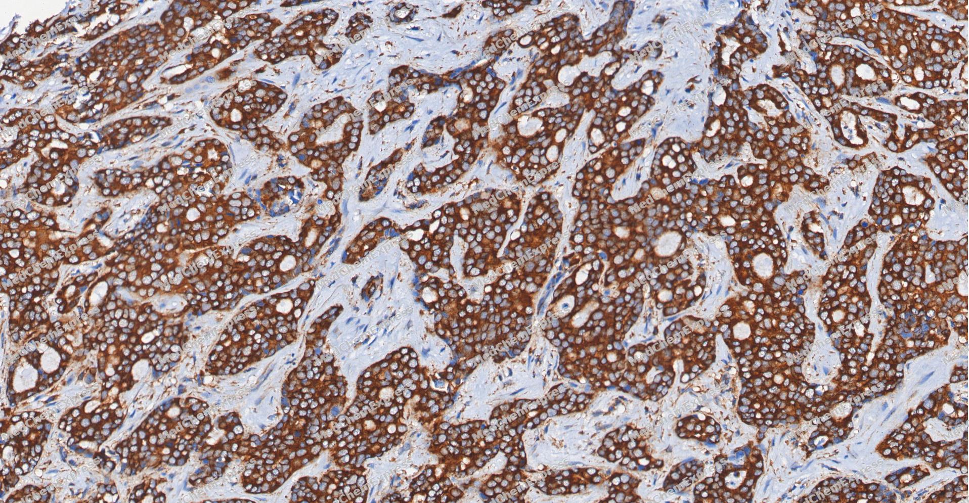
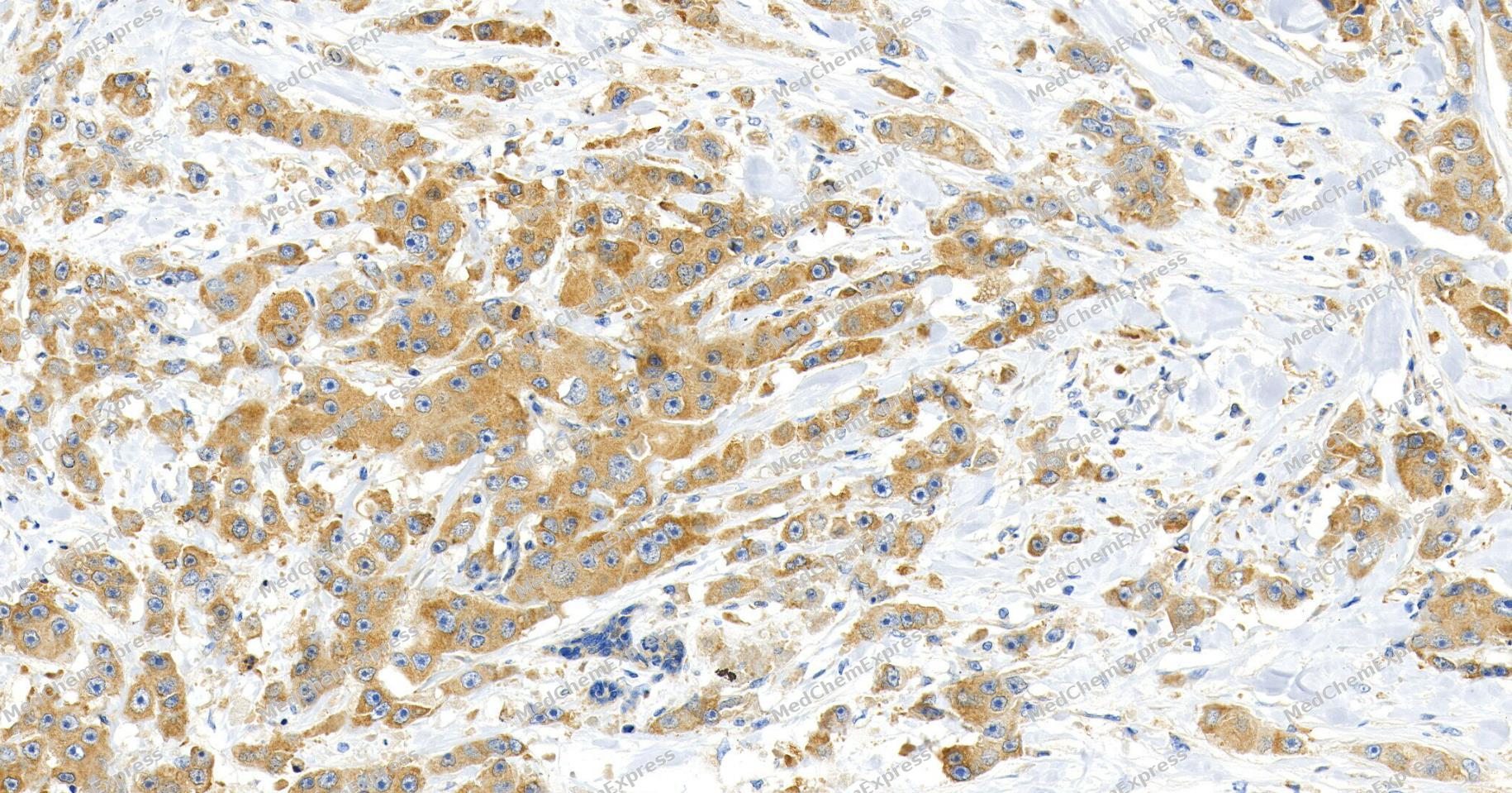 Immunohistochemical analysis of paraffin-embedded human breast cancer tissue using Bad Antibody (YA593). The section was pre-treated using heat mediated antigen retrieval with Tris/EDTA buffer (pH 9.0) for 20 minutes. The tissues were probed with the primary antibody (HY-P80025, 1/100) overnight at 4℃. The detection was performed using an HRP conjugated compact polymer system. DAB was used as the chromogen. Tissues were counterstained with hematoxylin and mounted with neutral balsam.
 -
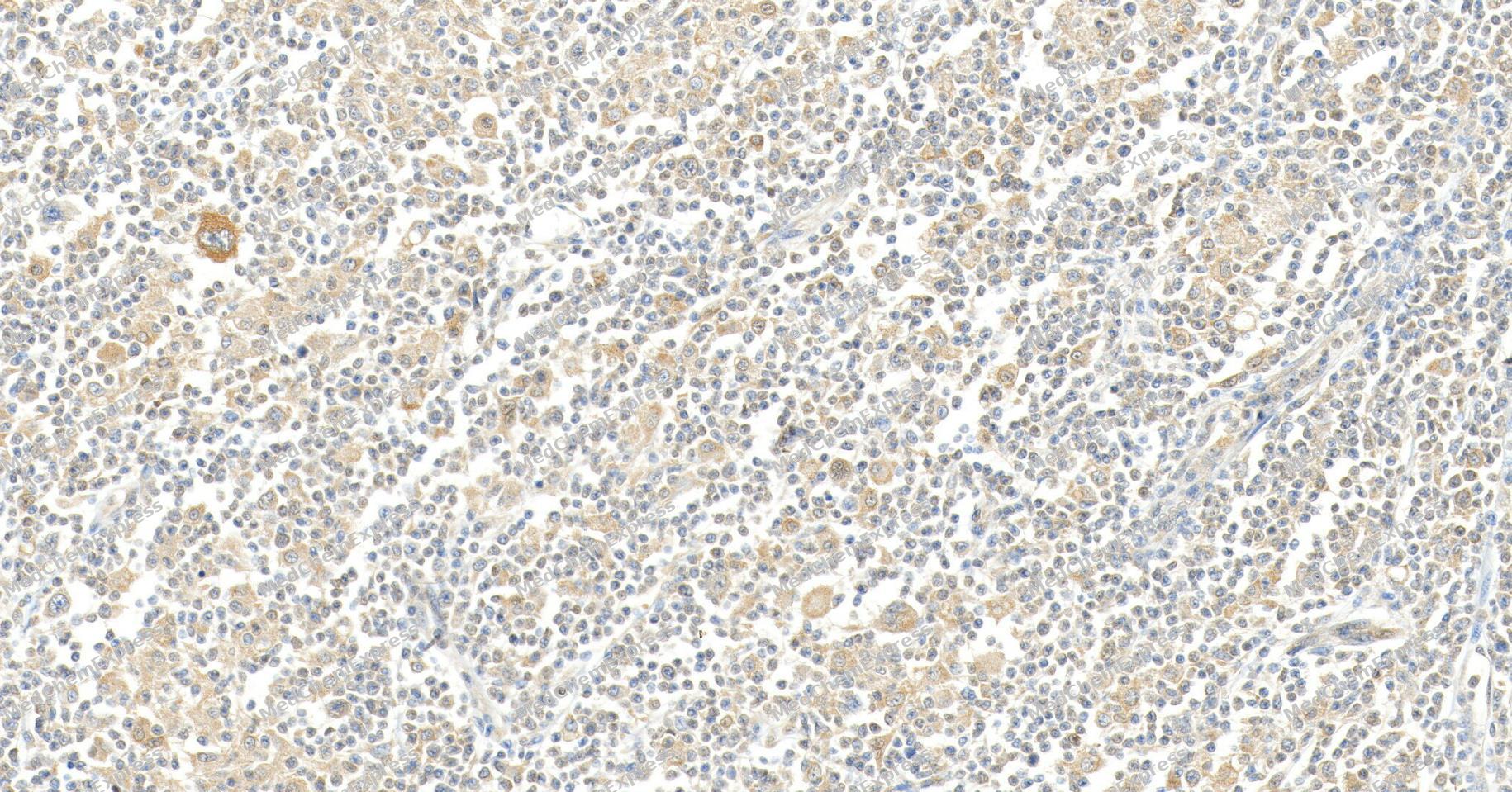 Immunohistochemical analysis of paraffin-embedded human lymphoma tissue using Bad Antibody (YA593). The section was pre-treated using heat mediated antigen retrieval with Tris/EDTA buffer (pH 9.0) for 20 minutes. The tissues were probed with the primary antibody (HY-P80025, 1/100) overnight at 4℃. The detection was performed using an HRP conjugated compact polymer system. DAB was used as the chromogen. Tissues were counterstained with hematoxylin and mounted with neutral balsam.
 -
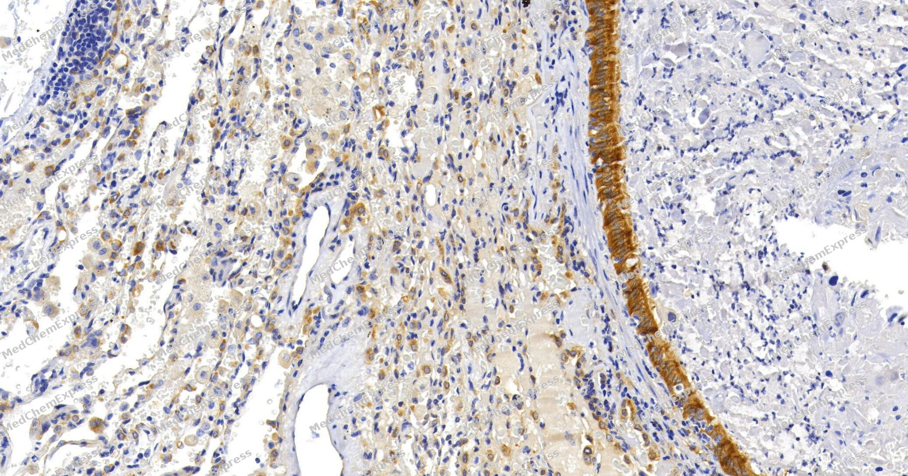 Immunohistochemical analysis of paraffin-embedded human lung cancer tissue using Bad Antibody (YA593). The section was pre-treated using heat mediated antigen retrieval with Tris/EDTA buffer (pH 9.0) for 20 minutes. The tissues were probed with the primary antibody (HY-P80025, 1/100) overnight at 4℃. The detection was performed using an HRP conjugated compact polymer system. DAB was used as the chromogen. Tissues were counterstained with hematoxylin and mounted with neutral balsam.


Background
-
Function
BAD is a pro-apoptotic BH3-only Bcl-2 family protein that promotes apoptosis by binding Bcl-xL and Bcl-2, displacing BAX, and reversing death-repressor activity[1]. Mechanistically, survival-factor signaling phosphorylates BAD, causing 14-3-3 binding rather than Bcl-xL binding, while Akt phosphorylation blocks BAD-induced neuronal death[2][3]. Therefore, BAD links growth-factor survival pathways to the intrinsic mitochondrial apoptosis machinery, where Bcl-2 family interactions control apoptotic commitment[3][4]. In disease-relevant metabolic models, BAD resides in a glucokinase-containing mitochondrial complex that integrates glycolysis and apoptosis, and BAD deficiency or non-phosphorylatable BAD mutants impair glucose homeostasis[5]. In pancreatic β-cells, BAD supports glucose-stimulated insulin secretion and β-cell survival, giving BAD practical value in diabetes-related apoptosis and metabolism studies[6]. Compared with activator isoforms such as BID, BIM, and PUMA, BAD functions mainly as a sensitizer that neutralizes anti-apoptotic Bcl-2 proteins rather than directly activating BAX-BAK[4]. For experimental applications, phospho-BAD BH3 helix mimetics activate glucokinase through a mechanism distinct from classical allosteric activators[7].
-
Subcellular Localization
Mitochondrion outer membrane; Cytoplasm
-
Expression
Tissue_specificity:Expression in multiple organizations -
Subunit
Forms heterodimers with the anti-apoptotic proteins, Bcl-X(L), Bcl-2 and Bcl-W.
-
SwissProt ID
-
Synonyms
Bbc6, Bad, Bcl2-associated agonist of cell death, BAD, Bcl-2-binding component 6, Bcl-xL/Bcl-2-associated death promoter, Bcl2 antagonist of cell death
-
Research Field
Cell Biology


Documentation
-
Data Sheet (261 KB)
-
SDS (251 KB)
- English - EN (251 KB)
- Français - FR (251 KB)
- Deutsch - DE (251 KB)
- Norwegian - NO (251 KB)
- Español - ES (251 KB)
- Swedish - SV (251 KB)
- Italian - IT (251 KB)
- Korean - KR (251 KB)
- Portuguese - PT (251 KB)
-
User Guide for Antibodies (1077 KB)
References
[1]. Yang E, et al. Bad, a heterodimeric partner for Bcl-XL and Bcl-2, displaces Bax and promotes cell death. Cell. 1995 Jan 27;80(2):285-91. [Content Brief]
[2]. Zha J, et al. Serine phosphorylation of death agonist BAD in response to survival factor results in binding to 14-3-3 not BCL-X(L). Cell. 1996 Nov 15;87(4):619-28. [Content Brief]
[3]. Datta SR, et al. Akt phosphorylation of BAD couples survival signals to the cell-intrinsic death machinery. Cell. 1997 Oct 17;91(2):231-41. [Content Brief]
[4]. Hongay CF, et al. Antisense transcription controls cell fate in Saccharomyces cerevisiae. Cell. 2006 Nov 17;127(4):735-45. [Content Brief]
[5]. Danial NN, et al. BAD and glucokinase reside in a mitochondrial complex that integrates glycolysis and apoptosis. Nature. 2003 Aug 21;424(6951):952-6. [Content Brief]
[6]. Danial NN, et al. Dual role of proapoptotic BAD in insulin secretion and beta cell survival. Nat Med. 2008 Feb;14(2):144-53. [Content Brief]
[7]. Szlyk B, et al. A phospho-BAD BH3 helix activates glucokinase by a mechanism distinct from that of allosteric activators. Nat Struct Mol Biol. 2014 Jan;21(1):36-42. [Content Brief]

 Powered by Bioz
