



BDNF Antibody
(Synonyms: BDNF; MGC34632; Abrineurin; ANON2; Brain Derived Neurotrophic Factor; Neurotrophin; BULN2)Based on 1 publication(s) in Google Scholar
BDNF Antibody is a Rabbit-derived and non-conjugated IgG polyclonal antibody, targeting to BDNF.
-
Host:
Rabbit
-
Isotype:
IgG
-
Application:
WB, IHC-P, ICC/IF
-
Reactivity :
Human, Mouse, Rat
-
Formulation:
Supplied in Rabbit IgG in 10mM phosphate buffered saline , pH 7.4, 150mM sodium chloride, 0.05% BSA, 0.02% sodium azide and 50% glycerol.
-
Conjugation:
Non-conjugated


To place orders, for customer services and technical support, please contact: MedChemExpress USA
Tel: 609-228-6898 E-mail: [email protected] [email protected]



Publications Citing Use of MedChemExpress (MCE) BDNF Antibody
More

Applications
| Application |
WB

WB: Western Blot
|
IHC-P
IHC-P: Immunohistochemistry-Paraffin
|
ICC/IF
ICC/IF: Immunocytochemistry/
Immunofluorescence |
|---|---|---|---|
| Dilution Ratio | 1:500-1:1000 | 1:50-1:100 | 1:50-1:200 |


Product Details
BDNF Antibody is a Rabbit-derived and non-conjugated IgG polyclonal antibody, targeting to BDNF.
-
Host Rabbit

-
Clonality Polyclonal

-
Species ReactivityHuman, Mouse, Rat
-
Observed Molecular WeightObserved band size: 32 kDa
Note: Due to possible protein modifications or aggregation, the molecular weight should be confirmed by actual measurement, and the predicted value is for reference only.

-
Calculated Molecular Weight Predicted band size: 28 kDa

Entrez Gene: 627 Human ; 12064 Mouse ; 24225 Rat
SwissProt: P23560 Human ; P21237 Mouse ; P23363 Rat
OMIM: 113505 Human
Synthetic peptide corresponding to Human BDNF aa150-200.
Endogenous
affinity purified
Non-conjugated
Unmodified
IgG


Product Properties
-
Appearance
Solution
-
Formulation
Supplied in Rabbit IgG in 10mM phosphate buffered saline , pH 7.4, 150mM sodium chloride, 0.05% BSA, 0.02% sodium azide and 50% glycerol.
-
Concentration
Batch-dependent, Please check the COA for the concentration of each lot. Check Lot Concentration
-
Storage & Stability
Stored at -20°C for 1 year. Avoid repeated freeze / thaw cycles.
-
Shipping
Shipping with blue ice.


Publications (1)
-
Journal Impact Factor
-
Most Recent
-
Neurotoxicol Teratol
Sevoflurane induced hippocampal neuron cell injury via the miR-132-5p/BDNF axis and further caused cognitive impairment in rats. [Abstract]2025 Oct 26:112:107567. PMID: 41151746


Verification Images
-
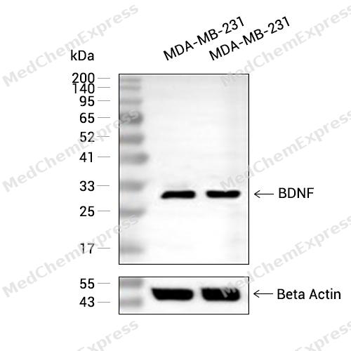 Western blot analysis of extracts from MDA-MB-231(lane 2(40μg) and MDA-MB-231(lane 3(80μg) using BDNF (HY-P80567) Rabbit mAb. Proteins were transferred to a PVDF membrane and blocked with 5% non-fat milk in TBST for 2 hour at room temperature. The primary antibody (1/1000) and Loading control antibody (Beta Actin, HY-P80438, 1/10000) was used in 5% non-fat milk in TBST at 4°C overnight. Goat Anti-Mouse/Rabbit IgG-HRP Secondary Antibody (1/10000) was used for 1 hour at room temperature.
 -
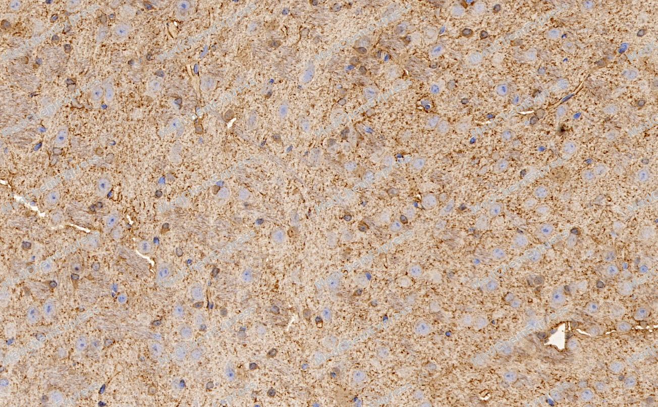 Immunohistochemical analysis of paraffin-embedded Mouse brain tissue using BDNF Antibody. The section was pre-treated using heat mediated antigen retrieval with Tris-EDTA buffer (pH 9.0) for 8 minutes. The tissues were blocked in QuickBlock for 20 minutes at room temperature, washed with ddH2O and PBS, and then probed with the primary antibody (HY-P80567, 1/100) in 4℃ overnight. The detection was performed using an HRP conjugated compact polymer system. DAB was used as the chromogen. Tissues were counterstained with hematoxylin and mounted with DPX.
 -
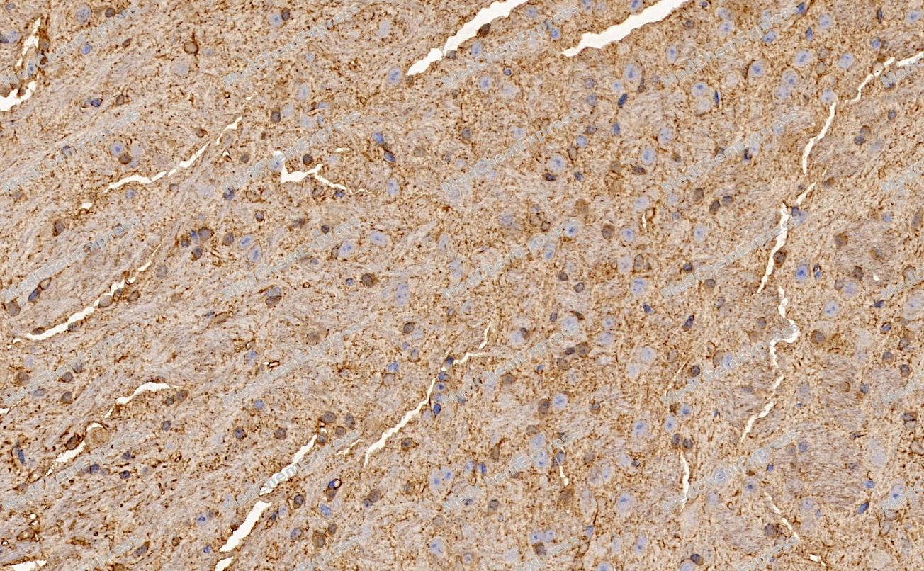 Immunohistochemical analysis of paraffin-embedded Mouse brain tissue using BDNF Antibody. The section was pre-treated using heat mediated antigen retrieval with Tris-EDTA buffer (pH 9.0) for 8 minutes. The tissues were blocked in QuickBlock for 20 minutes at room temperature, washed with ddH2O and PBS, and then probed with the primary antibody (HY-P80567, 1/100) in 4℃ overnight. The detection was performed using an HRP conjugated compact polymer system. DAB was used as the chromogen. Tissues were counterstained with hematoxylin and mounted with DPX.
 -
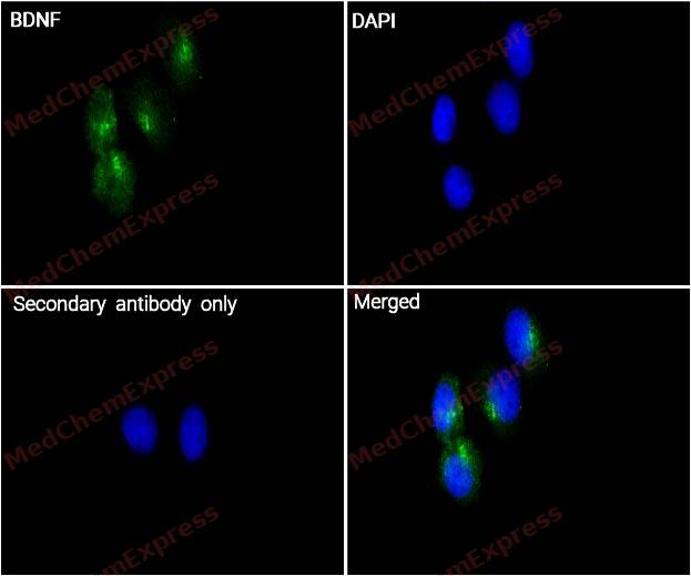 Immunocytochemistry analysis of Hela cells labeling BDNF with BDNF Antibody (HY-P80567) at 1/100 dilution. Cells were fixed in 4% paraformaldehyde for 15 minutes at room temperature, permeabilized with 0.1% Triton X-100 in PBS for 15 minutes at room temperature, then blocked with quick block buffer for 10 minutes at room temperature. Cells were then incubated with BDNF Antibody (HY-P80567) at 1/100 dilution in quick block buffer overnight at 4 ℃. AF488-conjugated Goat Anti-Rabbit IgG H&L(HY-P8002, Green) was used as the secondary antibody at 1/1,000 dilution. PBS instead of the primary antibody was used as the secondary antibody only control. The Nuclear counterstain was DAPI (Blue).
 -
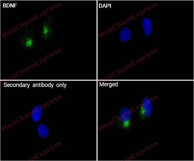 Immunocytochemistry analysis of Hela cells labeling BDNF with BDNF Antibody (HY-P80567) at 1/200 dilution. Cells were fixed in 4% paraformaldehyde for 15 minutes at room temperature, permeabilized with 0.1% Triton X-100 in PBS for 15 minutes at room temperature, then blocked with quick block buffer for 10 minutes at room temperature. Cells were then incubated with BDNF Antibody (HY-P80567) at 1/200 dilution in quick block buffer overnight at 4 ℃. AF488-conjugated Goat Anti-Rabbit IgG H&L(HY-P8002, Green) was used as the secondary antibody at 1/1,000 dilution. PBS instead of the primary antibody was used as the secondary antibody only control. The Nuclear counterstain was DAPI (Blue).


Background
-
Function
BDNF is important signaling molecule that activates signaling cascades downstream of NTRK2. During development, promotes the survival and differentiation of selected neuronal populations of the peripheral and central nervous systems. Participates in axonal growth, pathfinding and in the modulation of dendritic growth and morphology. Major regulator of synaptic transmission and plasticity at adult synapses in many regions of the CNS. The versatility of BDNF is emphasized by its contribution to a range of adaptive neuronal responses including long-term potentiation (LTP), long-term depression (LTD), certain forms of short-term synaptic plasticity, as well as homeostatic regulation of intrinsic neuronal excitability; Important signaling molecule that activates signaling cascades downstream of NTRK2. Activates signaling cascades via the heterodimeric receptor formed by NGFR and SORCS2. Signaling via NGFR and SORCS2 plays a role in synaptic plasticity and long-term depression (LTD). Binding to NGFR and SORCS2 promotes neuronal apoptosis. Promotes neuronal growth cone collapse[1][2][3].
-
Subcellular Localization
Secreted; Secreted
-
Expression
Tissue_specificity:Protein levels were detected in both plasma and saliva (PubMed: 11152678, PubMed: 19467646) . High expression was observed in the hippocampus, amygdala, cerebral cortex, and cerebellum of brain tissue. It was also expressed in the heart, lungs, skeletal muscle, testes, prostate, and placenta. -
Isoforms & Post-Translational Modification
P23560 has 5 isomers: P23560-1: 27818 Da (predicted); P23560-2: 28872 Da (predicted); P23560-3: 29817 Da (predicted); P23560-4: 37120 Da (predicted); P23560-5: 31116 Da (predicted).
N-glycosylated and glycosulfated, contrary to mature BDNF;Mature BDNF is produced by proteolytic removal of the propeptide, catalyzed by a FURIN family member. In addition, the precursor form is proteolytically cleaved within the propeptide, but this is not an obligatory intermediate for the production of mature BDNF (PubMed:11152678). Can be converted into mature BDNF by plasmin (PLG) (PubMed:19467646) -
Subunit
Monomers and homodimers. Binds to NTRK2/TRKB (PubMed:11152678, PubMed:8527932). Can form heterodimers with other neurotrophin family members, such as NTF3 and NTF4 (in vitro), but the physiological relevance of this is not clear (PubMed:10631974, PubMed:7703225). BDNF precursor form: interacts with the heterodimer formed by NGFR and SORCS2 (PubMed:24908487)
-
SwissProt ID
-
Synonyms
BDNF; MGC34632; Abrineurin; ANON2; Brain Derived Neurotrophic Factor; Neurotrophin; BULN2
-
Research Field
Neuroscience
Documentation
-
Data Sheet (261 KB)
-
SDS (252 KB)
- English - EN (252 KB)
- Français - FR (252 KB)
- Deutsch - DE (252 KB)
- Norwegian - NO (252 KB)
- Español - ES (252 KB)
- Swedish - SV (252 KB)
- Italian - IT (252 KB)
- Korean - KR (252 KB)
- Portuguese - PT (252 KB)
-
User Guide for Antibodies (1077 KB)
References
[1]. Mowla SJ, et al. Biosynthesis and post-translational processing of the precursor to brain-derived neurotrophic factor. J Biol Chem. 2001 Apr 20;276(16):12660-6. [Content Brief]
[2]. Glerup S, et al. SorCS2 regulates dopaminergic wiring and is processed into an apoptotic two-chain receptor in peripheral glia. Neuron. 2014 Jun 4;82(5):1074-87. [Content Brief]
[3]. Giza JI, et al. The BDNF Val66Met Prodomain Disassembles Dendritic Spines Altering Fear Extinction Circuitry and Behavior. Neuron. 2018 Jul 11;99(1):163-178.e6. [Content Brief]

 Powered by Bioz
