



beta Catenin Antibody (YA584)
(Synonyms: CTNNB, OK/SW-cl.35, PRO2286, CTNNB1, Catenin beta-1, Beta-catenin)Based on 1 publication(s) in Google Scholar
beta Catenin Antibody (YA584) is a Rabbit-derived and non-conjugated IgG monoclonal antibody, targeting to beta Catenin.
-
Host:
Rabbit
-
Isotype:
IgG
-
Application:
WB, IHC-P, ICC/IF, IP, mIHC
-
Reactivity :
Human, Mouse, Rat
-
Formulation:
Supplied in 1*TBS (pH7.4), 0.05% BSA and 40% Glycerol. Preservative: 0.05% Sodium Azide.
-
Conjugation:
Non-conjugated


To place orders, for customer services and technical support, please contact: MedChemExpress USA
Tel: 609-228-6898 E-mail: [email protected] [email protected]



-

HY-P80497
GSK3 beta Antibody (YA397) -
HY-P80626
c-Myc Antibody (YA497) -
HY-P8001
HRP-conjugated Goat Anti-Rabbit IgG H&L -
HY-P80820
Phospho-GSK3 beta (Ser9) Antibody (YA193) -
HY-P83694
ZO-1/TJP1 Antibody (YA3430) -
HY-P8004
HRP-conjugated Goat Anti-Mouse IgG H&L -
HY-P80450
GPX4 Antibody (YA399) -
HY-P80438
Beta Actin Antibody (YA823) -
HY-P80316
RUNX2 Antibody (YA085) -
HY-P80112
E-Cadherin Antibody (YA778) -
HY-P80951
FITC-conjugated Goat Anti-Rabbit IgG H&L -
HY-P83351
Phospho-beta Catenin (Ser33/Ser37) Antibody (YA3096)
Publications Citing Use of MedChemExpress (MCE) beta Catenin Antibody (YA584)
More

Applications
| Application |
WB

WB: Western Blot
|
IHC-P
IHC-P: Immunohistochemistry-Paraffin
|
ICC/IF
ICC/IF: Immunocytochemistry/
Immunofluorescence |
IP
IP: Immunoprecipitation
|
mIHC
mIHC: Multiplex Immunohistochemical
|
|---|---|---|---|---|---|
| Dilution Ratio | 1:1000-2000 | 1:200-1:1000 | 1:100 | Use at an assay dependent concentration | 1:2000 |


Product Details
beta Catenin Antibody (YA584) is a Rabbit-derived and non-conjugated IgG monoclonal antibody, targeting to beta Catenin.
-
Host Rabbit

-
Clonality Recombinant,Monoclonal

-
Species ReactivityHuman, Mouse, Rat
-
Observed Molecular WeightObserved band size: 100 kDa
Note: Due to possible protein modifications or aggregation, the molecular weight should be confirmed by actual measurement, and the predicted value is for reference only.

-
Calculated Molecular Weight Predicted band size: 85 kDa

Entrez Gene: 1499 Human ; 12387 Mouse ; 84353 Rat
SwissProt: P35222 Human ; Q02248 Mouse ; Q9WU82 Rat
OMIM: 114500 Human
Synthetic peptide corresponding to Human Beta-Catenin.AA range:30-70.
Endogenous
Protein A affinity purified.
Non-conjugated
Unmodified
IgG


Product Properties
-
Appearance
Solution
-
Formulation
Supplied in 1*TBS (pH7.4), 0.05% BSA and 40% Glycerol. Preservative: 0.05% Sodium Azide.
-
Concentration
Batch-dependent, Please check the COA for the concentration of each lot. Check Lot Concentration
-
Storage & Stability
Stored at -20°C for 1 year. Avoid repeated freeze / thaw cycles.
-
Shipping
Shipping with blue ice.


Publications (1)
-
Journal Impact Factor
-
Most Recent
-
Int J Nanomedicine
Engineered E. coli OMVs Carrying the Membrane-Binding hGC33 Fragment Precisely Target Liver Cancer and Effectively Treat Tumor. [Abstract]2025 May 22:20:6573-6590. PMID: 40433120


Verification Images
-
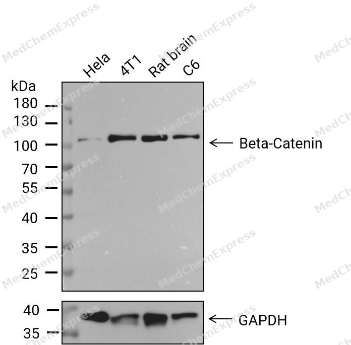 Western blot analysis of extracts from Hela(lane 1(20μg)) 、4T1(lane 2(20μg)) 、Rat brain(lane 3(20μg))and C6(lane 4(20μg) using Beta-Catenin Antibody(HY-P80488). Proteins were transferred to a PVDF membrane and blocked with 5%BSA in TBST for 1.5 hour at room temperature. The primary antibody ( 1/1000) and Loading control antibody (GAPDH, 1/1000) was used in 5% BSA in TBST at 4℃ overnight. Goat Anti-Rabbit IgG-HRP Secondary Antibody (1/10,000) was used for 1 hour at room temperature.
 -
 Western blot analysis of extracts from A431 (lane 2(20μg) and A431 (lane 3(40μg), HT-29 (lane 4(20μg) and HT-29 (lane 5(40μg) using beta Catenin (HY-P80488) Rabbit mAb. Proteins were transferred to a PVDF membrane and blocked with 5% non-fat milk in TBST for 2 hour at room temperature. The primary antibody (1/1000) and Loading control antibody (Beta Actin, HY-P80438, 1/10000) was used in 5% non-fat milk in TBST at 4°C overnight. Goat Anti-Mouse/Rabbit IgG-HRP Secondary Antibody (1/10000) was used for 1 hour at room temperature.
 -
 Immunohistochemical analysis of paraffin-embedded human appendix tissue using Beta-Catenin Antibody. The section was pre-treated using heat mediated antigen retrieval with Tris/EDTA buffer (pH 9.0) for 20 minutes. The tissues were probed with the primary antibody (HY-P80488, 1/100) overnight at 4℃. The detection was performed using an HRP conjugated compact polymer system. DAB was used as the chromogen. Tissues were counterstained with hematoxylin and mounted with neutral balsam.
 -
 Immunohistochemical analysis of paraffin-embedded human stomach tissue using Beta-Catenin Antibody. The section was pre-treated using heat mediated antigen retrieval with Tris/EDTA buffer (pH 9.0) for 20 minutes. The tissues were probed with the primary antibody (HY-P80488, 1/100) overnight at 4℃. The detection was performed using an HRP conjugated compact polymer system. DAB was used as the chromogen. Tissues were counterstained with hematoxylin and mounted with neutral balsam.
 -
 Immunohistochemical analysis of paraffin-embedded human colon cancer tissue using Beta-Catenin Antibody. The section was pre-treated using heat mediated antigen retrieval with Tris/EDTA buffer (pH 9.0) for 20 minutes. The tissues were probed with the primary antibody (HY-P80488, 1/100) overnight at 4℃. The detection was performed using an HRP conjugated compact polymer system. DAB was used as the chromogen. Tissues were counterstained with hematoxylin and mounted with neutral balsam.
 -
 Immunohistochemical analysis of paraffin-embedded human cervical cancer tissue using Beta-Catenin Antibody. The section was pre-treated using heat mediated antigen retrieval with Tris/EDTA buffer (pH 9.0) for 20 minutes. The tissues were probed with the primary antibody (HY-P80488, 1/100) overnight at 4℃. The detection was performed using an HRP conjugated compact polymer system. DAB was used as the chromogen. Tissues were counterstained with hematoxylin and mounted with neutral balsam.
 -
 Immunohistochemical analysis of paraffin-embedded mouse colon tissue using beta Catenin Antibody. The section was pre-treated using heat mediated antigen retrieval with sodium citrate buffer (pH 6.0) for 8 minutes. The tissues were blocked in QuickBlock for 20 minutes at room temperature, washed with ddH2O and PBS, and then probed with the primary antibody at 1/100 dilution in 4℃ overnight. The detection was performed using an HRP conjugated compact polymer system. DAB was used as the chromogen. Tissues were counterstained with hematoxylin and mounted with DPX.
 -
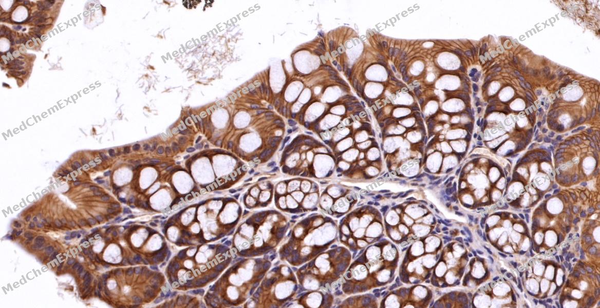 Immunohistochemical analysis of paraffin-embedded mouse colon tissue using beta Catenin Antibody. The section was pre-treated using heat mediated antigen retrieval with sodium citrate buffer (pH 6.0) for 8 minutes. The tissues were blocked in QuickBlock for 20 minutes at room temperature, washed with ddH2O and PBS, and then probed with the primary antibody at 1/100 dilution in 4℃ overnight. The detection was performed using an HRP conjugated compact polymer system. DAB was used as the chromogen. Tissues were counterstained with hematoxylin and mounted with DPX.
 -
 Immunocytochemistry analysis of Hela cells labeling beta Catenin with beta Catenin Antibody (HY-P80488) at 1/100 dilution. Cells were fixed in 4% paraformaldehyde for 15 minutes at room temperature, permeabilized with 0.1% Triton X-100 in PBS for 15 minutes at room temperature, then blocked with quick block buffer for 10 minutes at room temperature. Cells were then incubated with beta Catenin Antibody (HY-P80488) at 1/100 dilution in quick block buffer overnight at 4 ℃. AF488-conjugated Goat Anti-Rabbit IgG H&L(HY-P8002, Green) was used as the secondary antibody at 1/1,000 dilution. PBS instead of the primary antibody was used as the secondary antibody only control. The Nuclear counterstain was DAPI (Blue).
 -
 Immunocytochemistry analysis of Hela cells labeling beta Catenin with beta Catenin Antibody (HY-P80488) at 1/200 dilution. Cells were fixed in 4% paraformaldehyde for 15 minutes at room temperature, permeabilized with 0.1% Triton X-100 in PBS for 15 minutes at room temperature, then blocked with quick block buffer for 10 minutes at room temperature. Cells were then incubated with beta Catenin Antibody (HY-P80488) at 1/200 dilution in quick block buffer overnight at 4 ℃. AF488-conjugated Goat Anti-Rabbit IgG H&L(HY-P8002, Green) was used as the secondary antibody at 1/1,000 dilution. PBS instead of the primary antibody was used as the secondary antibody only control. The Nuclear counterstain was DAPI (Blue).


Background
-
Function
beta Catenin is key downstream component of the canonical Wnt signaling pathway. In the absence of Wnt, forms a complex with AXIN1, AXIN2, APC, CSNK1A1 and GSK3B that promotes phosphorylation on N-terminal Ser and Thr residues and ubiquitination of CTNNB1 via BTRC and its subsequent degradation by the proteasome. In the presence of Wnt ligand, CTNNB1 is not ubiquitinated and accumulates in the nucleus, where it acts as a coactivator for transcription factors of the TCF/LEF family, leading to activate Wnt responsive genes. Also acts as a coactivator for other transcription factors, such as NR5A2. Promotes epithelial to mesenchymal transition/mesenchymal to epithelial transition (EMT/MET) via driving transcription of CTNNB1/TCF-target genes. Involved in the regulation of cell adhesion, as component of an E-cadherin:catenin adhesion complex. Acts as a negative regulator of centrosome cohesion. Involved in the CDK2/PTPN6/CTNNB1/CEACAM1 pathway of insulin internalization. Blocks anoikis of malignant kidney and intestinal epithelial cells and promotes their anchorage-independent growth by down-regulating DAPK2. Disrupts PML function and PML-NB formation by inhibiting RANBP2-mediated sumoylation of PML. Promotes neurogenesis by maintaining sympathetic neuroblasts within the cell cycle. Involved in chondrocyte differentiation via interaction with SOX9: SOX9-binding competes with the binding sites of TCF/LEF within CTNNB1, thereby inhibiting the Wnt signaling. Acts as a positive regulator of odontoblast differentiation during mesenchymal tooth germ formation, via promoting the transcription of differentiation factors such as LEF1, BMP2 and BMP4. Activity is repressed in a MSX1-mediated manner at the bell stage of mesenchymal tooth germ formation which prevents premature differentiation of odontoblasts[1][2][3][4][5][6][7][8][9][10].
-
Subcellular Localization
Cytoplasm; Nucleus; Cytoplasm, cytoskeleton; Cell junction, adherens junction; Cell junction; Cell membrane; Cytoplasm, cytoskeleton, microtubule organizing center, centrosome; Cytoplasm, cytoskeleton, spindle pole; Synapse; Cytoplasm, cytoskeleton, cilium basal body
-
Expression
Tissue_specificity:Expressed in various hair follicle cell types: basal cells and peripheral stromal cells, as well as outer and inner root sheath cells. Expressed in the colon. Present in cortical neurons (protein level) . Expressed in breast cancer tissue (protein level) (PubMed:29367600) -
Subunit
Two separate complex-associated pools are found in the cytoplasm.
-
SwissProt ID
-
Synonyms
CTNNB, OK/SW-cl.35, PRO2286, CTNNB1, Catenin beta-1, Beta-catenin
-
Research Field
Signal Transduction
Documentation
-
Data Sheet (263 KB)
-
SDS (251 KB)
- English - EN (251 KB)
- Français - FR (251 KB)
- Deutsch - DE (251 KB)
- Norwegian - NO (251 KB)
- Español - ES (251 KB)
- Swedish - SV (251 KB)
- Italian - IT (251 KB)
- Korean - KR (251 KB)
- Portuguese - PT (251 KB)
-
User Guide for Antibodies (1077 KB)
References
[1]. Lillehoj EP, et al. MUC1 inhibits cell proliferation by a beta-catenin-dependent mechanism. Biochim Biophys Acta. 2007 Jul;1773(7):1028-38. [Content Brief]
[2]. Weiske J, et al. The tumor suppressor Fhit acts as a repressor of beta-catenin transcriptional activity. Proc Natl Acad Sci U S A. 2007 Dec 18;104(51):20344-9. [Content Brief]
[3]. Bahmanyar S, et al. beta-Catenin is a Nek2 substrate involved in centrosome separation. Genes Dev. 2008 Jan 1;22(1):91-105. [Content Brief]
[4]. Li H, et al. Down-regulation of death-associated protein kinase-2 is required for beta-catenin-induced anoikis resistance of malignant epithelial cells. J Biol Chem. 2009 Jan 23;284(4):2012-22. [Content Brief]
[5]. Fiset A, et al. Compartmentalized CDK2 is connected with SHP-1 and β-catenin and regulates insulin internalization. Cell Signal. 2011 May;23(5):911-9. [Content Brief]
[6]. Satow R, et al. β-catenin inhibits promyelocytic leukemia protein tumor suppressor function in colorectal cancer cells. Gastroenterology. 2012 Mar;142(3):572-81. [Content Brief]
[7]. Genovese G, et al. The tumor suppressor HINT1 regulates MITF and β-catenin transcriptional activity in melanoma cells. Cell Cycle. 2012 Jun 1;11(11):2206-15. [Content Brief]
[8]. Yu Y, et al. Kindlin 2 forms a transcriptional complex with β-catenin and TCF4 to enhance Wnt signalling. EMBO Rep. 2012 Aug;13(8):750-8. [Content Brief]
[9]. Yumoto F, et al. Structural basis of coactivation of liver receptor homolog-1 by β-catenin. Proc Natl Acad Sci U S A. 2012 Jan 3;109(1):143-8. [Content Brief]
[10]. Park J, et al. DSG2 Is a Functional Cell Surface Marker for Identification and Isolation of Human Pluripotent Stem Cells. Stem Cell Reports. 2018 Jul 10;11(1):115-127. [Content Brief]

 Powered by Bioz
