



FITC-conjugated Goat Anti-Rabbit IgG H&L
Based on 3 publication(s) in Google Scholar
FITC-conjugated Goat Anti-Rabbit IgG H&L is an FITC-conjugated,Goat-derived anti-Rabbit antibody. FITC-conjugated Goat Anti-Rabbit IgG H&L can binds to the IgG heavy and light chain of Rabbit antibodies.
-
Host:
Goat
-
Isotype:
IgG
-
Application:
ICC/IF, IF-Tissue, FC
-
Reactivity :
Rabbit
-
Formulation:
Supplied in PBS, pH7.4, containing 50% glycerol, 0.5% BSA and 0.02% sodium azide.
-
Conjugation:
FITC


To place orders, for customer services and technical support, please contact: MedChemExpress USA
Tel: 609-228-6898 E-mail: [email protected] [email protected]



-

HY-P80950
FITC-conjugated Goat Anti-Mouse IgG H&L -
HY-P81017
Cy3-conjugated Goat Anti-Rabbit IgG H&L -
HY-P81016
Cy3-conjugated Goat Anti-Mouse IgG H&L -
HY-P8002
AF488-conjugated Goat Anti-Rabbit IgG H&L -
HY-P80043
Calreticulin Antibody (YA571) -
HY-P8003
AF594-conjugated Goat Anti-Rabbit IgG H&L -
HY-D0814
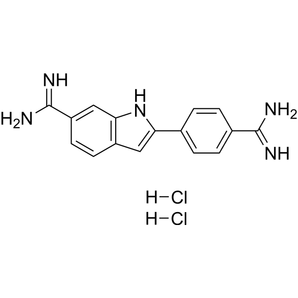
DAPI dihydrochlorideFluorescent DNA Stain -
HY-P8001
HRP-conjugated Goat Anti-Rabbit IgG H&L -
HY-P81013
AF647-conjugated Goat Anti-Mouse IgG H&L -
HY-P80599
CD31 Antibody (YA806) -
HY-P83739
alpha smooth muscle Actin Antibody(YA3468) -
HY-P80183
HMGB1 Antibody (YA361)
Publications Citing Use of MedChemExpress (MCE) FITC-conjugated Goat Anti-Rabbit IgG H&L
More
 3 Publications Citing Use of MCE FITC-conjugated Goat Anti-Rabbit IgG H&L (3)
-
IF

-
IF

-
IF


Applications
| Application |
ICC/IF

ICC/IF: Immunocytochemistry/
Immunofluorescence |
IF-Tissue
IF-Tissue: Immunofluorescence-Tissue
|
FC
FC: Flow Cytometry
|
|---|---|---|---|
| Dilution Ratio | 1:100-1000 | 1:100-1000 | 1:1000-4000 |


Product Details
FITC-conjugated Goat Anti-Rabbit IgG H&L is an FITC-conjugated,Goat-derived anti-Rabbit antibody. FITC-conjugated Goat Anti-Rabbit IgG H&L can binds to the IgG heavy and light chain of Rabbit antibodies.
-
Host Goat

-
Clonality Polyclonal

-
Species ReactivityRabbit
Synthetic peptide corresponding to Rabbit IgG.
Affinity Purified
FITC
Unmodified
IgG heavy and light chain
Whole IgG Antibodies
Human, Mouse
IgG


Product Properties
-
Appearance
Solution
-
Formulation
Supplied in PBS, pH7.4, containing 50% glycerol, 0.5% BSA and 0.02% sodium azide.
-
Concentration
Batch-dependent, Please check the COA for the concentration of each lot. Check Lot Concentration
-
Storage & Stability
Stored at -20°C for 1 year. Protect from light. Avoid repeated freeze / thaw cycles.
-
Shipping
Shipping with blue ice.


Publications (3)
-
Journal Impact Factor
-
Most Recent
-
Signal Transduct Target Ther
Family with sequence similarity 114 member A1 orchestrates immune evasion in triple-negative breast cancer. [Abstract]2025 Nov 18;10(1):373. PMID: 41249116
FITC-conjugated Goat Anti-Rabbit IgG H&L purchased from MedChemExpress. Usage Cited in: Signal Transduct Target Ther. 2025 Nov 18;10(1):373. [Abstract]
Cells with or without E2F4-GFP or E2F4-Myc plasmid transfection were fixed with 4% formaldehyde, blocked with 5% BSA, and subjected to IF staining. For cells not transfected or transfected with E2F4-Myc, anti-E2F4 (10923-1-AP, Proteintech, 1:20 dilution), anti-IgG (98136-1-RR, Proteintech, 1:50 dilution), and anti-Myc (16286-1-AP, Proteintech, 1:100 dilution) antibodies were used for staining. The primary antibody-incubated samples were incubated with goat anti-rabbit IgG H&L (FITC) (MCE, HY-P80951, 1:100 dilution).
-
Gut Microbes
Targeted modulation of intestinal barrier and mucosal immune-related microbiota attenuates IgA nephropathy progression. [Abstract]2025 Dec;17(1):2458184. PMID: 39875350
FITC-conjugated Goat Anti-Rabbit IgG H&L purchased from MedChemExpress. Usage Cited in: Gut Microbes. 2025 Dec;17(1):2458184. [Abstract]
Colon tissues fixed in 4% paraformaldehyde were embedded in paraffin and sectioned (4 μm). The sections were deparaffinized to water for antigen recovery. Sections were incubated at room temperature in the dark with 3% hydrogen peroxide solution for 25 minutes to block endogenous peroxidase activity. After blocking with 3% BSA, sections were incubated overnight at 4°C with rabbit anti-mouse IgA antibody (bs-0774 R, Bioss), rabbit polyclonal anti-C3 (21337–1-AP, Proteintech), and goat anti-mouse IgG H&L (FITC) (HY-P80950, MedChemExpress), then incubated with Goat Anti-Rabbit IgG H&L (FITC) (HY-P80951, MedChemExpress) at room temperature for 50 minutes.
-
FITC-conjugated Goat Anti-Rabbit IgG H&L purchased from MedChemExpress. Usage Cited in: SSRN. 2026 Mar 7.
Increased intestinal inflammation and weakened barrier function in adult zebrafish exposed to environmental concentrations of arecoline and arecaidine. Representative confocal immunofluorescence images.


Verification Images
-
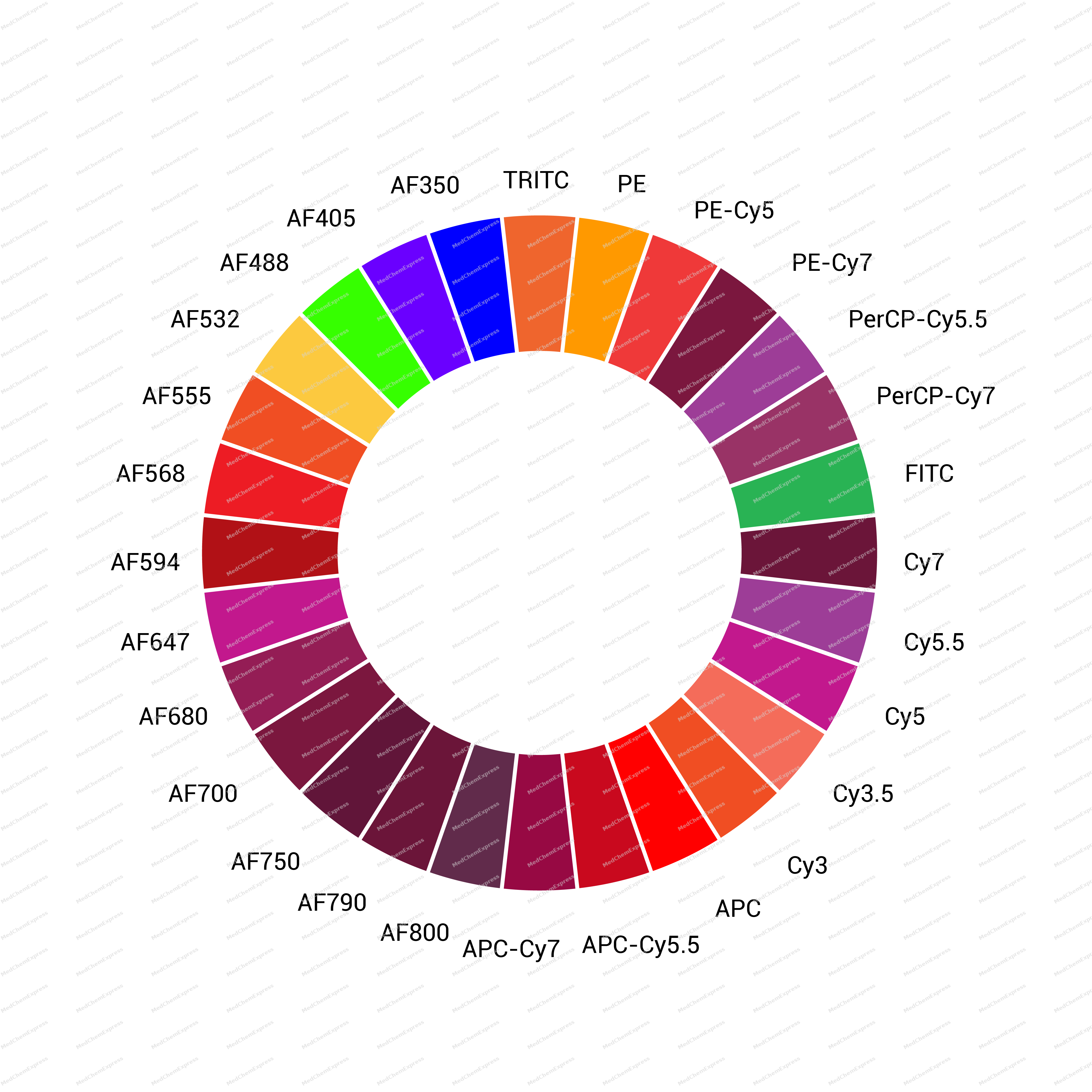 Please select the appropriate fluorescent dye based on your fluorescence imaging system. According to the species of the experimental sample you are studying and the host species of the primary antibody, choose the appropriate species of the secondary antibody.


Background
-
Function
Secondary antibodies are antibodies that bind to primary antibodies in immunoassays. They do not directly recognize the target antigen but rather the Fc region of the primary antibody or the entire immunoglobulin (Ig). Secondary antibodies are typically conjugated to fluorescent groups (such as Alexa Fluor), enzymes (such as HRP and alkaline phosphatase), or proteins (such as biotin).
-
Research Field
Immunology
Documentation
-
Data Sheet (230 KB)
-
SDS (252 KB)
- English - EN (252 KB)
- Français - FR (252 KB)
- Deutsch - DE (252 KB)
- Norwegian - NO (252 KB)
- Español - ES (252 KB)
- Swedish - SV (252 KB)
- Italian - IT (252 KB)
- Korean - KR (252 KB)
- Portuguese - PT (252 KB)
-
User Guide for Antibodies (1077 KB)

 Powered by Bioz
