



RUNX2 Antibody (YA085)
(Synonyms: AML3, CBFA1, OSF2, PEBP2A, RUNX2, Runt-related transcription factor 2, Acute myeloid leukemia 3 protein, Core-binding factor subunit alpha-1, Oncogene AML-3, Osteoblast-specific transcription factor 2, Polyomavirus enhancer-binding protein 2 alpha A subunit, SL3-3 enhancer factor 1 alpha A subunit, SL3/AKV core-binding factor alpha A subunit, CBF-alpha-1, OSF-2, PEA2-alpha A, PEBP2-alpha A)Based on 7 publication(s) in Google Scholar
RUNX2 Antibody (YA085) is a Rabbit-derived and non-conjugated IgG monoclonal antibody, targeting to RUNX2.
-
Host:
Rabbit
-
Isotype:
IgG
-
Application:
WB, IHC-P, ICC/IF, IP, FC, IF-Tissue, mIHC
-
Reactivity :
Human, Mouse, Rat
-
Formulation:
Supplied in 1*TBS (pH7.4), 0.05% BSA and 40% Glycerol. Preservative: 0.05% Sodium Azide.
-
Conjugation:
Non-conjugated


To place orders, for customer services and technical support, please contact: MedChemExpress USA
Tel: 609-228-6898 E-mail: [email protected] [email protected]



-

HY-P81217
Osteocalcin Antibody -
HY-P81227
Collagen I Antibody -
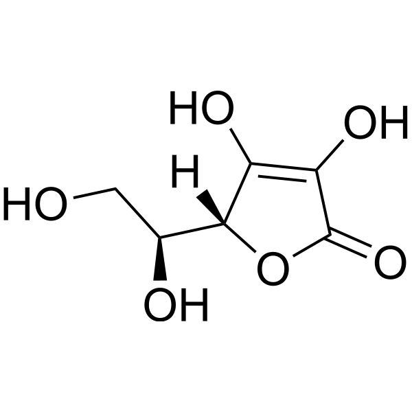
HY-B0166
L-Ascorbic acid -

HY-14648
Dexamethasone -
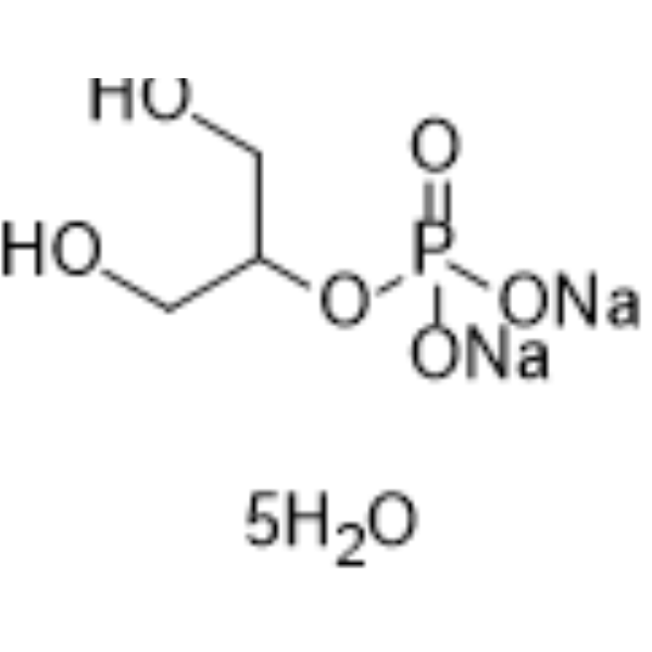
HY-D0886
β-Glycerophosphate disodium salt pentahydrate -
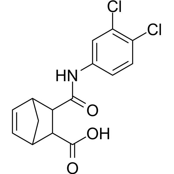
HY-107999
CADD522 -
HY-P80488
beta Catenin Antibody (YA584) -
HY-P80771
Osteopontin Antibody -
HY-P80839
Phospho-NF-KB p65 (Ser536) Antibody -
HY-P84129
CD31 Antibody (YA3826) -
HY-P81125
SP7/Osterix Antibody (YA3539) -
HY-P86653
Alkaline phosphatase Antibody (YA6345)
Publications Citing Use of MedChemExpress (MCE) RUNX2 Antibody (YA085)
More- Adv Funct Mater. 2025 Apr 01.
- J Nanobiotechnology. 2026 May 13.
- Adv Healthc Mater. 2025 Aug;14(20):e2500614. [Abstract]
- Appl Mater Today. 2024 Feb, 36, 102026.
- J Ethnopharmacol. 2026 Oct 28:369:121939. [Abstract]
- Clin Transl Oncol. 2026 Feb 25. [Abstract]
- Connect Tissue Res. 2024 Nov;65(6):433-446. [Abstract]


Applications
| Application |
WB

WB: Western Blot
|
ICC/IF
ICC/IF: Immunocytochemistry/
Immunofluorescence |
IF-Tissue
IF-Tissue: Immunofluorescence-Tissue
|
IHC-P
IHC-P: Immunohistochemistry-Paraffin
|
FC
FC: Flow Cytometry
|
IP
IP: Immunoprecipitation
|
mIHC
mIHC: Multiplex Immunohistochemical
|
|---|---|---|---|---|---|---|---|
| Dilution Ratio | 1:5000-1:10000 | 1:1000-1:5000 | 1:200-1:500 | 1:200-1:1000 | 1:5000 | 1-2 μg/sample | 1:2000 |


Product Details
RUNX2 Antibody (YA085) is a Rabbit-derived and non-conjugated IgG monoclonal antibody, targeting to RUNX2.
-
Host Rabbit

-
Clonality Recombinant,Monoclonal

-
Species ReactivityHuman, Mouse, Rat
-
Observed Molecular WeightObserved band size: 57 kDa
Note: Due to possible protein modifications or aggregation, the molecular weight should be confirmed by actual measurement, and the predicted value is for reference only.

-
Calculated Molecular Weight Predicted band size: 57 kDa

Entrez Gene: 860 Human ; 12393 Mouse ; 367218 Rat
SwissProt: Q13950 Human ; Q08775 Mouse ; Q9Z2J9 Rat
OMIM: 119600 Human
Synthetic peptide corresponding to Human RUNX2.AA range:300-450.
Endogenous
Protein A affinity purified.
Non-conjugated
Unmodified
IgG


Product Properties
-
Appearance
Solution
-
Formulation
Supplied in 1*TBS (pH7.4), 0.05% BSA and 40% Glycerol. Preservative: 0.05% Sodium Azide.
-
Concentration
Batch-dependent, Please check the COA for the concentration of each lot. Check Lot Concentration
-
Storage & Stability
Stored at -20°C for 1 year. Avoid repeated freeze / thaw cycles.
-
Shipping
Shipping with blue ice.


Publications (7)
-
Journal Impact Factor
-
Most Recent
-
Adv Healthc Mater
Cascade Regulation of Blood Clot Stabilization-Cell Migration-Osteogenic Differentiation by Hollow Hydrogels for Periodontal Bone Regeneration and Repair. [Abstract]2025 Aug;14(20):e2500614. PMID: 40357812 -
Clin Transl Oncol
Apigenin 7-glucoside reprograms tumor metabolism and enhances immunotherapy efficacy in colorectal cancer via DLX5. [Abstract]2026 Feb 25. PMID: 41739401 -
Connect Tissue Res
NEDD4L affects stability of the CHEK2/TP53 axis through ubiquitination modification to enhance osteogenic differentiation of periodontal ligament stem cells. [Abstract]2024 Nov;65(6):433-446. PMID: 39373023


Verification Images
-
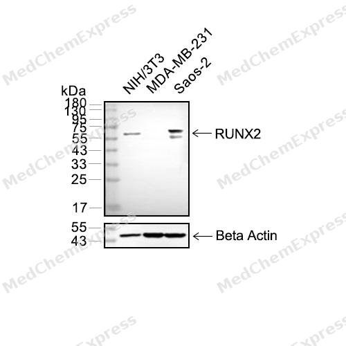 Western blot analysis of extracts from NIH3T3 (lane 2(20μg) , MDA-MB231(lane 3(20μg) and Saos-2(lane 4(20μg) using RUNX2(HY-P80316) Rabbit mAb. Proteins were transferred to a PVDF membrane and blocked with 5% non-fat milk in TBST for 2 hour at room temperature. The primary antibody (1/1000) and Loading control antibody (Beta Actin, HY-P80438, 1/10000) was used in 5% non-fat milk in TBST at 4°C overnight. Goat Anti-Mouse IgG-HRP Secondary Antibody (1/10000) was used for 1 hour at room temperature.
 -
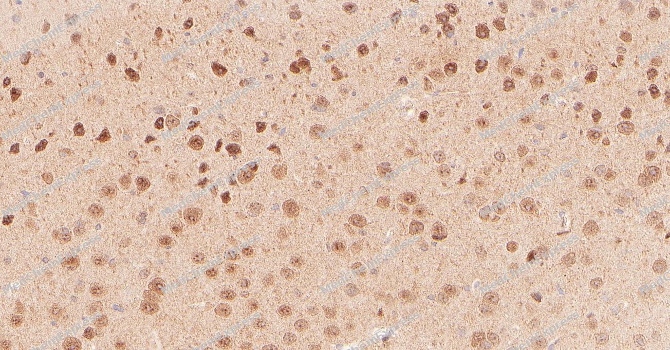 Immunohistochemical analysis of paraffin-embedded Mouse brain tissue using RUNX2 Antibody. The section was pre-treated using heat mediated antigen retrieval with sodium citrate buffer (pH 6.0) for 8 minutes. The tissues were blocked in QuickBlock for 20 minutes at room temperature, washed with ddH2O and PBS, and then probed with the primary antibody (HY-P80316, 1/500) in 4℃ overnight. The detection was performed using an HRP conjugated compact polymer system. DAB was used as the chromogen. Tissues were counterstained with hematoxylin and mounted with DPX.
 -
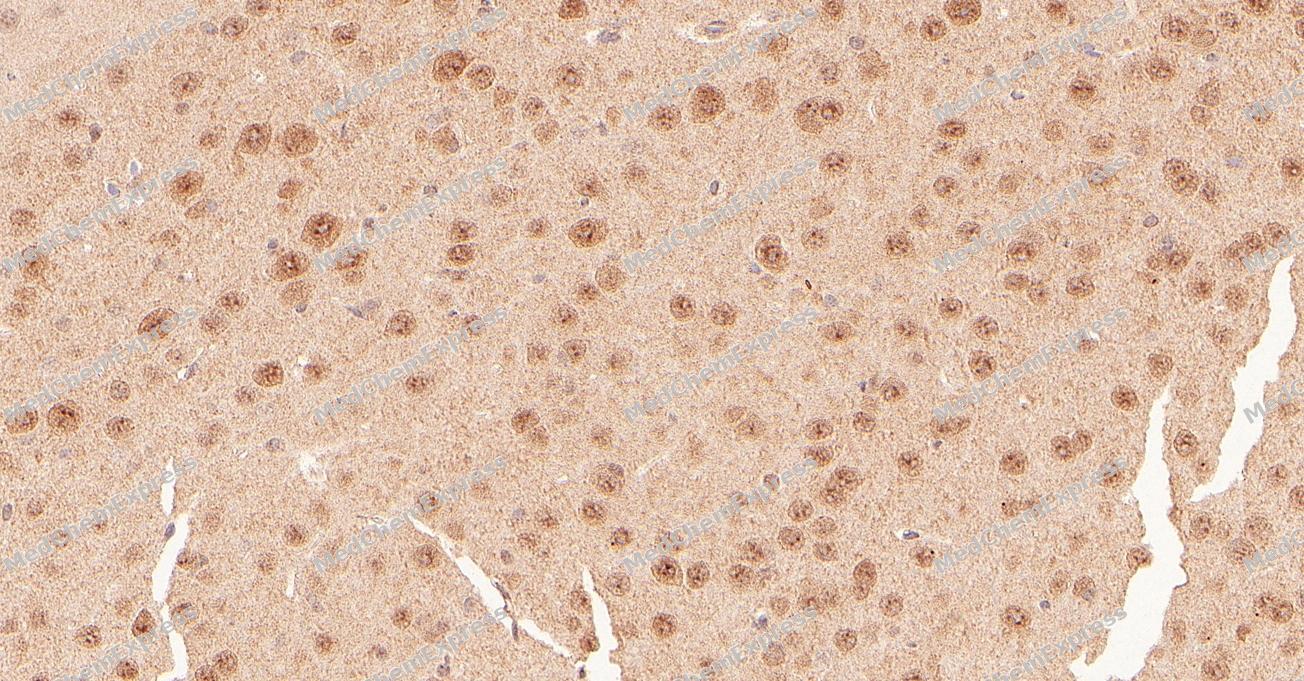 Immunohistochemical analysis of paraffin-embedded Mouse brain tissue using RUNX2 Antibody. The section was pre-treated using heat mediated antigen retrieval with sodium citrate buffer (pH 6.0) for 8 minutes. The tissues were blocked in QuickBlock for 20 minutes at room temperature, washed with ddH2O and PBS, and then probed with the primary antibody (HY-P80316, 1/500) in 4℃ overnight. The detection was performed using an HRP conjugated compact polymer system. DAB was used as the chromogen. Tissues were counterstained with hematoxylin and mounted with DPX.
 -
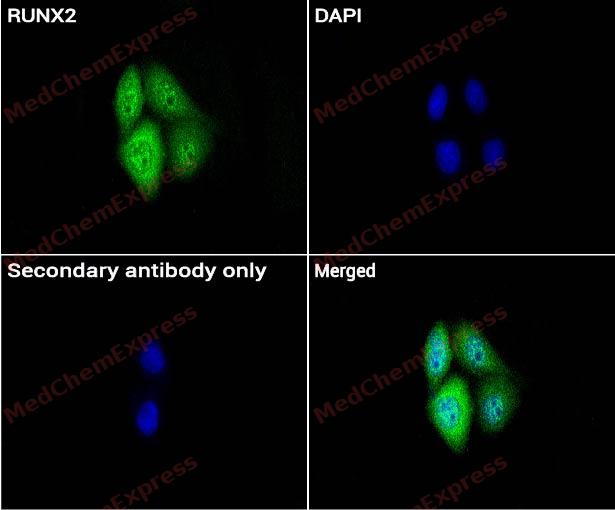 Immunocytochemistry analysis of HeLa cells labeling RUNX2 with RUNX2 Antibody (HY-P80316) at 1/100 dilution . Cells were fixed in 4% paraformaldehyde for 15 minutes at room temperature, permeabilized with 0.1% Triton X-100 in PBS for 15 minutes at room temperature, then blocked with quick block buffer for 10 minutes at room temperature. Cells were then incubated with RUNX2 Antibody (HY-P80316) at 1/100 dilution in quick block buffer overnight at 4 ℃. AF488-conjugated Goat Anti-Rabbit IgG H&L(HY-P8002, Green) was used as the secondary antibody at 1/1,000 dilution. PBS instead of the primary antibody was used as the secondary antibody only control. The Nuclear counterstain was DAPI (Blue).
 -
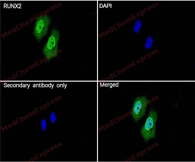 Immunocytochemistry analysis of HeLa cells labeling RUNX2 with RUNX2 Antibody (HY-P80316) at 1/200 dilution . Cells were fixed in 4% paraformaldehyde for 15 minutes at room temperature, permeabilized with 0.1% Triton X-100 in PBS for 15 minutes at room temperature, then blocked with quick block buffer for 10 minutes at room temperature. Cells were then incubated with RUNX2 Antibody (HY-P80316) at 1/200 dilution in quick block buffer overnight at 4 ℃. AF488-conjugated Goat Anti-Rabbit IgG H&L(HY-P8002, Green) was used as the secondary antibody at 1/1,000 dilution. PBS instead of the primary antibody was used as the secondary antibody only control. The Nuclear counterstain was DAPI (Blue).


Background
-
Function
RUNX2 (Runt-related transcription factor 2) is a master transcription factor that governs osteoblast differentiation, bone formation, and skeletal development by regulating osteogenic gene expression programs[1][2]. RUNX2 functions early during mesenchymal stem cell commitment to the osteoblast lineage and remains essential for progression through multiple stages of osteogenesis, while its expression is tightly controlled during osteoblast maturation[3][4]. Mechanistically, RUNX2 integrates signals from developmental and intracellular pathways, including MAPK-mediated activation and BMP-2/SMAD signaling, to coordinate transcriptional programs required for bone matrix production and mineralization[2][5]. RUNX2 also participates in chondrocyte maturation and endochondral bone development, highlighting its central role in skeletal tissue formation beyond osteoblast biology[3][6]. In disease and experimental models, loss of RUNX2 function causes profound skeletal abnormalities, and RUNX2 deficiency in mice results in the absence of osteoblasts and impaired bone formation, establishing RUNX2 as an indispensable regulator of skeletal development[3][7]. In humans, heterozygous mutations in RUNX2 cause cleidocranial dysplasia, a hereditary skeletal disorder characterized by defects in bone development[7][8]. Compared with related RUNX family members, RUNX2 exhibits a specialized role in osteoblast lineage specification and bone-forming activity, although partial functional overlap with RUNX3 has been reported during chondrocyte maturation[3]. Two major RUNX2 isoforms have been described, with evidence indicating distinct contributions to osteoblast development, where type I is associated with early osteoblastogenesis and type II contributes to later stages of osteoblastic maturation[9]. For experimental applications, RUNX2 activity is widely used as a molecular indicator of osteogenic differentiation, and modulation of BMP-2 signaling can alter RUNX2 expression in osteoblast models, providing a useful framework for studying bone formation mechanisms[5].
-
Subcellular Localization
Nucleus; Cytoplasm
-
Expression
Tissue_specificity:Specifically expressed in osteoblasts -
Subunit
Heterodimer of an alpha and a beta subunit. The alpha subunit binds DNA as a monomer and through the Runt domain. DNA-binding is increased by heterodimerization. Interacts with XRCC6 (Ku70) and XRCC5 (Ku80). Interacts with HIVEP3. Interacts with IFI204. Interaction with SATB2; the interaction results in enhanced DNA binding and transactivation by these transcription factors. Binds to HIPK3. Interacts with FOXO1 (via a C-terminal region); the interaction inhibits RUNX2 transcriptional activity towards BGLAP. This interaction is prevented on insulin or IGF1 stimulation as FOXO1 is exported from the nucleus (By similarity). Interacts with CCNB1, KAT6A and KAT6B. Interacts with FOXP3. Interacts with TMEM119 (By similarity). Interacts with OLFM2 (By similarity). Interacts with IPO7; the interaction inhibits RUNX2 nuclear translocation in osteoblasts (By similarity)
-
SwissProt ID
-
Synonyms
AML3, CBFA1, OSF2, PEBP2A, RUNX2, Runt-related transcription factor 2, Acute myeloid leukemia 3 protein, Core-binding factor subunit alpha-1, Oncogene AML-3, Osteoblast-specific transcription factor 2, Polyomavirus enhancer-binding protein 2 alpha A subunit, SL3-3 enhancer factor 1 alpha A subunit, SL3/AKV core-binding factor alpha A subunit, CBF-alpha-1, OSF-2, PEA2-alpha A, PEBP2-alpha A
-
Research Field
Epigenetics and Nuclear Signaling
Documentation
-
Data Sheet (262 KB)
-
SDS (254 KB)
- English - EN (254 KB)
- Français - FR (254 KB)
- Deutsch - DE (254 KB)
- Norwegian - NO (254 KB)
- Español - ES (254 KB)
- Swedish - SV (254 KB)
- Italian - IT (254 KB)
- Korean - KR (254 KB)
- Portuguese - PT (254 KB)
-
User Guide for Antibodies (1077 KB)
References
[1]. Vimalraj S, et al. Runx2: Structure, function, and phosphorylation in osteoblast differentiation. Int J Biol Macromol. 2015;78:202-8. [Content Brief]
[2]. Franceschi RT, et al. Regulation of the osteoblast-specific transcription factor, Runx2: responsiveness to multiple signal transduction pathways. J Cell Biochem. 2003 Feb 15;88(3):446-54. [Content Brief]
[3]. Komori T. Regulation of Proliferation, et al. Regulation of Proliferation, Differentiation and Functions of Osteoblasts by Runx2. Int J Mol Sci. 2019 Apr 4;20(7):1694. [Content Brief]
[4]. Zhu S, et al. Cell signaling and transcriptional regulation of osteoblast lineage commitment, differentiation, bone formation, and homeostasis. Cell Discov. 2024 Jul 2;10(1):71. [Content Brief]
[6]. Liu TM, et al. Transcriptional regulatory cascades in Runx2-dependent bone development. Tissue Eng Part B Rev. 2013 Jun;19(3):254-63. [Content Brief]
[7]. Baniwal SK, et al. Runx2 promotes both osteoblastogenesis and novel osteoclastogenic signals in ST2 mesenchymal progenitor cells. Osteoporos Int. 2012 Apr;23(4):1399-413. [Content Brief]
[8]. RUNX2 gene information from NCBI.
[9]. Bruderer M, et al. Role and regulation of RUNX2 in osteogenesis. Eur Cell Mater. 2014 Oct 23;28:269-86. [Content Brief]

 Powered by Bioz
