



Phospho-MSK1 (Ser376) Antibody (YA172)
(Synonyms: RPS6KA5; MSK1; Ribosomal protein S6 kinase alpha-5; S6K-alpha-5; 90 kDa ribosomal protein S6 kinase 5; Nuclear mitogen- and stress-activated protein kinase 1; RSK-like protein kinase; RSKL)
Based on 1 Customer Validation
Phospho-MSK1 (Ser376) Antibody (YA172) is a Rabbit-derived and non-conjugated IgG monoclonal antibody, targeting to Phospho-MSK1 (Ser376).
-
Host:
Rabbit
-
Isotype:
IgG
-
Application:
WB, IHC-P, IP
-
Reactivity :
Human, Rat
-
Formulation:
Supplied in 50 mM Tris-Glycine (pH 7.4), 0.15 M NaCl, 40% Glycerol and 0.05% BSA. Preservative: 0.01% Sodium azide
-
Conjugation:
Non-conjugated


To place orders, for customer services and technical support, please contact: MedChemExpress USA
Tel: 609-228-6898 E-mail: [email protected] [email protected]





Applications
| Application |
WB

WB: Western Blot
|
IHC-P
IHC-P: Immunohistochemistry-Paraffin
|
IP
IP: Immunoprecipitation
|
|---|---|---|---|
| Dilution Ratio | 1:500-1:1000 | 1:50-1:100 | 1:20 |


Product Details
Phospho-MSK1 (Ser376) Antibody (YA172) is a Rabbit-derived and non-conjugated IgG monoclonal antibody, targeting to Phospho-MSK1 (Ser376).
-
Host Rabbit

-
Clonality Recombinant,Monoclonal

-
Species ReactivityHuman, Rat
-
Observed Molecular WeightObserved band size: 90 kDa
Note: Due to possible protein modifications or aggregation, the molecular weight should be confirmed by actual measurement, and the predicted value is for reference only.

-
Calculated Molecular Weight Predicted band size: 90 kDa

Synthetic phosphopeptide corresponding to residues surrounding Ser376 of Human MSK1.The exact sequence is proprietary to MCE.
Endogenous
affinity purified
Non-conjugated
Phosphorylated
IgG


Product Properties
-
Appearance
Solution
-
Formulation
Supplied in 50 mM Tris-Glycine (pH 7.4), 0.15 M NaCl, 40% Glycerol and 0.05% BSA. Preservative: 0.01% Sodium azide
-
Concentration
Batch-dependent, Please check the COA for the concentration of each lot. Check Lot Concentration
-
Storage & Stability
Stored at -20°C for 1 year. Avoid repeated freeze / thaw cycles.
-
Shipping
Shipping with blue ice.


Verification Images
-
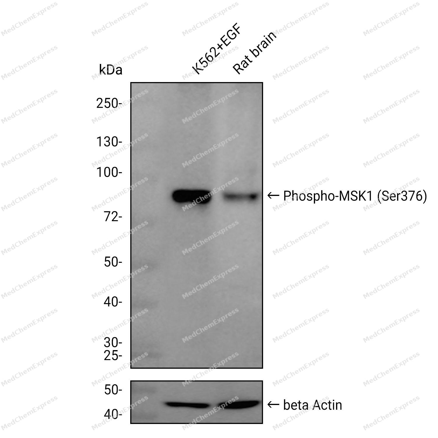
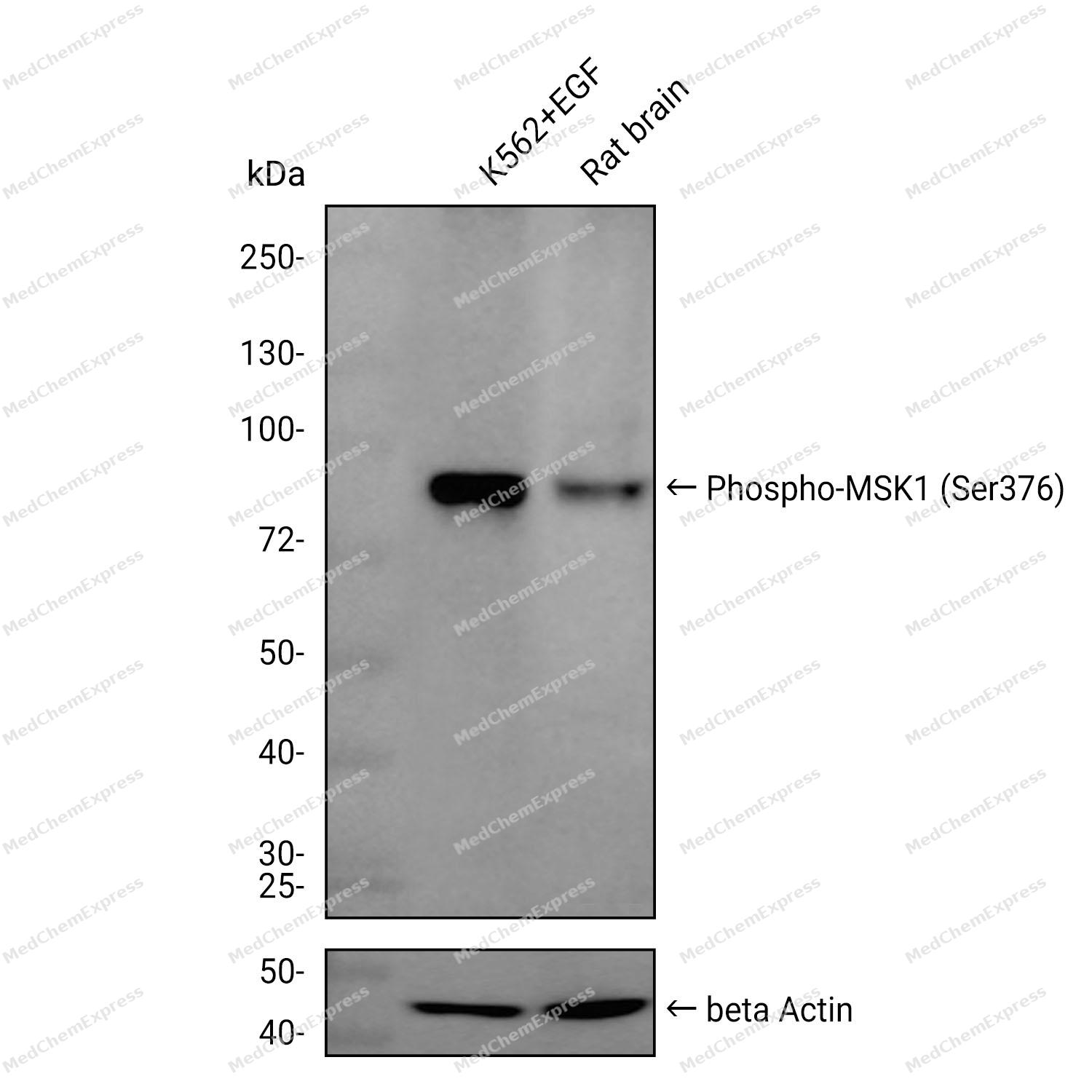 Western blot analysis was performed on extracts from K562+EGF (lane 1, 15 μg) and Rat brain (lane 2, 15 μg) using Phospho-MSK1 (Ser376) Rabbit mAb.Proteins were transferred to a PVDF membrane and blocked with 5% non-fat milk in TBST at 4°C overnight.The primary antibody (1:1000 dilution) and the loading control antibody (beta-Actin, HY-P80438, 1:20000 dilution) were incubated in 5% non-fat milk in TBST for 1 hour at 37°C.Goat Anti-Rabbit IgG-HRP Secondary Antibody (1:20000 dilution) was then applied for 40 minutes at 37°C.
 -
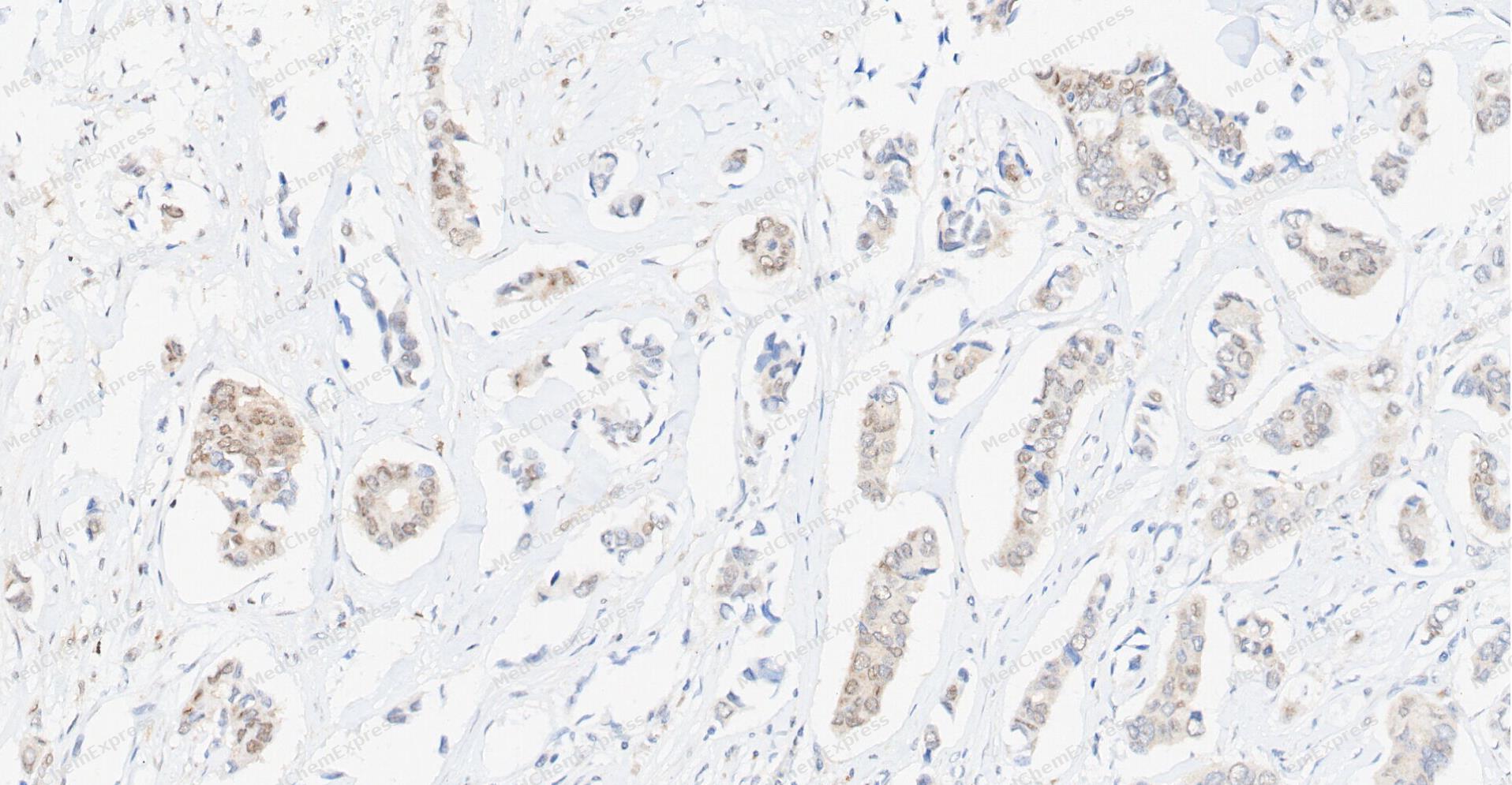 Immunohistochemical analysis of paraffin-embedded human Breast Cancer tissue using Phospho-MSK1 antibody was performed. The section was pretreated using high-temperature and high-pressure mediated EDTA antigen retrieval buffer (pH 9.0), for 5 minutes. The tissues were incubated with primary antibody (HY-P80836, 1:100 dilution) at room temperature for 60 minutes. Detection was performed using an HRP conjugated compact polymer system. DAB was used as the chromogen. The tissues were counterstained with hematoxylin and mounted with neutral balsam mounting medium.
 -
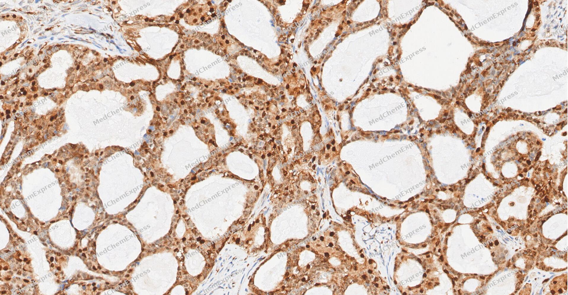 Immunohistochemical analysis of paraffin-embedded human Ovarian Cancer tissue using Phospho-MSK1 antibody was performed. The section was pretreated using high-temperature and high-pressure mediated EDTA antigen retrieval buffer (pH 9.0), for 5 minutes. The tissues were incubated with primary antibody (HY-P80836, 1:100 dilution) at room temperature for 60 minutes. Detection was performed using an HRP conjugated compact polymer system. DAB was used as the chromogen. The tissues were counterstained with hematoxylin and mounted with neutral balsam mounting medium.
 -
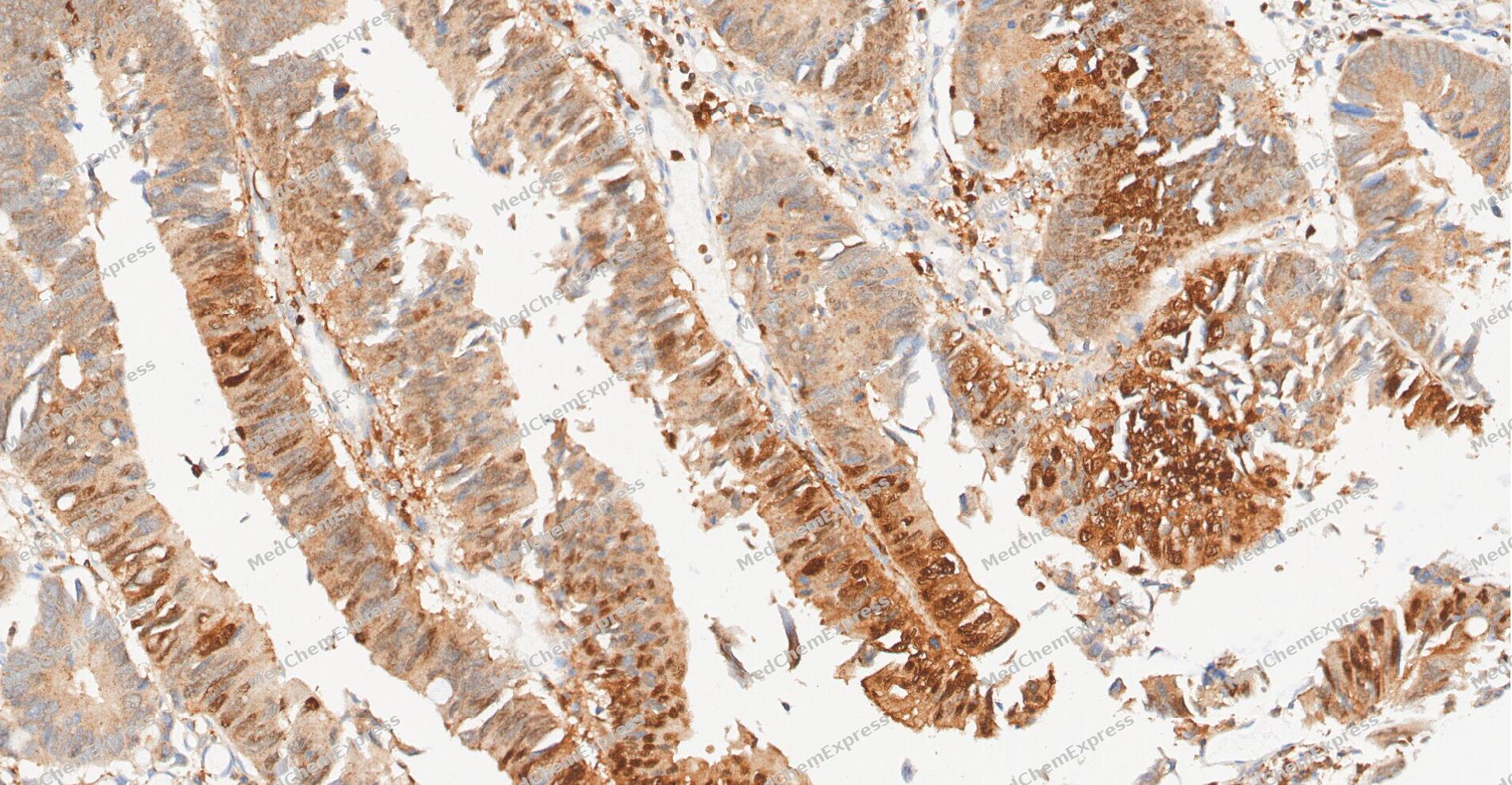 Immunohistochemical analysis of paraffin-embedded human Colon cancer tissue using Phospho-MSK1 antibody was performed. The section was pretreated using high-temperature and high-pressure mediated EDTA antigen retrieval buffer (pH 9.0), for 5 minutes. The tissues were incubated with primary antibody (HY-P80836, 1:100 dilution) at room temperature for 60 minutes. Detection was performed using an HRP conjugated compact polymer system. DAB was used as the chromogen. The tissues were counterstained with hematoxylin and mounted with neutral balsam mounting medium.
 -
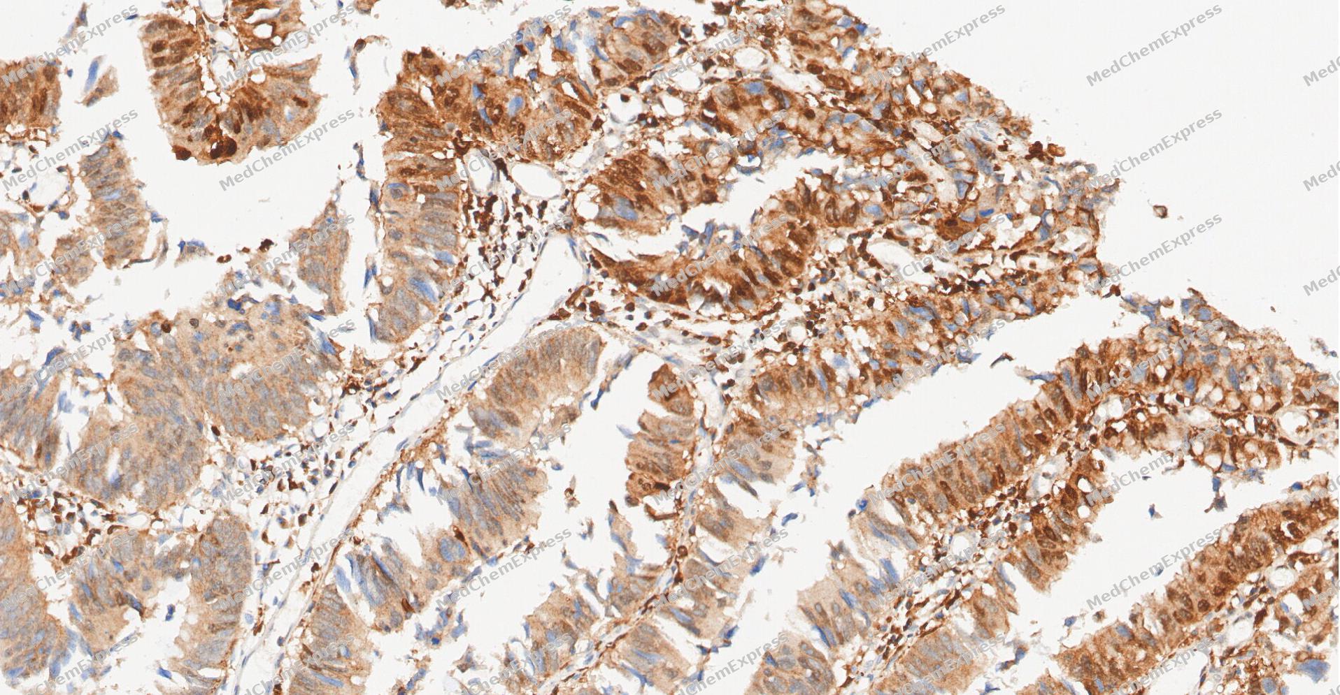 Immunohistochemical analysis of paraffin-embedded human Colon cancer tissue using Phospho-MSK1 antibody was performed. The section was pretreated using high-temperature and high-pressure mediated EDTA antigen retrieval buffer (pH 9.0), for 5 minutes. The tissues were incubated with primary antibody (HY-P80836, 1:100 dilution) at room temperature for 60 minutes. Detection was performed using an HRP conjugated compact polymer system. DAB was used as the chromogen. The tissues were counterstained with hematoxylin and mounted with neutral balsam mounting medium.
 -
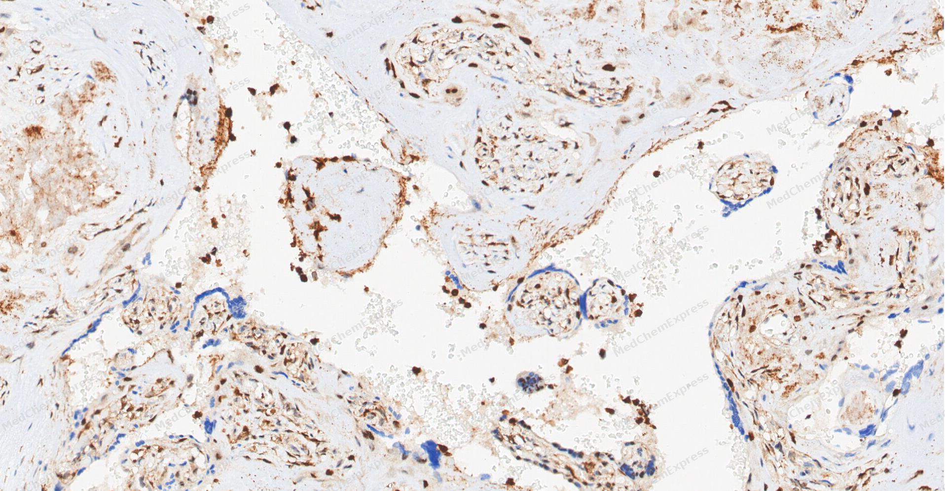 Immunohistochemical analysis of paraffin-embedded human Placenta tissue using Phospho-MSK1 antibody was performed. The section was pretreated using high-temperature and high-pressure mediated EDTA antigen retrieval buffer (pH 9.0), for 5 minutes. The tissues were incubated with primary antibody (HY-P80836, 1:100 dilution) at room temperature for 60 minutes. Detection was performed using an HRP conjugated compact polymer system. DAB was used as the chromogen. The tissues were counterstained with hematoxylin and mounted with neutral balsam mounting medium.
 -
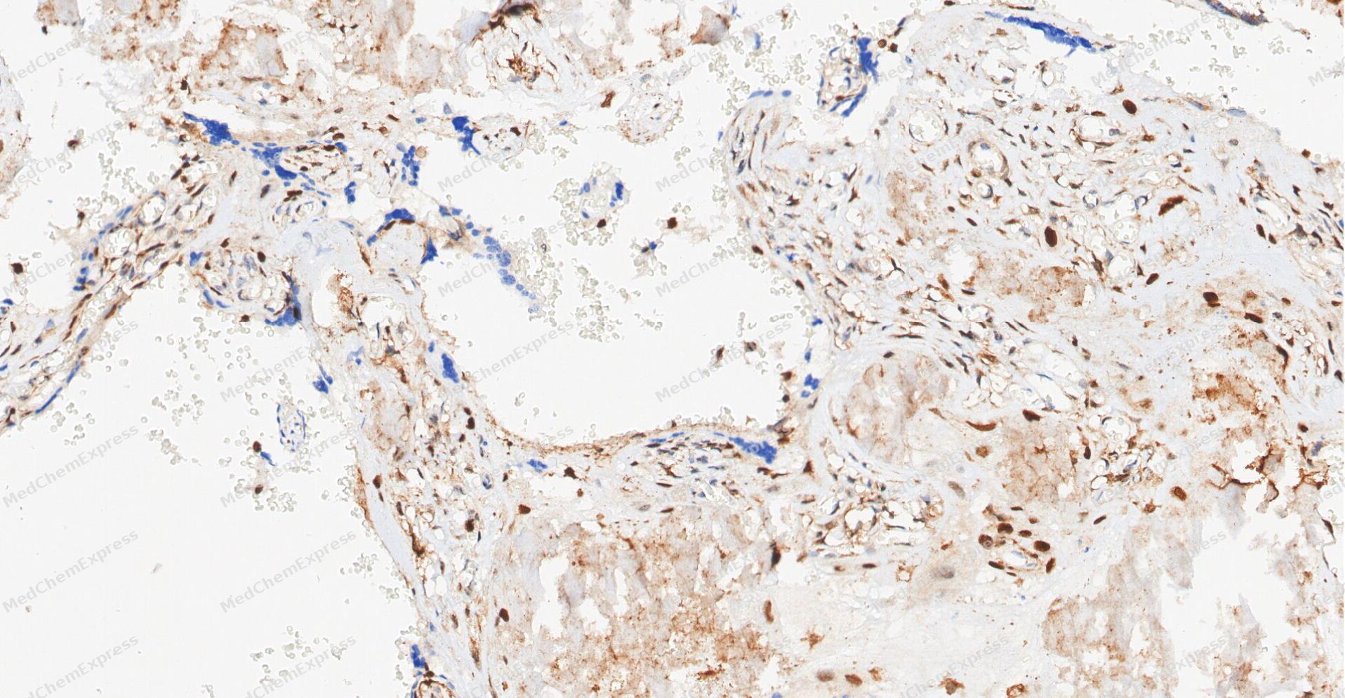 Immunohistochemical analysis of paraffin-embedded human Placenta tissue using Phospho-MSK1 antibody was performed. The section was pretreated using high-temperature and high-pressure mediated EDTA antigen retrieval buffer (pH 9.0), for 5 minutes. The tissues were incubated with primary antibody (HY-P80836, 1:100 dilution) at room temperature for 60 minutes. Detection was performed using an HRP conjugated compact polymer system. DAB was used as the chromogen. The tissues were counterstained with hematoxylin and mounted with neutral balsam mounting medium.
 -
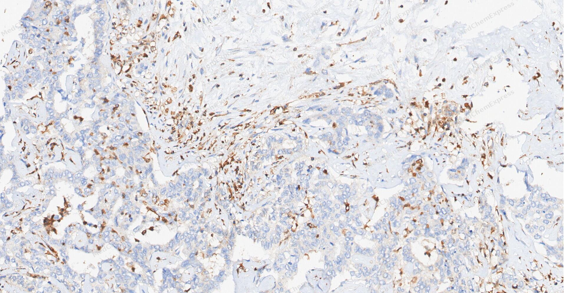 Immunohistochemical analysis of paraffin-embedded human cholangiocarcinoma tissue using Phospho-MSK1 antibody was performed. The section was pretreated using high-temperature and high-pressure mediated EDTA antigen retrieval buffer (pH 9.0), for 5 minutes. The tissues were incubated with primary antibody (HY-P80836, 1:100 dilution) at room temperature for 60 minutes. Detection was performed using an HRP conjugated compact polymer system. DAB was used as the chromogen. The tissues were counterstained with hematoxylin and mounted with neutral balsam mounting medium.
 -
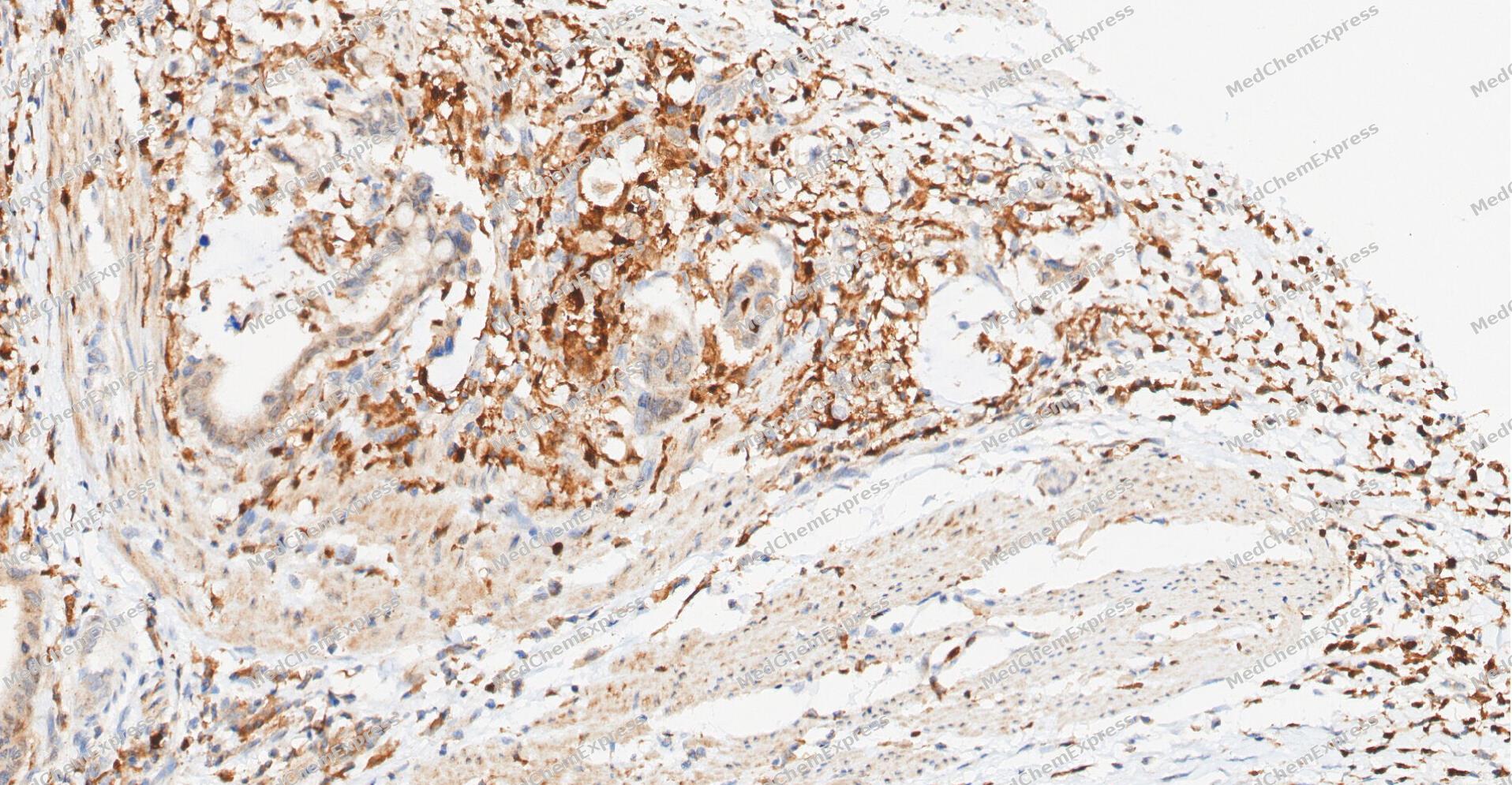 Immunohistochemical analysis of paraffin-embedded human Gastric Cancer tissue using Phospho-MSK1 antibody was performed. The section was pretreated using high-temperature and high-pressure mediated EDTA antigen retrieval buffer (pH 9.0), for 5 minutes. The tissues were incubated with primary antibody (HY-P80836, 1:100 dilution) at room temperature for 60 minutes. Detection was performed using an HRP conjugated compact polymer system. DAB was used as the chromogen. The tissues were counterstained with hematoxylin and mounted with neutral balsam mounting medium.
 -
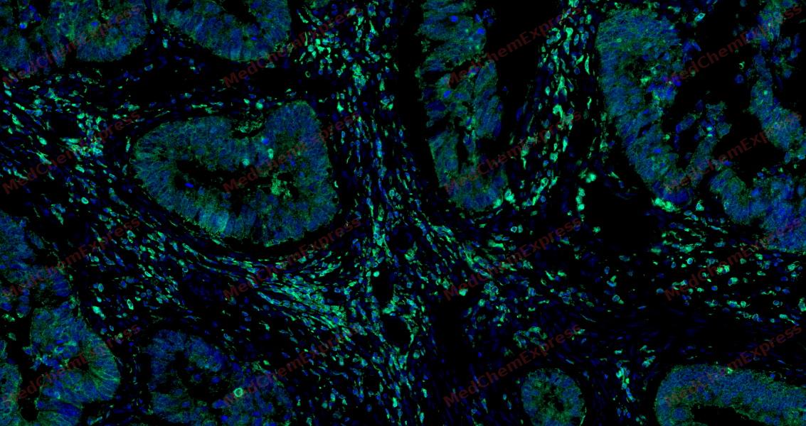 Tyramide signaling amplification based immunofluorescence was performed on paraffin-embedded human Colon cancer tissue using Phospho-MSK1 (Ser376) antibody. Antigen retrieval was performed in EDTA buffer pH 9.0 (95 °C, 20 min) followed by cooling to RT. Then incubated with primary antibody (HY-P80836, 1:100 dilution) at room temperature for 60 minutes and HRP conjugated secondary antibody for 10 minutes. Fluorescence was then developed with Vari Fluor 532 TSA (200×)(HY-D1832). The tissues were counterstained with DAPI and mounted with Anti-fade mounting medium.
 -
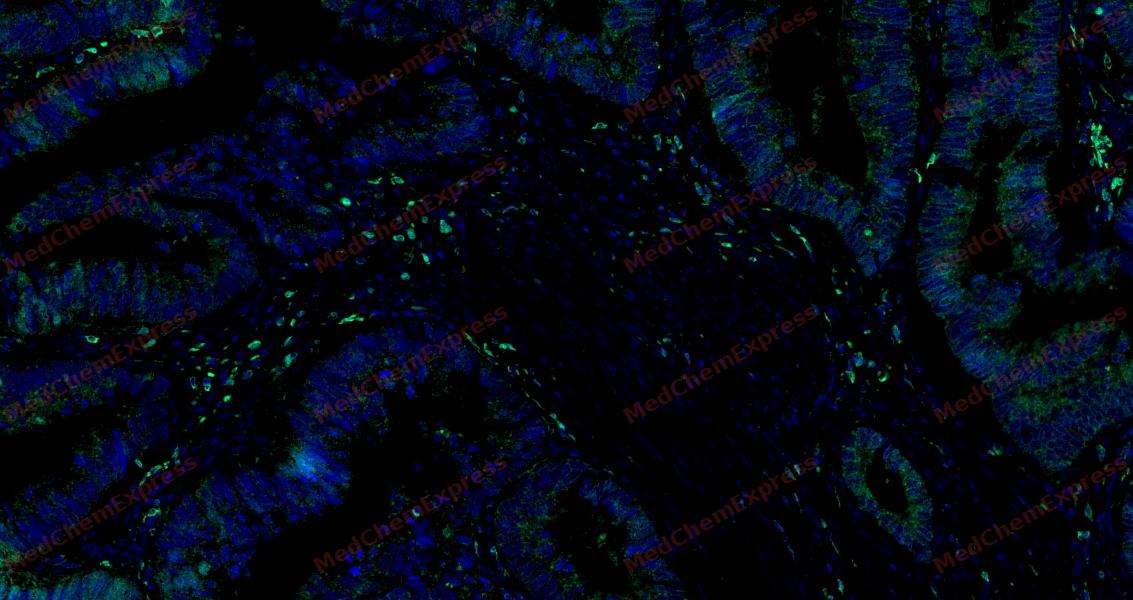 Tyramide signaling amplification based immunofluorescence was performed on paraffin-embedded human Colon cancer tissue using Phospho-MSK1 (Ser376) antibody. Antigen retrieval was performed in EDTA buffer pH 9.0 (95 °C, 20 min) followed by cooling to RT. Then incubated with primary antibody (HY-P80836, 1:100 dilution) at room temperature for 60 minutes and HRP conjugated secondary antibody for 10 minutes. Fluorescence was then developed with Vari Fluor 532 TSA (200×)(HY-D1832). The tissues were counterstained with DAPI and mounted with Anti-fade mounting medium.
 -
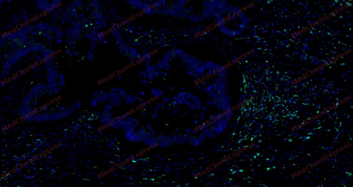 Tyramide signaling amplification based immunofluorescence was performed on paraffin-embedded human Colon cancer tissue using Phospho-MSK1 (Ser376) antibody. Antigen retrieval was performed in EDTA buffer pH 9.0 (95 °C, 20 min) followed by cooling to RT. Then incubated with primary antibody (HY-P80836, 1:100 dilution) at room temperature for 60 minutes and HRP conjugated secondary antibody for 10 minutes. Fluorescence was then developed with Vari Fluor 532 TSA (200×)(HY-D1832). The tissues were counterstained with DAPI and mounted with Anti-fade mounting medium.
 -
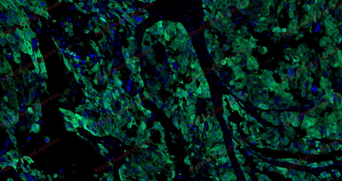 Tyramide signaling amplification based immunofluorescence was performed on paraffin-embedded human ovarian carcinoma tissue using Phospho-MSK1 (Ser376) antibody. Antigen retrieval was performed in EDTA buffer pH 9.0 (95 °C, 20 min) followed by cooling to RT. Then incubated with primary antibody (HY-P80836, 1:100 dilution) at room temperature for 60 minutes and HRP conjugated secondary antibody for 10 minutes. Fluorescence was then developed with Vari Fluor 532 TSA (200×)(HY-D1832). The tissues were counterstained with DAPI and mounted with Anti-fade mounting medium.
 -
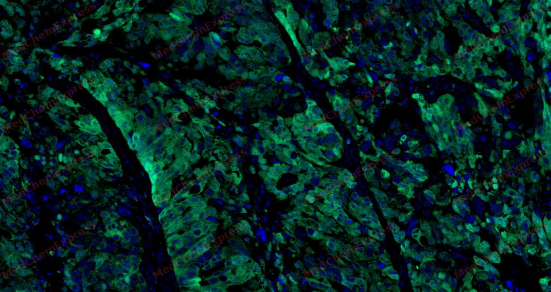 Tyramide signaling amplification based immunofluorescence was performed on paraffin-embedded human ovarian carcinoma tissue using Phospho-MSK1 (Ser376) antibody. Antigen retrieval was performed in EDTA buffer pH 9.0 (95 °C, 20 min) followed by cooling to RT. Then incubated with primary antibody (HY-P80836, 1:100 dilution) at room temperature for 60 minutes and HRP conjugated secondary antibody for 10 minutes. Fluorescence was then developed with Vari Fluor 532 TSA (200×)(HY-D1832). The tissues were counterstained with DAPI and mounted with Anti-fade mounting medium.
 -
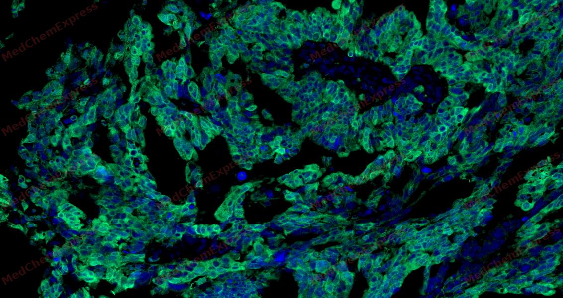 Tyramide signaling amplification based immunofluorescence was performed on paraffin-embedded human ovarian carcinoma tissue using Phospho-MSK1 (Ser376) antibody. Antigen retrieval was performed in EDTA buffer pH 9.0 (95 °C, 20 min) followed by cooling to RT. Then incubated with primary antibody (HY-P80836, 1:100 dilution) at room temperature for 60 minutes and HRP conjugated secondary antibody for 10 minutes. Fluorescence was then developed with Vari Fluor 532 TSA (200×)(HY-D1832). The tissues were counterstained with DAPI and mounted with Anti-fade mounting medium.


Background
-
Function
MSK1 (mitogen- and stress-activated protein kinase 1) is a predominantly nuclear serine/threonine kinase that functions downstream of the ERK1/2 and p38 MAPK pathways, integrating mitogenic and stress signals into transcriptional responses through phosphorylation-dependent activation mechanisms[1][2]. MSK1 regulates gene expression by phosphorylating key nuclear substrates, including CREB, ATF1, and histone H3, thereby controlling immediate-early gene induction and chromatin-associated transcriptional programs[2][3]. Mechanistically, activation of upstream MAPKs promotes sequential phosphorylation and autophosphorylation events within MSK1, enabling its N-terminal kinase domain to phosphorylate downstream targets involved in stimulus-responsive gene regulation[1][2]. In disease-relevant systems, MSK1 contributes to anti-inflammatory transcriptional networks, regulates cytokine expression, and participates in neuronal proliferation, synaptic plasticity, and neurogenesis-related processes[2]. MSK1 also functions as a chromatin-associated transcriptional coactivator, directly interacting with p53 and promoting p21 gene expression through p53-dependent histone H3 phosphorylation at target promoters[3]. Compared with the closely related isoform MSK2, MSK1 shares substantial functional overlap and is often considered functionally redundant in many MAPK-dependent signaling contexts, although isoform-specific expression patterns and experimental models continue to support investigation of distinct biological contributions[2]. For experimental applications, pharmacological inhibitors such as H89 and dimethylfumarate have been used to suppress MSK1/MSK2 signaling and evaluate downstream transcriptional and inflammatory responses in cellular and disease models[2].
-
Subcellular Localization
Nucleus; Cytoplasm
-
Expression
Tissue_specificity:It is widely expressed in the heart, brain, and placenta, at high levels. It is present in lower levels in the lungs, kidneys, and liver. -
Isoforms & Post-Translational Modification
O75582 has 3 isomers: O75582-1: 89865 Da (predicted); O75582-2: 61769 Da (predicted); O75582-3: 81780 Da (predicted).
Ser-376 and Thr-581 phosphorylation is required for kinase activity. Ser-376 and Ser-212 are autophosphorylated by the C-terminal kinase domain, and their phosphorylation is essential for the catalytic activity of the N-terminal kinase domain. Phosphorylated at Ser-360, Thr-581 and Thr-700 by MAPK1/ERK2, MAPK3/ERK1 and MAPK14/p38-alpha. Autophosphorylated at Ser-750, Ser-752 and Ser-758 by the N-terminal kinase domain;Ubiquitinated -
Subunit
Forms a complex with either MAPK1/ERK2 or MAPK3/ERK1 in quiescent cells which transiently dissociates following mitogenic stimulation. Also associates with MAPK14/p38-alpha. Activated RPS6KA5 associates with and phosphorylates the NF-kappa-B p65 subunit RELA. Interacts with CREBBP and EP300
-
SwissProt ID
-
Synonyms
RPS6KA5; MSK1; Ribosomal protein S6 kinase alpha-5; S6K-alpha-5; 90 kDa ribosomal protein S6 kinase 5; Nuclear mitogen- and stress-activated protein kinase 1; RSK-like protein kinase; RSKL
-
Research Field
Signal Transduction


Documentation
-
Data Sheet (260 KB)
-
SDS (252 KB)
- English - EN (252 KB)
- Français - FR (252 KB)
- Deutsch - DE (252 KB)
- Norwegian - NO (252 KB)
- Español - ES (252 KB)
- Swedish - SV (252 KB)
- Italian - IT (252 KB)
- Korean - KR (252 KB)
- Portuguese - PT (252 KB)
-
User Guide for Antibodies (1077 KB)
References
[1]. McCoy CE, et al. MSK1 activity is controlled by multiple phosphorylation sites. Biochem J. 2005 Apr 15;387(Pt 2):507-17. [Content Brief]
[2]. Reyskens KM, et al. Emerging Roles of the Mitogen and Stress Activated Kinases MSK1 and MSK2. Front Cell Dev Biol. 2016 Jun 10;4:56. [Content Brief]
[3]. Ahn J, et al. MSK1 functions as a transcriptional coactivator of p53 in the regulation of p21 gene expression. Exp Mol Med. 2018 Oct 10;50(10):1-12. [Content Brief]

 Powered by Bioz
