



CCF642
Based on 1 publication(s) in Google Scholar
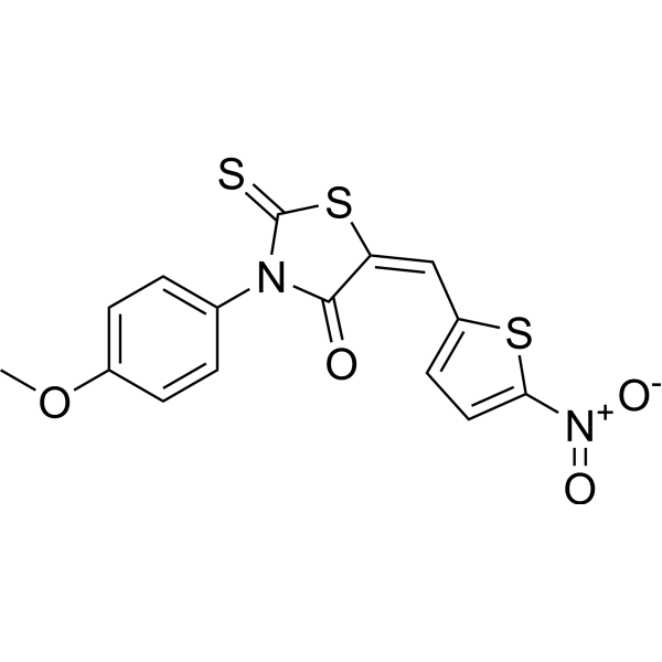
CCF642 is a potent protein disulfide isomerases (PDI) inhibitor with an IC50 of 2.9 μM. CCF642 causes acute endoplasmic reticulum (ER) stress in multiple myeloma cells accompanied by apoptosis-inducing calcium release. CCF642 has broad anti-multiple myeloma activity.
Nur für Forschungszwecke. Wir verkaufen nicht an Patienten.
- Reinheit : 98.0%
- CAS. Nr.: 346640-08-2
- Formel: C15H10N2O4S3
- Molecular Weight:378.45
-
Speicherung:Powder -20°C, 3 years , 4°C, 2 years ; In solvent -80°C, 2 years , -20°C, 1 year

To place orders, for customer services and technical support, please contact: MedChemExpress USA
Tel: 609-228-6898 E-mail: [email protected] [email protected]


-
 Biologische Aktivität
-
Chemical Information
-
Lösungsmittel & Löslichkeit
- Protokoll
- Reinheit & Dokumentation
- Verweise
-
 Help & FAQs


-
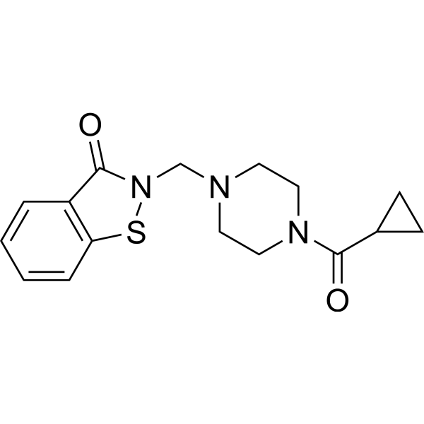
HY-100432
LOC14PDI Inhibitor -
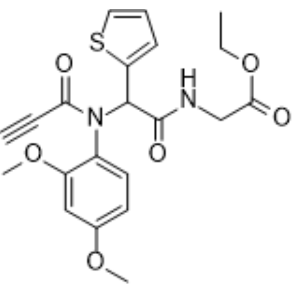
HY-100433
PACMA 31Protein Disulfide Isomerase Inhibitor -
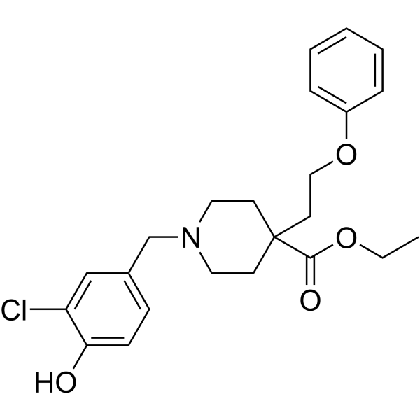
HY-114086
ML359PDI Inhibitor -
HY-19696
TauroursodeoxycholateEndoplasmic Reticulum Stress Inhibitor -
HY-12861
CB-5083p97 AAA ATPase/VCP Inhibitor -

HY-100430R
CCF642 (Standard)PDI Inhibitor -
HY-W107464
G6PDi-1G6PD Inhibitor -
HY-19707
4μ8CIRE1 Inhibitor -
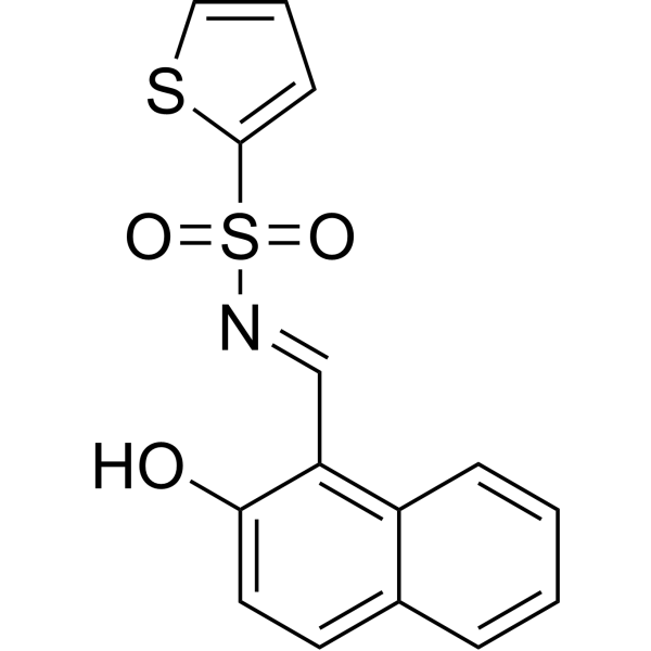
HY-15845
STF-083010IRE1 Inhibitor -
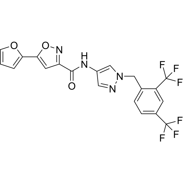
HY-108434
Ceapin-A7ATF6α Signaling Blocker -
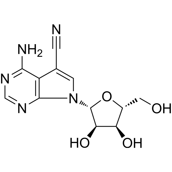
HY-103248
ToyocamycinIRE1/CDK Inhibitor -
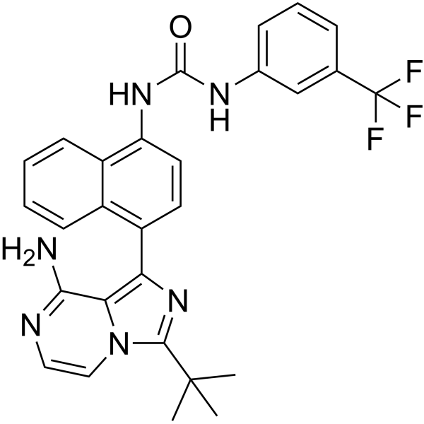
HY-19708
KIRA6IRE1α RNase Inhibitor
Publications Citing Use of MedChemExpress (MCE) CCF642
More

Biologische Aktivität
Beschreibung
IC50 & Target
IC50: 2.9 μM (PDI)[1]
Cellular Effect
|
Cell Line

|
Type | Value | Description | References |
|---|---|---|---|---|
| A549 | IC50 |
12.97 μM
Compound: 2; CCF642
|
Cytotoxicity against human A549 cells assessed as reduction in cell viability incubated for 24 hrs by resazurin assay
Cytotoxicity against human A549 cells assessed as reduction in cell viability incubated for 24 hrs by resazurin assay
|
[PMID: 38381886] |
| EA.hy 926 | IC50 |
5.57 μM
Compound: 2; CCF642
|
Cytotoxicity against human EA.hy 926 cells assessed as reduction in cell viability incubated for 24 hrs by resazurin assay
Cytotoxicity against human EA.hy 926 cells assessed as reduction in cell viability incubated for 24 hrs by resazurin assay
|
[PMID: 38381886] |
| MDA-MB-231 | IC50 |
6.42 μM
Compound: 2; CCF642
|
Cytotoxicity against human MDA-MB-231 cells assessed as reduction in cell viability incubated for 24 hrs by resazurin assay
Cytotoxicity against human MDA-MB-231 cells assessed as reduction in cell viability incubated for 24 hrs by resazurin assay
|
[PMID: 38381886] |
| RPMI-8226 | IC50 |
8.77 μM
Compound: 2; CCF642
|
Cytotoxicity against human RPMI-8226 cells assessed as reduction in cell viability incubated for 24 hrs by resazurin assay
Cytotoxicity against human RPMI-8226 cells assessed as reduction in cell viability incubated for 24 hrs by resazurin assay
|
[PMID: 38381886] |
| U-87MG ATCC | IC50 |
>20 μM
Compound: 2; CCF642
|
Cytotoxicity against human U87 cells assessed as reduction in cell viability incubated for 24 hrs by resazurin assay
Cytotoxicity against human U87 cells assessed as reduction in cell viability incubated for 24 hrs by resazurin assay
|
[PMID: 38381886] |
In Vitro
CCF642 (3 μM; 0.5-6 hours) increases PERK dimerization by phosphorylation and IRE1-α oligomerization within 30 minutes in KMS-12-PE confirming accumulation of misfolded ER proteins[1].
CCF642, a bone marrow-sparing compound, exhibits a submicromolar IC50 in 10 of 10 multiple myeloma cell lines (MM1.S, MM1.R, KMS-12-PE, KMS-12-BM, NCI-H929, U266, RPMI 8226, JJN-3, HRMM.09-luc, 5TGM1-luc)[1].
MedChemExpress (MCE) has not independently confirmed the accuracy of these methods. They are for reference only.
-
Cell Line:MM1.S cells
-
Concentration:3 μM
-
Incubation Time:0.5, 1, 2, 4, 6 hours
-
Result:Increased PERK dimerization by phosphorylation and IRE1-α oligomerization within 30 minutes in KMS-12-PE confirming accumulation of misfolded ER proteins.
In Vivo
MedChemExpress (MCE) has not independently confirmed the accuracy of these methods. They are for reference only.
-
Animal Model:C57BL/KaLwRij mice of 6 to 8 weeks of age with 5TGM1-luc[1]
-
Dosage:10 mg/kg
-
Administration:i.p.; three times a week; for 24 days
-
Result:Significantly prolonged life of 5TGM1-luc–bearing mice and suppressed 5TGM1-luc growth as determined by life imaging.


Chemical Information
-
CAS. Nr. 346640-08-2
-
Appearance Solid
-
Molecular Weight 378.45
-
Formel C15H10N2O4S3
-
Color Yellow to orange
-
SMILES
O=C1N(C2=CC=C(OC)C=C2)C(S/C1=C/C3=CC=C([N+]([O-])=O)S3)=S
-
Versand
Room temperature in continental US; may vary elsewhere.
-
Speicherung
Powder -20°C 3 years 4°C 2 years In solvent -80°C 2 years -20°C 1 year


Publications (1)
-
Journal Impact Factor
-
Most Recent


Lösungsmittel & Löslichkeit
In Vitro:
DMSO : ≥ 30 mg/mL (79.27 mM; Hygroscopic DMSO has a significant impact on the solubility of product, please use newly opened DMSO)
* "≥" means soluble, but saturation unknown.
Please refer to the solubility information to select the appropriate solvent. Once prepared, please aliquot and store the solution to prevent product inactivation from repeated freeze-thaw cycles.
Storage method and period of stock solution: -80°C, 2 years; -20°C, 1 year. When stored at -80°C, please use it within 2 years. When stored at -20°C, please use it within 1 year.
Please refer to the solubility information to select the appropriate solvent. Once prepared, please aliquot and store the solution to prevent product inactivation from repeated freeze-thaw cycles.
Storage method and period of stock solution: -80°C, 2 years; -20°C, 1 year. When stored at -80°C, please use it within 2 years. When stored at -20°C, please use it within 1 year.
Konzentration (Stammlösung) × Volumen (Stammlösung) = Konzentration (Ziellösung) × Volumen (Ziellösung)
In Vivo:
Select the appropriate dissolution method based on your experimental animal and administration route.
- For the following dissolution methods, please ensure to first prepare a clear stock solution using an In Vitro approach and then sequentially add co-solvents:
- To ensure reliable experimental results, the clarified stock solution can be appropriately stored based on storage conditions. As for the working solution for In Vivo experiments, it is recommended to prepare freshly and use it on the same day.
- The percentages shown for the solvents indicate their volumetric ratio in the final prepared solution. If precipitation or phase separation occurs during preparation, heat and/or sonication can be used to aid dissolution.
Add each solvent one by one: 10% DMSO 40% PEG300 5% Tween-80 45% Saline
Solubility: 0.62 mg/mL (1.64 mM); Suspended solution; Need ultrasonic
This protocol yields a suspended solution of 0.62 mg/mL. Suspended solution can be used for oral and intraperitoneal injection.
Taking 1 mL working solution as an example, add 100 μL DMSO stock solution (6.2 mg/mL) to 400 μL PEG300, and mix evenly; then add 50 μL Tween-80 and mix evenly; then add 450 μL Saline to adjust the volume to 1 mL.
Preparation of Saline: Dissolve 0.9 g sodium chloride in ddH₂O and dilute to 100 mL to obtain a clear Saline solution.
In Vivo Dissolution Calculator
Please enter the basic information of animal experiments:
-
-
-
-
Recommended: Prepare an additional quantity of animals to account for potential losses during experiments.
Please enter your animal formula composition:
-
%DMSO +
Recommended: Keep the proportion of DMSO in working solution below 2% if your animal is weak.
-
%+
-
+%Tween-80 + +
-
%Saline +
The co-solvents required include: DMSO, . All of co-solvents are available by MedChemExpress (MCE). , Tween 80. All of co-solvents are available by MedChemExpress (MCE).
Working solution concentration: 0.22 mg/mL
Method for preparing stock solution: mg drug dissolved in μL DMSO. Stock solution concentration: mg/mL.
1. Take μL DMSO stock solution;
2. Add μL .
μL , mix evenly;
3. Then add μL Tween 80, mix evenly;
4. Then add μL
Please ensure that the stock solution in the first step is dissolved to a clear state, and add co-solvents in sequence. You can use ultrasonic heating (ultrasonic cleaner, recommended frequency 20-40 kHz), vortexing, etc. to assist dissolution.


Reinheit & Dokumentation
-
Data Sheet (276 KB)
-
SDS (393 KB)
- English - EN (393 KB)
- Français - FR (393 KB)
- Deutsch - DE (393 KB)
- Norwegian - NO (393 KB)
- Español - ES (393 KB)
- Swedish - SV (393 KB)
- Italian - IT (393 KB)
- Korean - KR (393 KB)
- Portuguese - PT (393 KB)
-
Handling Instructions (2659 KB)
Verweise


Complete Stock Solution Preparation Table
Please refer to the solubility information to select the appropriate solvent. Once prepared, please aliquot and store the solution to prevent product inactivation from repeated freeze-thaw cycles.
Storage method and period of stock solution: -80°C, 2 years; -20°C, 1 year. When stored at -80°C, please use it within 2 years. When stored at -20°C, please use it within 1 year.
| Optional Solvent | Concentration Solvent Mass | 1 mg | 5 mg | 10 mg | 25 mg |
|---|---|---|---|---|---|
| DMSO | 1 mM | 2.6424 mL | 13.2118 mL | 26.4236 mL | 66.0589 mL |
| 5 mM | 0.5285 mL | 2.6424 mL | 5.2847 mL | 13.2118 mL | |
| 10 mM | 0.2642 mL | 1.3212 mL | 2.6424 mL | 6.6059 mL | |
| 15 mM | 0.1762 mL | 0.8808 mL | 1.7616 mL | 4.4039 mL | |
| 20 mM | 0.1321 mL | 0.6606 mL | 1.3212 mL | 3.3029 mL | |
| 25 mM | 0.1057 mL | 0.5285 mL | 1.0569 mL | 2.6424 mL | |
| 30 mM | 0.0881 mL | 0.4404 mL | 0.8808 mL | 2.2020 mL | |
| 40 mM | 0.0661 mL | 0.3303 mL | 0.6606 mL | 1.6515 mL | |
| 50 mM | 0.0528 mL | 0.2642 mL | 0.5285 mL | 1.3212 mL | |
| 60 mM | 0.0440 mL | 0.2202 mL | 0.4404 mL | 1.1010 mL |


CCF642 Related Classifications
HY-100430 Related Classifications
 Powered by Bioz