



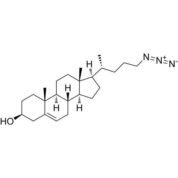
Chol-N3
Chol-N3 is a bioorthogonal-based chol probe. Chol-N3 can combine with super-resolution fluorescence microscopy (SRM), providing direct visualization of nanoscale lipid heterogeneity in the cell surface of resting living cells. Chol-N3 is a click chemistry reagent, it contains an Azide group and can undergo copper-catalyzed azide-alkyne cycloaddition reaction (CuAAc) with molecules containing Alkyne groups. It can also undergo strain-promoted alkyne-azide cycloaddition (SPAAC) reactions with molecules containing DBCO or BCN groups.
Nur für Forschungszwecke. Wir verkaufen nicht an Patienten.
- CAS. Nr.: 2106868-12-4
- Formel: C24H39N3O
- Molecular Weight:385.59
-
Speicherung:
Please store the product under the recommended conditions in the Certificate of Analysis.

To place orders, for customer services and technical support, please contact: MedChemExpress USA
Tel: 609-228-6898 E-mail: [email protected] [email protected]


-
 Biologische Aktivität
-
Chemical Information
- Protokoll
- Reinheit & Dokumentation
- Verweise
-
 Help & FAQs




Biologische Aktivität
Beschreibung
In Vitro
Chol-N3 (25 μM; 16 h; living SH-SY5Y cells) distribution in living cells clearly shows the presence of nanoscopic domains in the PM[1].
Chol-N3 (15 μM; 16 h; living SH-SY5Y cells) has nanoscale spatiotemporal diffusion dynamics in SH-SY5Y cells[1].
MedChemExpress (MCE) has not independently confirmed the accuracy of these methods. They are for reference only.


Chemical Information
-
CAS. Nr. 2106868-12-4
-
Molecular Weight 385.59
-
Formel C24H39N3O
-
SMILES
C[C@@]12[C@](CC[C@]2([H])[C@H](C)CCCN=[N+]=[N-])([H])[C@@]3([H])[C@@](CC1)([H])[C@@]4(C(C[C@H](CC4)O)=CC3)C
-
Versand
Room temperature in continental US; may vary elsewhere.
-
Speicherung
Please store the product under the recommended conditions in the Certificate of Analysis.


Reinheit & Dokumentation
Verweise

Calculators
Konzentration (Stammlösung) × Volumen (Stammlösung) = Konzentration (Ziellösung) × Volumen (Ziellösung)


Chol-N3 Related Classifications
HY-152901 Related Classifications
 Powered by Bioz