



Eriocitrin
Based on 6 publication(s) in Google Scholar
Eriocitrin is a flavonoid isolated from lemons that is a powerful antioxidant. Eriocitrin inhibits the proliferation of liver cancer cells by arresting the cell cycle in the S phase by upregulating p53, cyclin A, cyclin D3 and CDK6. Eriocitrin triggers apoptosis by activating intrinsic signaling pathways involving mitochondria.
For research use only. We do not sell to patients.
- Purity : 99.84%
- CAS No.: 13463-28-0
- Formula: C27H32O15
- Molecular Weight:596.53
-
Storage:
4°C, protect from light
* In solvent : -80°C, 6 months; -20°C, 1 month (protect from light)

To place orders, for customer services and technical support, please contact: MedChemExpress USA
Tel: 609-228-6898 E-mail: [email protected] [email protected]


-
 Biological Activity
-
Chemical Information
-
Solvent & Solubility
- Protocols
- Purity & Documentation
- References
-
 Help & FAQs


-
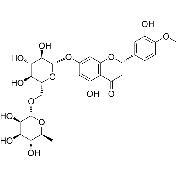
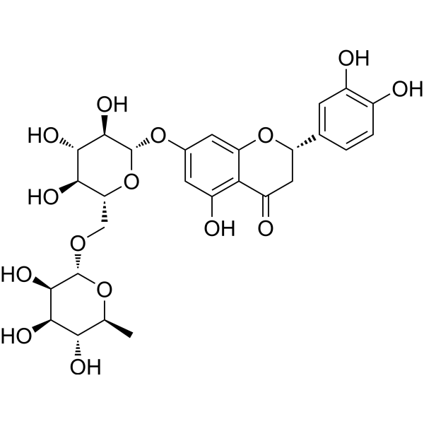
HY-15337
HesperidinAntioxidant -
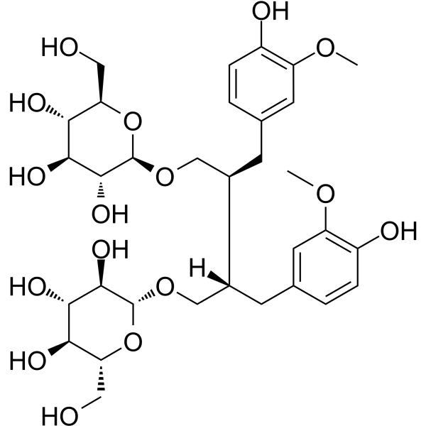
HY-105008
Secoisolariciresinol diglucosideAnti-inflammatory Drug -

HY-13259
MG-132Proteasome Inhibitor -
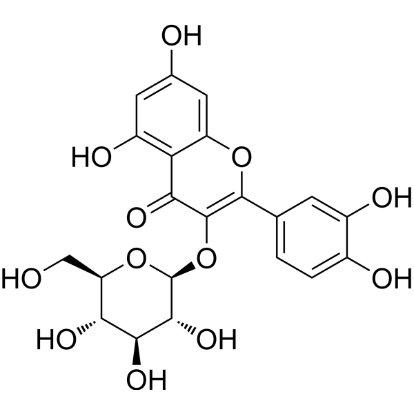
HY-N1445
IsoquercitrinNO Synthase Inhibitor -
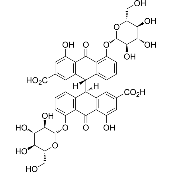
HY-N0366
Sennoside BPDGFR Inhibitor -
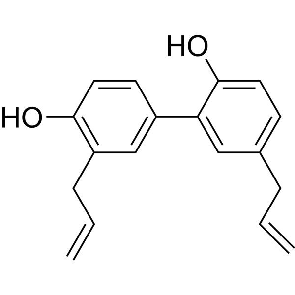
HY-N0003
HonokiolAutophagy Inducer -
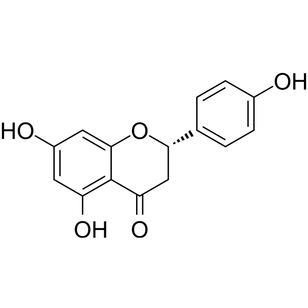
HY-N0100
NaringeninFlavonoid -
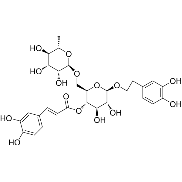
HY-N0028
Forsythiaside AAnti-Inflammatory Agent -
HY-N0178
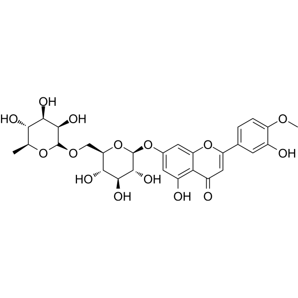
DiosminAryl Hydrocarbon Receptor Agonist -
HY-N0637
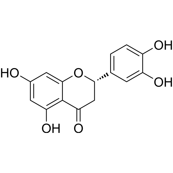
EriodictyolEndogenous Metabolite -
HY-N0021
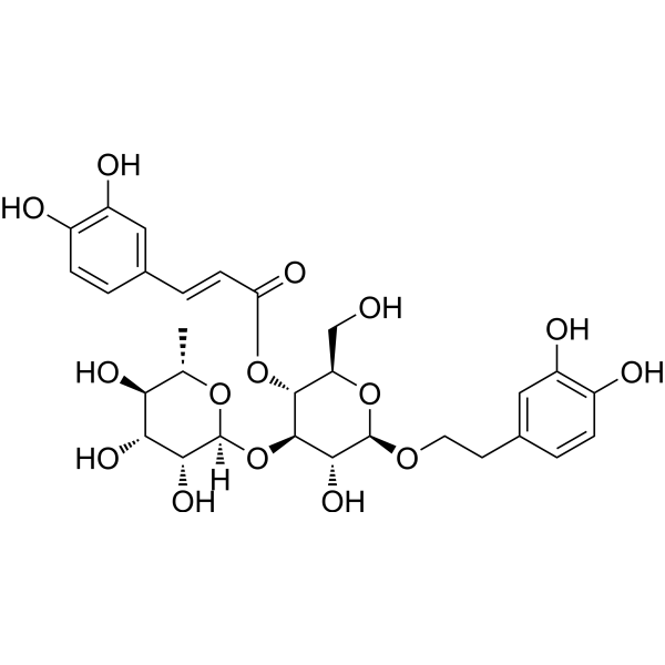
VerbascosideBacterial Inhibitor -
HY-N0148
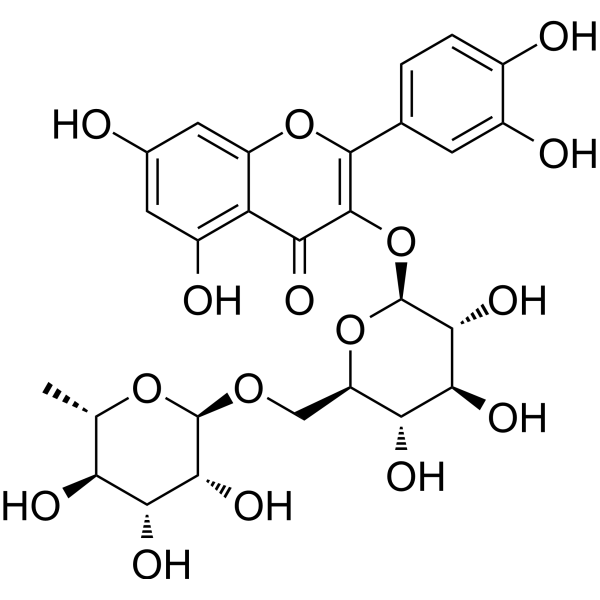
RutinCBR1 Inhibitor
Publications Citing Use of MedChemExpress (MCE) Eriocitrin
More Customer Validation & Images

-
Cell Proliferation/Viability Assay
-
Cell Proliferation/Viability Assay
-
Cell Migration/Invasion Assay
-
Bio/Physico-chemical Assay
-
WB


Biological Activity
Description
In Vitro
Eriocitrin (25-75 μM; 24 h) arrests the cell cycle in the S phase, (5-10 μM; 24 h) and inhibits proliferation and focus formation of liver cancer cell lines HepG2 and Huh7[1].
Eriocitrin (25-75 μM; 12 h) upregulates p53, cyclin A, cyclin D3, and CDK6 levels in HepG2 cells[1].
Eriocitrin (25-75 μM; 48 h) triggers apoptosis by activating mitochondria-related intrinsic signaling pathways[1].
MedChemExpress (MCE) has not independently confirmed the accuracy of these methods. They are for reference only.
In Vivo
MedChemExpress (MCE) has not independently confirmed the accuracy of these methods. They are for reference only.
- EdU Incorporation Assay (Click Chemistry-Based DNA Synthesis Measurement)
- Detection of Apoptosis
- TUNEL staining for apoptotic DNA fragmentation
- Flow cytometric DNA-content cell-cycle staining
- Annexin V plus membrane-impermeant dye apoptosis staining
- BrdU Incorporation Assay
- Apoptosis Solutions
- Protocol for Cell Cycle
- Liver Cancer Modeling
- Cell Viability Determination by MTT Colorimetric Assay
Clinical Trial
| NCT Number | Sponsor | Condition | Start Date |
Phase

|
|---|---|---|---|---|
| NCT01329991 | 2011-05 | PHASE1 |


Chemical Information
-
CAS No. 13463-28-0
-
Appearance Solid
-
Molecular Weight 596.53
-
Formula C27H32O15
-
Color Light yellow to yellow
-
SMILES
O=C(C[C@@H](C1=CC(O)=C(O)C=C1)OC2=CC(O[C@@H]([C@@H]([C@@H](O)[C@@H]3O)O)O[C@@H]3CO[C@H](O[C@@H](C)[C@H](O)[C@H]4O)[C@@H]4O)=C5)C2=C5O
-
Synonyms
Eriodictyol 7-rutinoside; Eriodictyol 7-O-rutinoside
-
Structure Classification
-
Initial Source
-
Shipping
Room temperature in continental US; may vary elsewhere.
-
Storage
4°C, protect from light
* In solvent : -80°C, 6 months; -20°C, 1 month (protect from light)


Publications (6)
-
Journal Impact Factor
-
Most Recent
-
Food Chem
Effects of sun drying combined with baking processes on the flavor quality of Chongqing Tuocha raw tea. [Abstract]2025 Dec 30:497:146992. PMID: 41285060 -
Food Chem
Flavonoid-mediated metabolic underpinning quality variation in red bud-sport pear mutants. [Abstract]2025 Oct 15:489:144992. PMID: 40466530 -
J Agric Food Chem
High-Pressure and Thermal Pasteurization Applied to Smoothies Enhances (Poly)Phenol Bioaccessibility along the Gastrointestinal Tract. [Abstract]2025 Jun 25;73(25):15561-15578. PMID: 40497562 -
Foods
Design of a Plant-Based Smoothie: Exploiting Ingredient Complementarity for a Diversified (Poly)phenolic Profile Quantified by Targeted LC-MS/MS Analysis. [Abstract]2026 Apr 9;15(8):1293. PMID: 42073181 -
BMC Cancer
Identification and validation of CDK1 as a promising therapeutic target for Eriocitrin in colorectal cancer: a combined bioinformatics and experimental approach. [Abstract]2025 Jan 13;25(1):76. PMID: 39806333
Eriocitrin purchased from MedChemExpress. Usage Cited in: BMC Cancer. 2025 Jan 13;25(1):76. [Abstract]
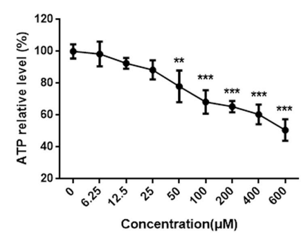
ATP assay analysis of the cell viability of HCT116 after Eriocitrin (6.25–600 μM; 24 h) treatment. Eriocitrin significantly inhibited the tumorigenic properties of CRC cells, as evidenced by a notable reduction in HCT116 cell proliferation.
Eriocitrin purchased from MedChemExpress. Usage Cited in: BMC Cancer. 2025 Jan 13;25(1):76. [Abstract]
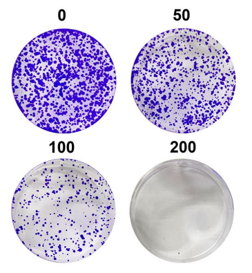
Colony formation assay analysis of the proliferation ability of HCT116 cells after Eriocitrin (50-200 μM; 24 h) treatment.
Eriocitrin purchased from MedChemExpress. Usage Cited in: BMC Cancer. 2025 Jan 13;25(1):76. [Abstract]
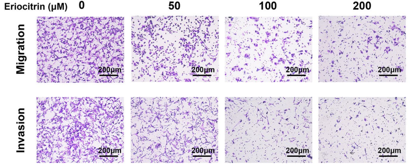
Transwell assays analysis of the migration and invasion abilities of HCT116 cells after Eriocitrin (50-200 μM; 24 h) treatment. The results revealed that the invasion and migration abilities of HCT116 cells were attenuated by Eriocitrin intervention in a significant dose-dependent manner.
Eriocitrin purchased from MedChemExpress. Usage Cited in: BMC Cancer. 2025 Jan 13;25(1):76. [Abstract]
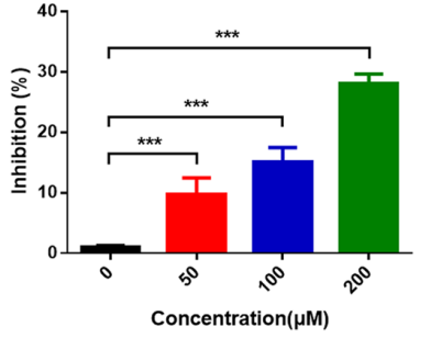
Detection of CDK1 activity in HCT116 cells after Eriocitrin (50-200 μM; 24 h) treatment. The results showed that Eriocitrin inhibited the activity of CDK1 in a dose-dependent manner.
Eriocitrin purchased from MedChemExpress. Usage Cited in: BMC Cancer. 2025 Jan 13;25(1):76. [Abstract]
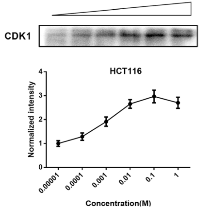
Validation of the specific binding of Eriocitrin (0.00001-1 M; 24 h) to CDK1 by ITDR assay. The results illustrated that as the concentration of Eriocitrin increased, the thermal stability of CDK1 progressively enhanced.
Eriocitrin purchased from MedChemExpress. Usage Cited in: BMC Cancer. 2025 Jan 13;25(1):76. [Abstract]
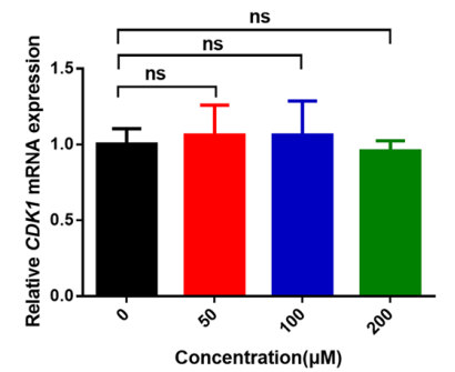
qRT-PCR analysis of the expression of CDK1 in HCT116 cells after Eriocitrin (50-200 μM; 24 h) treatment. The results showed that mRNA expression of CDK1 changed with the increase of Eriocitrin concentration.
-
Microbiol Spectr
Potential quorum-sensing inhibitor of Hafnia alvei H4-theaflavin-3,3´-digallate analyzed by virtual screening and molecular simulation. [Abstract]2023 Sep 21;11(5):e0267123. PMID: 37732782


Solvent & Solubility
In Vitro:
DMSO : 100 mg/mL (167.64 mM; Need ultrasonic; Hygroscopic DMSO has a significant impact on the solubility of product, please use newly opened DMSO)
Methanol : 31.25 mg/mL (52.39 mM; Need ultrasonic)
Please refer to the solubility information to select the appropriate solvent. Once prepared, please aliquot and store the solution to prevent product inactivation from repeated freeze-thaw cycles.
Storage method and period of stock solution: -80°C, 6 months; -20°C, 1 month (protect from light). When stored at -80°C, please use it within 6 months. When stored at -20°C, please use it within 1 month.
Please refer to the solubility information to select the appropriate solvent. Once prepared, please aliquot and store the solution to prevent product inactivation from repeated freeze-thaw cycles.
Storage method and period of stock solution: -80°C, 6 months; -20°C, 1 month (protect from light). When stored at -80°C, please use it within 6 months. When stored at -20°C, please use it within 1 month.
Concentration (start) × Volume (start) = Concentration (final) × Volume (final)
In Vivo:
Select the appropriate dissolution method based on your experimental animal and administration route.
- For the following dissolution methods, please ensure to first prepare a clear stock solution using an In Vitro approach and then sequentially add co-solvents:
- To ensure reliable experimental results, the clarified stock solution can be appropriately stored based on storage conditions. As for the working solution for In Vivo experiments, it is recommended to prepare freshly and use it on the same day.
- The percentages shown for the solvents indicate their volumetric ratio in the final prepared solution. If precipitation or phase separation occurs during preparation, heat and/or sonication can be used to aid dissolution.
Add each solvent one by one: 10% DMSO 40% PEG300 5% Tween-80 45% Saline
Solubility: ≥ 2.08 mg/mL (3.49 mM); Clear solution
This protocol yields a clear solution of ≥ 2.08 mg/mL (saturation unknown).
Taking 1 mL working solution as an example, add 100 μL DMSO stock solution (20.8 mg/mL) to 400 μL PEG300, and mix evenly; then add 50 μL Tween-80 and mix evenly; then add 450 μL Saline to adjust the volume to 1 mL.
Preparation of Saline: Dissolve 0.9 g sodium chloride in ddH₂O and dilute to 100 mL to obtain a clear Saline solution.
Add each solvent one by one: 10% DMSO 90% (20% SBE-β-CD in Saline)
Solubility: ≥ 2.08 mg/mL (3.49 mM); Clear solution
This protocol yields a clear solution of ≥ 2.08 mg/mL (saturation unknown).
Taking 1 mL working solution as an example, add 100 μL DMSO stock solution (20.8 mg/mL) to 900 μL 20% SBE-β-CD in Saline, and mix evenly.
Preparation of 20% SBE-β-CD in Saline (4°C, storage for one week): 2 g SBE-β-CD powder is dissolved in 10 mL Saline, completely dissolve until clear.
In Vivo Dissolution Calculator
Please enter the basic information of animal experiments:
-
-
-
-
Recommended: Prepare an additional quantity of animals to account for potential losses during experiments.
Please enter your animal formula composition:
-
%DMSO +
Recommended: Keep the proportion of DMSO in working solution below 2% if your animal is weak.
-
%+
-
+%Tween-80 + +
-
%Saline +
The co-solvents required include: DMSO, . All of co-solvents are available by MedChemExpress (MCE). , Tween 80. All of co-solvents are available by MedChemExpress (MCE).
Working solution concentration: 0.22 mg/mL
Method for preparing stock solution: mg drug dissolved in μL DMSO. Stock solution concentration: mg/mL. * In solvent : -80°C, 6 months; -20°C, 1 month (protect from light)
1. Take μL DMSO stock solution;
2. Add μL .
μL , mix evenly;
3. Then add μL Tween 80, mix evenly;
4. Then add μL
Please ensure that the stock solution in the first step is dissolved to a clear state, and add co-solvents in sequence. You can use ultrasonic heating (ultrasonic cleaner, recommended frequency 20-40 kHz), vortexing, etc. to assist dissolution.


Purity & Documentation
-
Data Sheet (278 KB)
-
SDS (393 KB)
- English - EN (393 KB)
- Français - FR (393 KB)
- Deutsch - DE (393 KB)
- Norwegian - NO (393 KB)
- Español - ES (393 KB)
- Swedish - SV (393 KB)
- Italian - IT (393 KB)
- Korean - KR (393 KB)
- Portuguese - PT (393 KB)
-
Handling Instructions (2659 KB)
References
[1]. Wang Z, et al. Eriocitrin from lemon suppresses the proliferation of human hepatocellular carcinoma cells through inducing apoptosis and arresting cell cycle. Cancer Chemother Pharmacol. 2016 Dec;78(6):1143-1150. [Content Brief]
[2]. Xu J, et al. Eriocitrin attenuates ischemia reperfusion-induced oxidative stress and inflammation in rats with acute kidney injury by regulating the dual-specificity phosphatase 14 (DUSP14)-mediated Nrf2 and nuclear factor-κB (NF-κB) pathways. Ann Transl Med. 2021 Feb;9(4):350. [Content Brief]


Complete Stock Solution Preparation Table
Please refer to the solubility information to select the appropriate solvent. Once prepared, please aliquot and store the solution to prevent product inactivation from repeated freeze-thaw cycles.
Storage method and period of stock solution: -80°C, 6 months; -20°C, 1 month (protect from light). When stored at -80°C, please use it within 6 months. When stored at -20°C, please use it within 1 month.
| Optional Solvent | Concentration Solvent Mass | 1 mg | 5 mg | 10 mg | 25 mg |
|---|---|---|---|---|---|
| Methanol / DMSO | 1 mM | 1.6764 mL | 8.3818 mL | 16.7636 mL | 41.9090 mL |
| 5 mM | 0.3353 mL | 1.6764 mL | 3.3527 mL | 8.3818 mL | |
| 10 mM | 0.1676 mL | 0.8382 mL | 1.6764 mL | 4.1909 mL | |
| 15 mM | 0.1118 mL | 0.5588 mL | 1.1176 mL | 2.7939 mL | |
| 20 mM | 0.0838 mL | 0.4191 mL | 0.8382 mL | 2.0955 mL | |
| 25 mM | 0.0671 mL | 0.3353 mL | 0.6705 mL | 1.6764 mL | |
| 30 mM | 0.0559 mL | 0.2794 mL | 0.5588 mL | 1.3970 mL | |
| 40 mM | 0.0419 mL | 0.2095 mL | 0.4191 mL | 1.0477 mL | |
| 50 mM | 0.0335 mL | 0.1676 mL | 0.3353 mL | 0.8382 mL | |
| DMSO | 60 mM | 0.0279 mL | 0.1397 mL | 0.2794 mL | 0.6985 mL |
| 80 mM | 0.0210 mL | 0.1048 mL | 0.2095 mL | 0.5239 mL | |
| 100 mM | 0.0168 mL | 0.0838 mL | 0.1676 mL | 0.4191 mL |


Eriocitrin Related Classifications
HY-N0636 Related Classifications
 Powered by Bioz